
Article Figures & Data
Figures
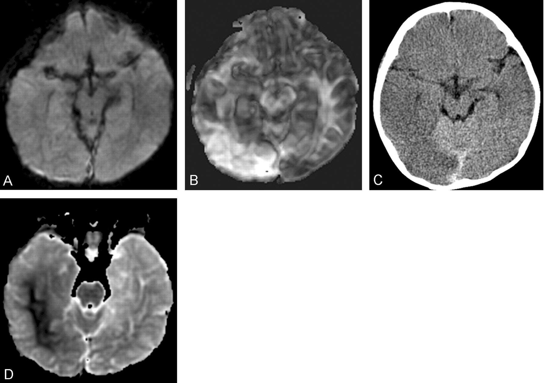
- Fig 1.
Images obtained in a 14-month-old male infant with traumatic brain swelling.
A, Conventional diffusion-weighted image (three encoding directions) obtained 18 hours after admission shows no evidence of parenchymal injury. The accompanying ADC map as well as conventional T2-weighted fast spin-echo and T2-weighted fluid-attenuated inversion recovery images (not shown) were equally unremarkable.
B, Fractional anisotropy map from full-tensor acquisition obtained 18 hours after admission (concurrent with A) shows readily apparent parenchymal abnormality characterized by increased FA in right occipital and posterior temporal lobes. An accompanying full-tensor ADC map (not shown) revealed minimally increased diffusivity in the affected region.
C, Follow-up CT scan obtained 93 hours after admission shows region of parenchymal edema characterized by ill-defined hypoattenuation and poor gray matter–white matter discrimination corresponding to earlier diffusion tensor abnormality (compare with B).
D, Conventional ADC map obtained 135 hours after admission shows a large area of reduced diffusivity in the affected region of the right hemisphere, primarily in white matter but also involving some cortex. Conventional T2-weighted fluid-attenuated inversion recovery images (not shown) revealed diffuse cortical swelling in affected areas.

In this issue


Jump to section
Related Articles
Cited By...
- Volumetric and Diffusion Tensor Imaging biomarkers indicating long lasting post-concussion abnormalities in a youth pig model of mild Traumatic Brain Injury
- Correlation of Quantitative Diffusion Tensor Tractography with Clinical Grades of Subacute Sclerosing Panencephalitis
- Diffusion tensor imaging of acute mild traumatic brain injury in adolescents