Advertisement

Main menu

  • Home
  • Content
    • Current Issue
    • Accepted Manuscripts
    • Article Preview
    • Past Issue Archive
    • Video Articles
    • AJNR Case Collection
    • Case of the Week Archive
    • Case of the Month Archive
    • Classic Case Archive
  • Special Collections
    • Advancing NeuroMRI with High-Relaxivity Contrast Agents
    • AJNR Awards
    • Low-Field MRI
    • Alzheimer Disease
    • ASNR Foundation Special Collection
    • Photon-Counting CT
    • View All
  • Multimedia
    • AJNR Podcasts
    • AJNR SCANtastic
    • Trainee Corner
    • MRI Safety Corner
    • Imaging Protocols
  • For Authors
    • Submit a Manuscript
    • Submit a Video Article
    • Submit an eLetter to the Editor/Response
    • Manuscript Submission Guidelines
    • Statistical Tips
    • Fast Publishing of Accepted Manuscripts
    • Graphical Abstract Preparation
    • Imaging Protocol Submission
    • Author Policies
  • About Us
    • About AJNR
    • Editorial Board
    • Editorial Board Alumni
  • More
    • Become a Reviewer/Academy of Reviewers
    • Subscribers
    • Permissions
    • Alerts
    • Feedback
    • Advertisers
    • ASNR Home

User menu

  • Alerts
  • Log in

Search

  • Advanced search
American Journal of Neuroradiology

American Journal of Neuroradiology

ASHNR American Society of Functional Neuroradiology ASHNR American Society of Pediatric Neuroradiology ASSR
  • Alerts
  • Log in

Advanced Search

  • Home
  • Content
    • Current Issue
    • Accepted Manuscripts
    • Article Preview
    • Past Issue Archive
    • Video Articles
    • AJNR Case Collection
    • Case of the Week Archive
    • Case of the Month Archive
    • Classic Case Archive
  • Special Collections
    • Advancing NeuroMRI with High-Relaxivity Contrast Agents
    • AJNR Awards
    • Low-Field MRI
    • Alzheimer Disease
    • ASNR Foundation Special Collection
    • Photon-Counting CT
    • View All
  • Multimedia
    • AJNR Podcasts
    • AJNR SCANtastic
    • Trainee Corner
    • MRI Safety Corner
    • Imaging Protocols
  • For Authors
    • Submit a Manuscript
    • Submit a Video Article
    • Submit an eLetter to the Editor/Response
    • Manuscript Submission Guidelines
    • Statistical Tips
    • Fast Publishing of Accepted Manuscripts
    • Graphical Abstract Preparation
    • Imaging Protocol Submission
    • Author Policies
  • About Us
    • About AJNR
    • Editorial Board
    • Editorial Board Alumni
  • More
    • Become a Reviewer/Academy of Reviewers
    • Subscribers
    • Permissions
    • Alerts
    • Feedback
    • Advertisers
    • ASNR Home
  • Follow AJNR on Twitter
  • Visit AJNR on Facebook
  • Follow AJNR on Instagram
  • Join AJNR on LinkedIn
  • RSS Feeds

AJNR Awards, New Junior Editors, and more. Read the latest AJNR updates


Improved Turnaround Times | Median time to first decision: 12 days

Research ArticleNeurointervention

Visualization of Intraaneurysmal Flow Patterns with Transluminal Flow Images of 3D MR Angiograms in Conjunction with Aneurysmal Configurations

Toru Satoh, Keisuke Onoda and Shoji Tsuchimoto
American Journal of Neuroradiology August 2003, 24 (7) 1436-1445;
Toru Satoh
aDepartment of Neurological Surgery, Ryofukai Satoh Neurosurgical Hospital, Hiroshima, Japan
  • Find this author on Google Scholar
  • Find this author on PubMed
  • Search for this author on this site
Keisuke Onoda
bDepartment of Neurological Surgery, Onomichi Municipal Hospital, Hiroshima, Japan
  • Find this author on Google Scholar
  • Find this author on PubMed
  • Search for this author on this site
Shoji Tsuchimoto
bDepartment of Neurological Surgery, Onomichi Municipal Hospital, Hiroshima, Japan
  • Find this author on Google Scholar
  • Find this author on PubMed
  • Search for this author on this site
  • Article
  • Figures & Data
  • Info & Metrics
  • Responses
  • References
  • PDF
Loading

Article Figures & Data

Figures

  • Fig 1.
    • Download figure
    • Open in new tab
    • Download powerpoint
    Fig 1.

    Schematic illustration of selective opacity curves used in the opacity chart of MR signal intensities: increasing curve for the conventional parallel volume-rendered imaging (top), spiked peak curve for transluminal imaging (middle), and spiked peak curve with a stepwise square curve for transluminal flow imaging (bottom). Serial observation of transluminal flow images represents the intraaneurysmal flow patterns by superimposing stepwise extracted intraluminal volume data, with signal intensities ranging 350–500, 320–500, 290–500, 260–500, 230–500, and 200–500, respectively, onto the corresponding transluminal images with aneurysmal configurations.
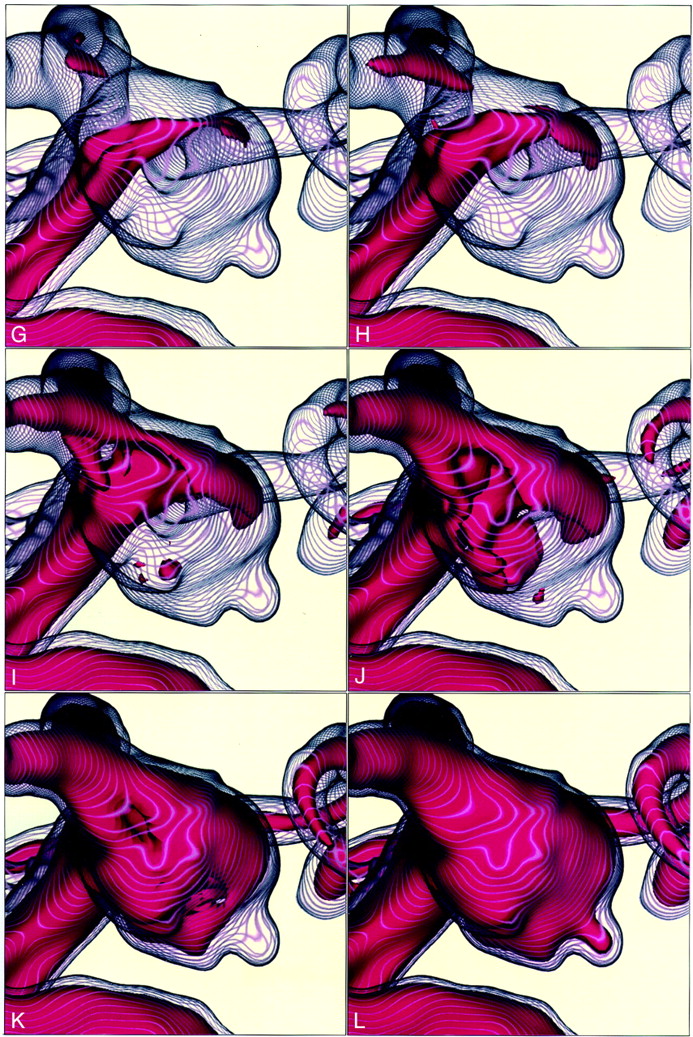
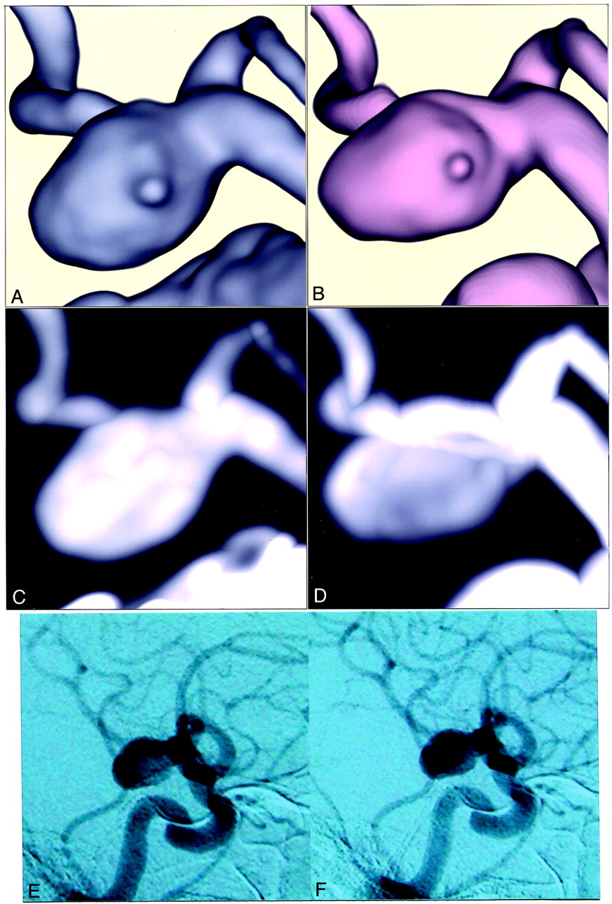
  • Fig 2.
    • Download figure
    • Open in new tab
    • Download powerpoint
    Fig 2.
    • Download figure
    • Open in new tab
    • Download powerpoint
    Fig 2.

    Case 1. 75-year-old woman with an unruptured left internal carotid-posterior communicating artery aneurysm.

    A and B, Three-dimensional volume-rendered CT and MR angiograms, respectively, superoinferior projection.

    C and D, magnified images of CT and MR angiograms, respectively, left lateral projection.

    E and F, Maximum intensity projection images of CT and MR angiograms, respectively. Figure 2 continues.

    Figure 2 continued. G–L, Transluminal flow images of 3D MR angiogram with stepwise intraluminal contents, with signal intensities ranging 350–500, 320–500, 290–500, 260–500, and 200–500, respectively, demonstrate blood flow as the three-dimensional distribution of signal intensities.

  • Fig 3.
    • Download figure
    • Open in new tab
    • Download powerpoint
    Fig 3.
    • Download figure
    • Open in new tab
    • Download powerpoint
    Fig 3.

    Case 2. 66-year-old man with an unruptured left internal carotid-posterior communicating artery aneurysm.

    A and B, Magnified volume-rendered CT and MR angiograms, respectively, right lateral projection.

    C and D, Maximum intensity projection images of CT and MR angiograms, respectively. E and F, Digital subtraction angiograms, lateral projection, obtained in the early and late arterial phases, respectively. Figure 3 continues.

    Figure 3 continued. G–L, Transluminal flow images of 3D MR angiogram with stepwise intraluminal contents, with signal intensities ranging 350–500, 320–500, 290–500, 260–500, 230–350, and 200–500, respectively.

  • Fig 4.
    • Download figure
    • Open in new tab
    • Download powerpoint
    Fig 4.

    Case 3. 63-year-old woman with an unruptured left internal carotid-ophthalmic artery aneurysm.

    A, Magnified volume-rendered MR angiogram and B, coordinated maximum intensity projection image, anteroposterior projection.

    C–F, Transluminal flow images of 3D MR angiogram with stepwise intraluminal contents, with signal intensities ranging 350–500, 320–500, 290–350, and 260–500, respectively.

PreviousNext
Back to top

In this issue

American Journal of Neuroradiology: 24 (7)
American Journal of Neuroradiology
Vol. 24, Issue 7
1 Aug 2003
  • Table of Contents
  • Index by author
Advertisement
Print
Download PDF
Email Article

Thank you for your interest in spreading the word on American Journal of Neuroradiology.

NOTE: We only request your email address so that the person you are recommending the page to knows that you wanted them to see it, and that it is not junk mail. We do not capture any email address.

Enter multiple addresses on separate lines or separate them with commas.
Visualization of Intraaneurysmal Flow Patterns with Transluminal Flow Images of 3D MR Angiograms in Conjunction with Aneurysmal Configurations
(Your Name) has sent you a message from American Journal of Neuroradiology
(Your Name) thought you would like to see the American Journal of Neuroradiology web site.
CAPTCHA
This question is for testing whether or not you are a human visitor and to prevent automated spam submissions.
Cite this article
Toru Satoh, Keisuke Onoda, Shoji Tsuchimoto
Visualization of Intraaneurysmal Flow Patterns with Transluminal Flow Images of 3D MR Angiograms in Conjunction with Aneurysmal Configurations
American Journal of Neuroradiology Aug 2003, 24 (7) 1436-1445;

Citation Manager Formats

  • BibTeX
  • Bookends
  • EasyBib
  • EndNote (tagged)
  • EndNote 8 (xml)
  • Medlars
  • Mendeley
  • Papers
  • RefWorks Tagged
  • Ref Manager
  • RIS
  • Zotero
0 Responses
Respond to this article
Share
Bookmark this article
Visualization of Intraaneurysmal Flow Patterns with Transluminal Flow Images of 3D MR Angiograms in Conjunction with Aneurysmal Configurations
Toru Satoh, Keisuke Onoda, Shoji Tsuchimoto
American Journal of Neuroradiology Aug 2003, 24 (7) 1436-1445;
del.icio.us logo Twitter logo Facebook logo Mendeley logo
  • Tweet Widget
  • Facebook Like
  • Google Plus One

Jump to section

  • Article
    • Abstract
    • Methods
    • Results
    • Discussion
    • Conclusion
    • Footnotes
    • References
  • Figures & Data
  • Info & Metrics
  • Responses
  • References
  • PDF

Related Articles

  • No related articles found.
  • PubMed
  • Google Scholar

Cited By...

  • Usefulness of high-resolution three-dimensional proton density-weighted turbo spin-echo MRI in distinguishing a junctional dilatation from an intracranial aneurysm of the posterior communicating artery: a pilot study
  • Visualization of Aneurysmal Neck and Dome after Coiling with 3D Multifusion Imaging of Silent MRA and FSE-MR Cisternography
  • Aneurysm outflow angle at MRA as discriminant for accurate diagnosis and differentiation between small sidewall cerebral aneurysms and infundibula
  • Identification of the Inflow Zone of Unruptured Cerebral Aneurysms: Comparison of 4D Flow MRI and 3D TOF MRA Data
  • Crossref
  • Google Scholar

This article has not yet been cited by articles in journals that are participating in Crossref Cited-by Linking.

More in this TOC Section

  • Prophylactic Endovascular Treatments for CBS
  • Smoking & Aneurysm Outcomes: A Meta-Analysis
  • “Buddy-wire anchoring” technique for TVE
Show more NEUROINTERVENTION

Similar Articles

Advertisement

Indexed Content

  • Current Issue
  • Accepted Manuscripts
  • Article Preview
  • Past Issues
  • Editorials
  • Editor's Choice
  • Fellows' Journal Club
  • Letters to the Editor
  • Video Articles

Cases

  • Case Collection
  • Archive - Case of the Week
  • Archive - Case of the Month
  • Archive - Classic Case

More from AJNR

  • Trainee Corner
  • Imaging Protocols
  • MRI Safety Corner
  • Book Reviews

Multimedia

  • AJNR Podcasts
  • AJNR Scantastics

Resources

  • Turnaround Time
  • Submit a Manuscript
  • Submit a Video Article
  • Submit an eLetter to the Editor/Response
  • Manuscript Submission Guidelines
  • Statistical Tips
  • Fast Publishing of Accepted Manuscripts
  • Graphical Abstract Preparation
  • Imaging Protocol Submission
  • Evidence-Based Medicine Level Guide
  • Publishing Checklists
  • Author Policies
  • Become a Reviewer/Academy of Reviewers
  • News and Updates

About Us

  • About AJNR
  • Editorial Board
  • Editorial Board Alumni
  • Alerts
  • Permissions
  • Not an AJNR Subscriber? Join Now
  • Advertise with Us
  • Librarian Resources
  • Feedback
  • Terms and Conditions
  • AJNR Editorial Board Alumni

American Society of Neuroradiology

  • Not an ASNR Member? Join Now

© 2025 by the American Society of Neuroradiology All rights, including for text and data mining, AI training, and similar technologies, are reserved.
Print ISSN: 0195-6108 Online ISSN: 1936-959X

Powered by HighWire