
Article Figures & Data
Figures
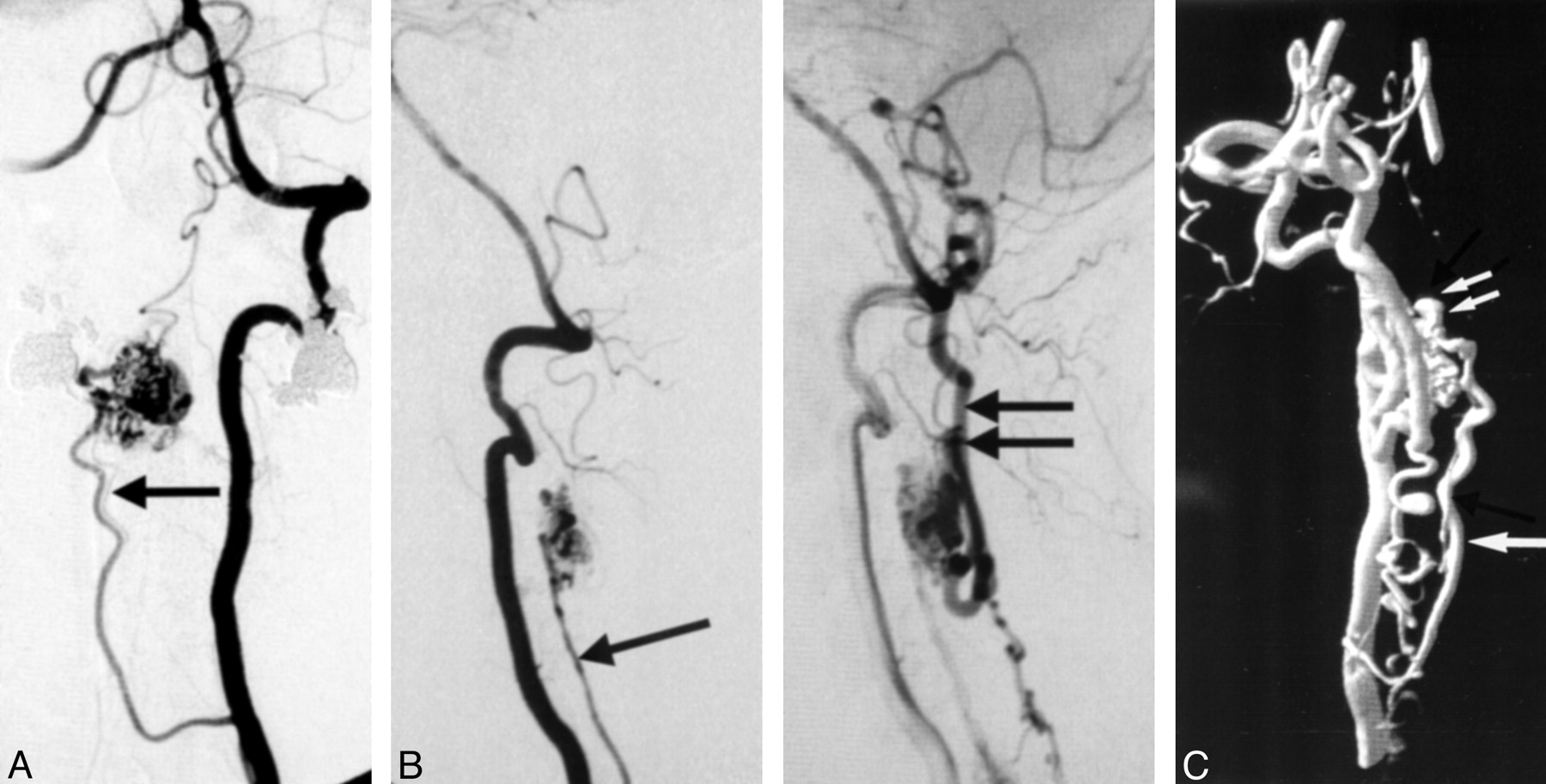
- Fig 1.
Patient 8, 54-year-old male patient with spinal dural AVF.
A, Unsubtracted anteroposterior (AP) (left) and subtracted lateral (right) spinal angiograms of left T12 intercostal artery show the spinal dural fistula. (arrow [AP projection], angiographic catheter; double arrow [AP projection], site of fistula at the dural sleeve; arrowhead [AP and lateral projection], anterior draining spinal vein).
B, 3D reconstruction of T12 intercostal rotational injection with partial opacification of the spinal column shows the site of the fistula (double arrow) in relation to the venous drainage (arrowhead).
C, Computer-generated rotation of the reconstructed image with a thin region of interest allows for a better demonstration of the fistula entering the intervertebral foramen.
D, Lateral projection of the 3D-reconstructed rotational image after successful obliteration of the fistula.
E, Similar view as in C, with the glue cast seen entering the spinal canal via the intervertebral foramen (double arrow).
- Fig 2.
Patient 4, 26-year-old female patient with spinal cord AVM.
A, AP (left) and lateral (right) subtracted spinal angiograms from the left T10 intercostal artery, demonstrating the supply to this conus AVM from the anterior spinal artery (arrow [AP projection]). Lazorthe’s basket is identified at the tip of the conus (double arrows [AP and lateral projections]).
B, 3D-RSA in the lateral projection shows several feeders to this primarily superficial pial malformation (arrow).
- Fig 3.
Patient 1, 20-year-old male patient with spinal cord AVM.
A, Lateral spinal angiogram of the left vertebral artery for this cervical AVM with contribution from multiple cervical levels.
B, 3D-RSA in the same projection as the prior study with enhanced detail in the course of the various feeding vessels. Note the depth of field created by the shadowing techniques provided in the software.
- Fig 4.
Patient 13, 51-year-old female patient with spinal cord AVM.
A, AP vertebral injection demonstrating supply to this cervical AVM from anterior and posterior spinal arteries. Note the duplication of the anterior spinal artery revealed by this view (arrow).
B, Lateral projection in the early (left) and late (right) arterial phases demonstrating the anterior spinal artery (arrow [early phase]) and the posterior draining vein (double arrows [late phase]).
C, 3D-RSA of the vertebral artery injection demonstrating resolution of fine details as depicted by the presence of the duplicated anterior spinal artery (arrow). Note the presence of the nidal aneurysm (double arrow).


Tables
Patient characteristics
Patient (no.) Age (y)/Sex Diagnosis Location Feeders Catheter Location 3D-RSA Benefits* 1 20/M Spinal cord AVM Cervical C3–C5 (ASA); right DA (radiculomedullary); left C6 (PSA); right VA (LSA) Left VA 1, 2 2 10/M Spinal cord AVM Cervical Bilateral VA (ASA and PSA) Right VA 2 3 14/M Spinal cord AVM Cervical Right DA (ASA) Right DA 1, 2 4 26/F Spinal cord AVM Conus Left T10 (ASA); right L2 (PSA); left T11 (PSA) Left T10 IA 2, 3 5 26/F Spinal cord AVM Cervical Left VA (ASA); right DA (ASA and PSA) Left VA 1, 2 6 18/M Spinal cord AVM Cervical C1–C2 (ASA); right C2 (LSA) Left VA 1, 2, 3 7 34/M Perimedullary spinal AVM Cervical Left C6 (ASA) Left VA 1, 2, 3 8 54/M Spinal dural AVF Thoracic Left T12 Left T12 IA 3, 4 9 45/F Spinal cord AVM Cervical Right C4 (ASA); left C5 (ASA) Right VA; left T4 IA 2 10 69/F Spinal dural AVF Thoracic Right T7 Right T7 IA 2, 3, 4 11 49/M Spinal cord AVM Cervical Right SA (ASA) Right SA 1, 2 12 63/M Nerve root AVM Cervical Left C5; right C5 (PSA) Left C5 RA 2, 3, 4 13 51/F Spinal cord AVM Cervical Right C3 (ASA); left PICA (LSA) Right VA 1, 2 14 46/M Spinal dural AVF Lumbar Left L1 Left L1 artery 2, 3, 4 Note.—AVM signifies arteriovenous malformation; ASA, anterior spinal artery; PSA, posterior spinal artery; VA, vertebral artery; LSA, lateral spinal artery; DA, dorsovervical artery; IA, intercostals artery; AVF, arteriovenous fistula; SA, subclavian artery; RA, radicular artery; and PICA, posterior inferior cerebellar artery.
↵* 1, visualization of aneurysm; 2, definition of angioarchitecture; 3, visualization of relationship to surrounding structures; and 4, need for CT to confirm location of embolic agent obviated.
In this issue


Jump to section
Related Articles
Cited By...
- Diagnostic accuracy of three-dimensional-rotational angiography and heavily T2-weighted volumetric magnetic resonance fusion imaging for the diagnosis of spinal arteriovenous shunts
- Management of Brain Arteriovenous Malformations: A Scientific Statement for Healthcare Professionals From the American Heart Association/American Stroke Association
- Flat Panel Catheter Angiotomography of the Spinal Venous System: An Enhanced Venous Phase for Spinal Digital Subtraction Angiography
- Efficacy of DynaCT Digital Angiography in the Detection of the Fistulous Point of Dural Arteriovenous Fistulas
- Three-dimensional CT scanning: a new diagnostic modality in congenital heart disease