
Article Figures & Data
Figures
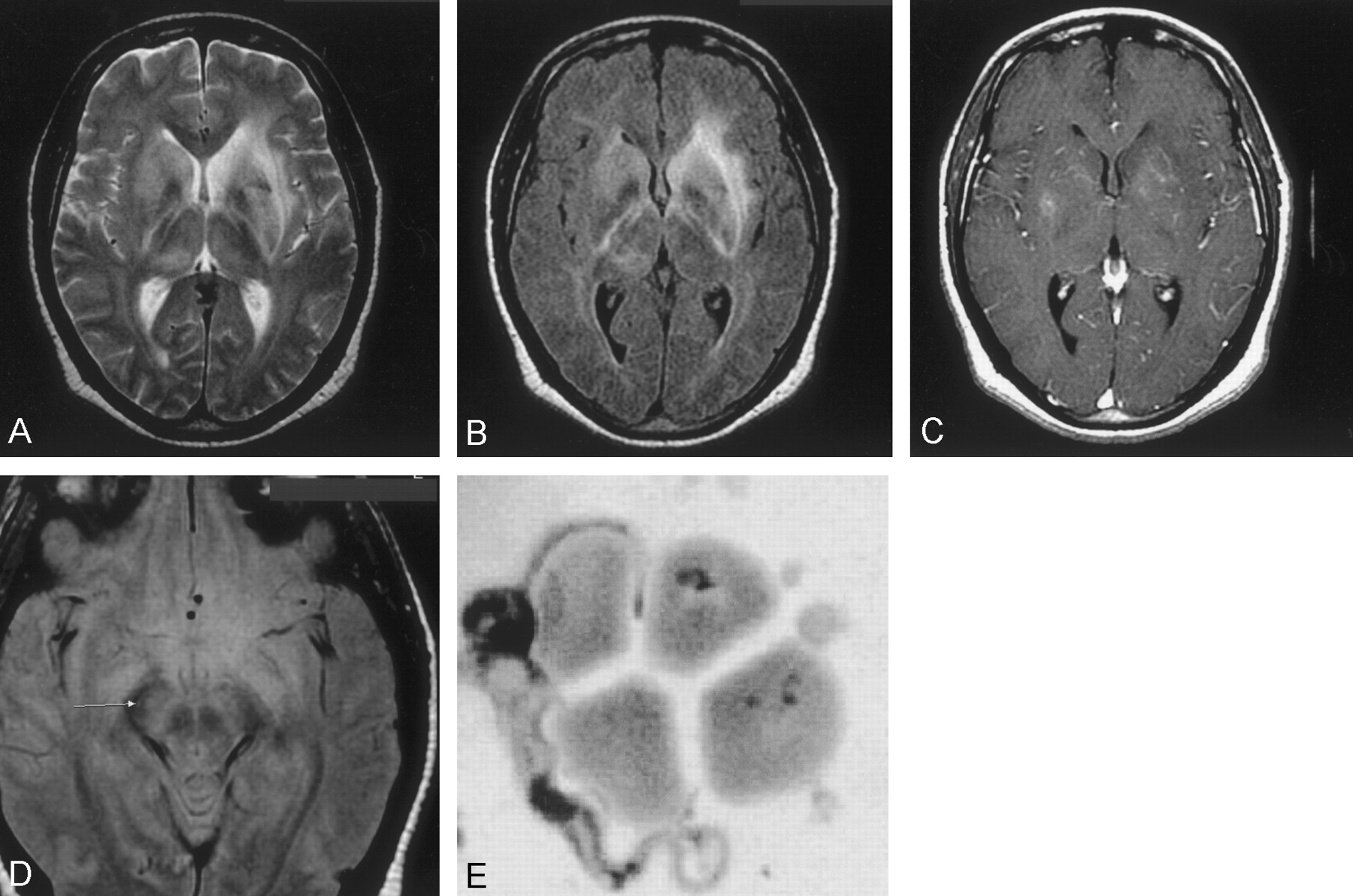
- Fig 1.
Images obtained at time of admission to the hospital.
A, Axial T2-weighted image (TR/TE/NEX, 3000/98/1; section thickness, 5 mm) shows diffuse hyperintensity in both basal ganglia and along the internal, external, and extreme capsules.
B, Axial FLAIR sequence (TR/TE/NEX, 10,002/162.5/1; section thickness, 5 mm) shows bilateral increased signal intensity involving the internal and external capsules bilaterally and optic radiations.
C, Axial T1-weighted gadolinium-enhanced image (TR/TE/NEX, 417/20/2; section thickness, 5 mm) shows minimal enhancement in the basal ganglia bilaterally.
Sahlas DJ, MacLean JD, Janevski J, Detsky AS. Out of Africa. N Engl J Med 2002; 347:749–753. Copyright 2002 Massachusetts Medical Society. All rights reserved.
D, Axial proton density–weighted (TR/TE/NEX, 3000/14/1; section thickness, 5 mm) image shows increased signal intensity in the midbrain. E, Trypanosome adjacent to red blood cells is seen after analysis of the CSF. (Sahlas DJ, MacLean JD, Janevski J, Detsky AS. Out of Africa. N Engl J Med 2002; 347:749–753. Copyright 2002 Massachusetts Medical Society. All rights reserved.) (GIEMSA stain; magnification, ×3000)
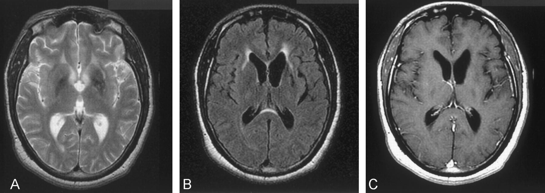
- Fig 2.
Follow-up images obtained 1 year after initial presentation.
A, Axial T2-weighted image (TR/TE/NEX, 3000/105/1; section thickness, 6 mm) shows notable decrease in signal intensity in the basal ganglia, whereas moderate hyperintensity persists in the left external capsule.
B, Axial FLAIR image (TR/TE/NEX, 10,002/175/1; section thickness, 5 mm) shows minimal hyperintensity extending to both external capsules and demonstrating significant increase in size of the lateral ventricles.
C, Axial T1-weighted gadolinium-enhanced (TR/TE/NEX, 500/20/2; section thickness, 5 mm) image shows no abnormal enhancement; however, notable ventricular enlargement is evident compared with that of initial MR images.


