
Article Figures & Data
Figures
- Fig 1.
Sketch of the Hyperform balloon microcatheter illustrates sealing of the aneurysm neck and both bifurcated branches.
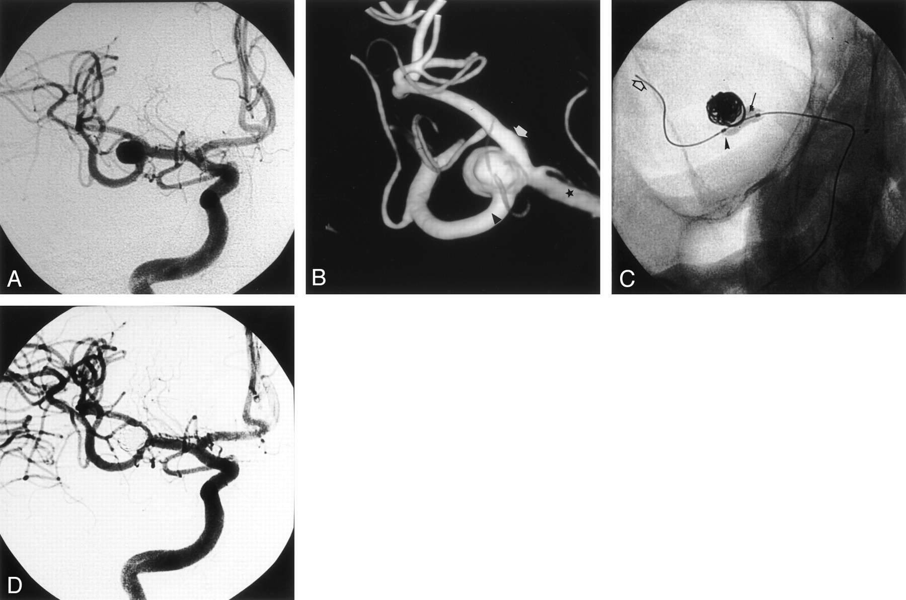
- Fig 2.
Case 1.
A, Right internal carotid artery (ICA) angiogram, best projection, shows the MCA bifurcation aneurysm.
B, Three-dimensional image of the right MCA bifurcation aneurysm shows the upper trunk (white arrow), the lower trunk (arrowhead), and the M1 segment (star) of the MCA. The lower trunk is clearly emerging from the neck of the aneurysm.
C, Radiograph of the inflated Hyperform balloon into the MCA bifurcation. The balloon is bulging into the origin of the upper trunk (arrow), and its distal tip is positioned in the origin of the lower trunk (arrowhead). The Silver Speed (Silver Speed 10; Micro Therapeutics) guidewire is secured distally into the posterior temporal artery (open arrow).
D, Right ICA control angiogram acquired at the end of the procedure shows total aneurysm occlusion and patency of upper and lower MCA trunks.
- Fig 3.
Case 2.
A, Left ICA angiogram shows a bilobulated MCA bifurcation aneurysm (arrow).
B, Three-dimensional angiogram shows the wide-neck aneurysm. The upper trunk origin appears dysplastic and enlarged (star). The lower trunk is indicated by the arrow.
C, Radiograph of the inflated Hyperform balloon. The balloon bulges into the origin of the upper trunk (thin arrow), and its distal tip is positioned in the origin of the lower trunk (arrowhead). The Silver Speed guidewire is secured distally into the posterior temporal artery (thick arrow).
D, Left ICA control angiogram acquired at the end of the procedure shows total aneurysm occlusion, with patency of both MCA upper and lower trunks.
- Fig 4.
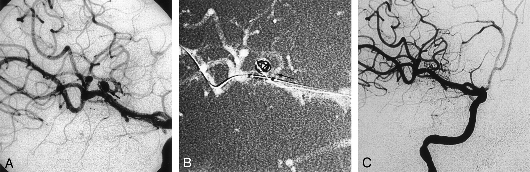
Case 3.
A, Right ICA angiogram, best projection, shows the MCA aneurysm with the upper trunk (arrow) emerging from the sac.
B, Right ICA angiogram, best projection, shows the Hyperform balloon inflated into the right MCA. The proximal part of the balloon is bulging into the origin of the upper trunk (arrow).
C, Right ICA angiogram acquired at the end of the procedure shows that aneurysm occlusion is partial to preserve the patency of the upper trunk.
- Fig 5.
Case 4.
A, Left vertebral angiogram, best frontal projection, shows the basilar tip aneurysm with unfavorable neck-to-fundus ratio.
B, Radiograph of the inflated Hyperform balloon. The balloon is inflated across the neck. The distal part (thick arrow) of the Hyperform balloon is positioned into the left P1 segment, and its proximal part (thin arrow) is positioned into the basilar artery. The balloon conforms easily to both diameters of the basilar and the P1 segment of the left posterior cerebral artery.
C, Left vertebral angiogram acquired after the procedure shows occlusion of the aneurysm with patency of both P1 segments.


In this issue


Jump to section
Related Articles
Cited By...
- Endovascular treatment of wide-necked intracranial aneurysms using the novel Contour Neurovascular System: a single-center safety and feasibility study
- Angiographic and clinical outcomes of balloon remodeling versus unassisted coil embolization in the ruptured aneurysm cohort of the GEL THE NEC study
- Y-Configuration Stent Placement (Crossing and Kissing) for Endovascular Treatment of Wide-Neck Cerebral Aneurysms Located at 4 Different Bifurcation Sites
- Balloon assisted treatment of intracranial aneurysms: the conglomerate coil mass technique
- Interventional Neuroradiology