
Article Figures & Data
Figures
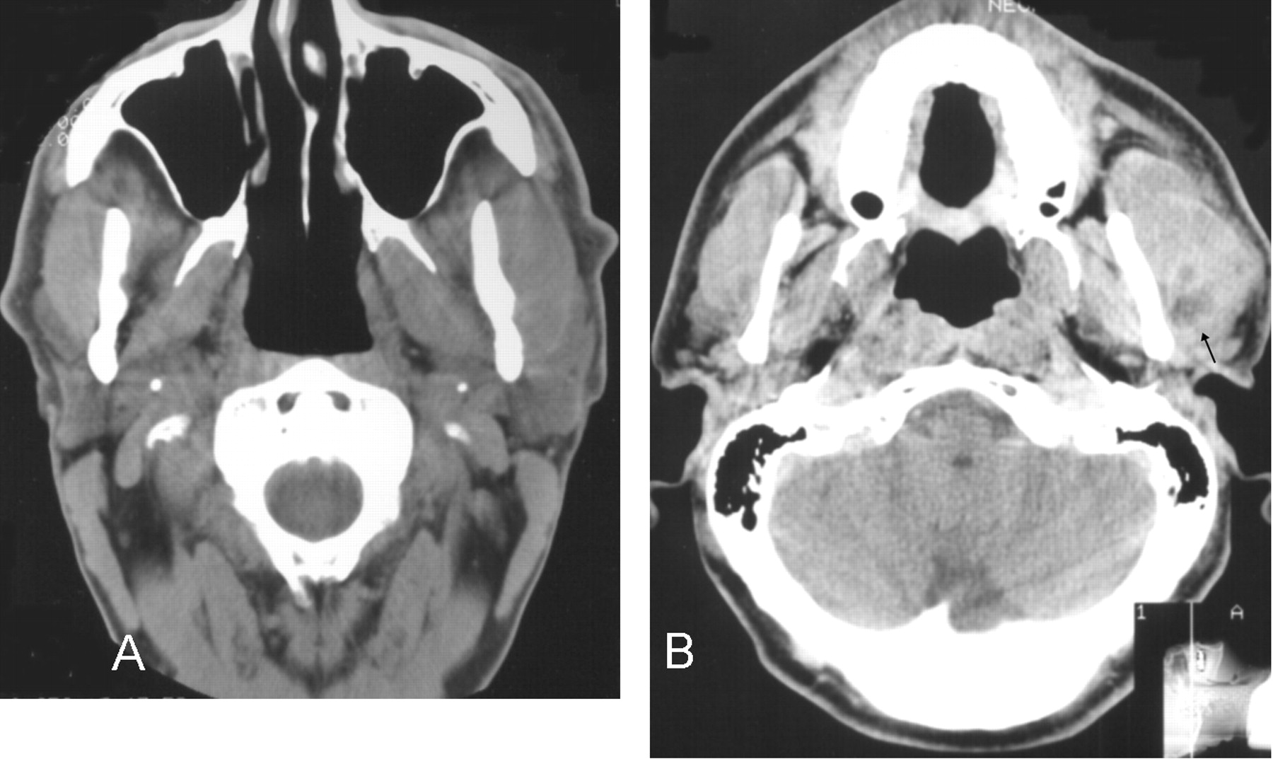
- Fig 1.
Patient 1.
A, Noncontrast axial CT image shows myositis of the left masseter muscle with diffuse swelling, but without significant surrounding inflammatory change or definite fluid collection. The parotid gland is normal.
B, Six months later, postcontrast axial CT image shows increased masseteric swelling and mandibular sclerosis with a discrete area of low attenuation (black arrow) with probable rim enhancement, suggesting the presence of an abscess.
- Fig 2.
Patient 2. Noncontrast axial CT image, soft-tissue view, shows diffuse thickening of the left masseter muscle with a suggestion of cortical thickening of the left mandibular ramus.
- Fig 3.
Patient 3. Postcontrast axial CT image shows diffuse thickening of the left masseter muscle with a small, low-attenuation area, but without clear rim enhancement, possibly representing an abscess. Bony sclerosis of the mandibular ramus is present with a focus of cortical disruption of the mandible laterally (better seen on bone windows) at the level of the abscess.
- Fig 4.
Patient 4.
A, Axial spin-echo T1-weighted image (500/20 [TR/TE]; number of signals averaged [NSA], 2256 × 224; section thickness, 5 mm) shows enlargement of the right masseter muscle with a subjacent curvilinear collection of low signal intensity believed to represent pus.
B, Axial fast spin-echo T2-weighted image (7100/125; NSA, 2; matrix, 256 × 224; section thickness, 5 mm) shows an elliptical mild high signal intensity zone lateral to the mandible corresponding to the low signal intensity seen on the T1 image and consistent with pus. Higher signal intensity between this collection and the masseter muscle probably represents edema and myositis.
C, Coronal spin-echo T1-weighted image (450/20; NSA, 2; matrix, 256 × 256; section thickness, 5 mm) shows thickening of the right mandibular ramus with low signal intensity within the marrow cavity consistent with cortical thickening and sclerosis of the marrow space.
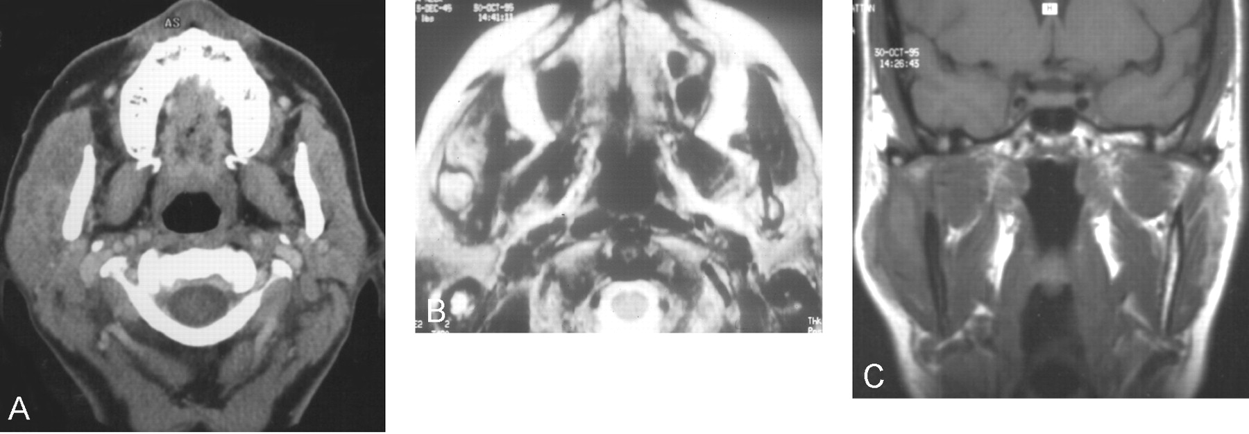
- Fig 5.
Patient 5.
A, Postcontrast axial CT image shows diffuse thickening of the right masseter muscle with a region of mildly low attenuation posteromedially.
B, T2-weighted axial MR imaging shows a more definite fluid collection posteriorly as well as a region of high signal intensity anteriorly within the right masseter muscle compatible with edema and myositis.
C, Noncontrast coronal T1-weighted image shows T1 shortening within the collection, which is mildly hyperintense relative to muscle. This probably represents proteinaceous fluid.
- Fig 6.
Original drawing by Bransby-Zachary illustrating the insertions of the masseter (reproduced with the permission of British Dental Journal 1948; 84:10–13).


