
Article Figures & Data
Figures
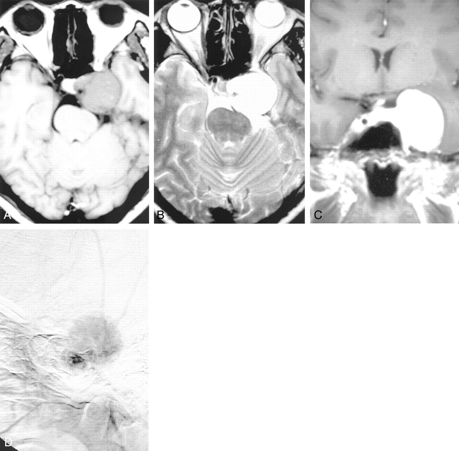
- Fig 1.
A 37-year-old woman presented with diplopia for 2 months. MR images show that the mass produces low signal intensity on axial T1-weighted (A) and a homogeneous and markedly high signal intensity on axial T2-weighted images (B) and is strongly enhanced after contrast material administration (C). The mass is located in the left cavernous sinus, extends to the middle cranial fossa, and encircles the left ICA (arrow). Angiography of the lateral projection of the external carotid artery reveals some vascular blush that is supplied by the middle meningeal artery in the venous phase (D).
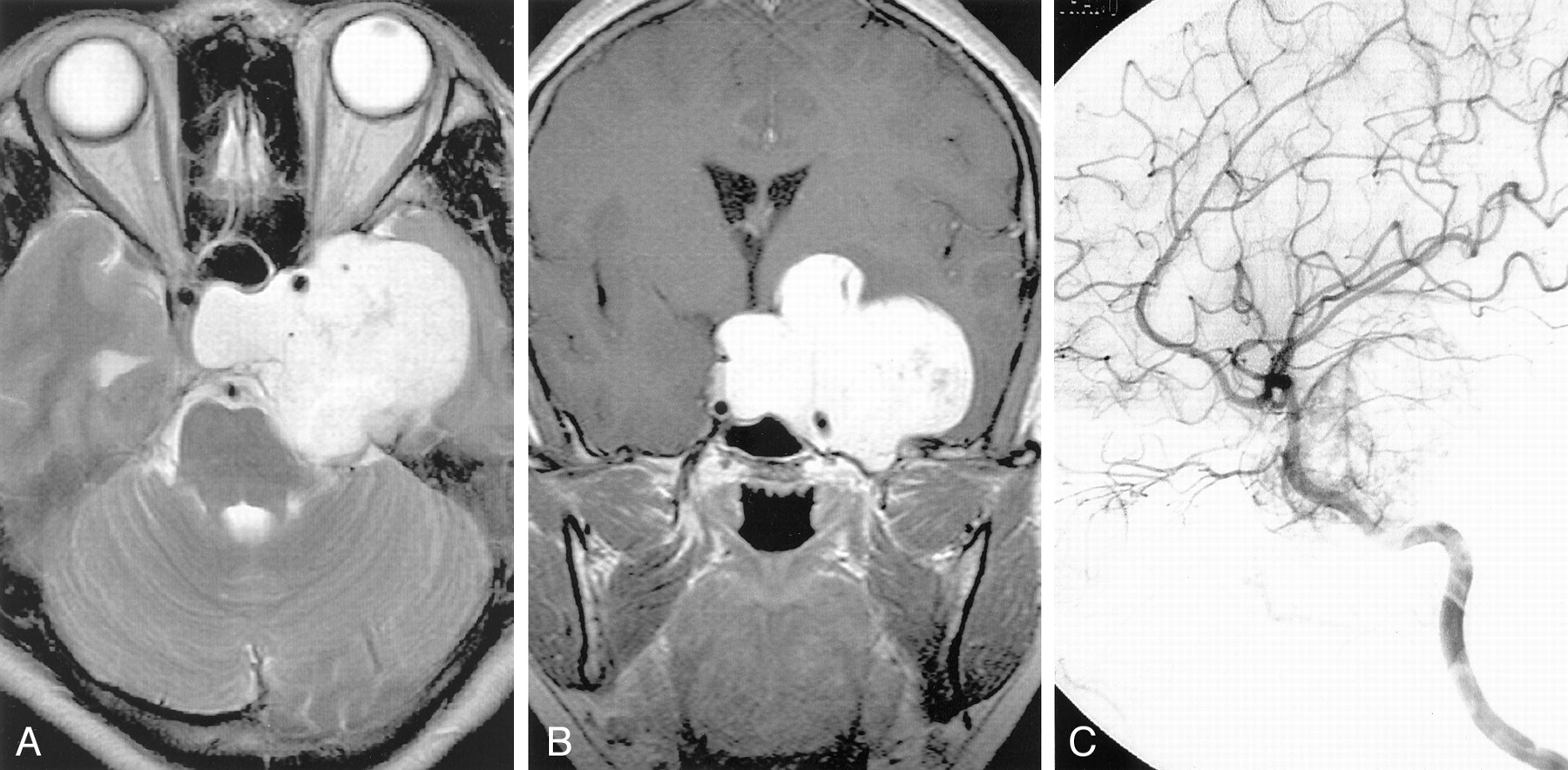
- Fig 2.
A 36-year-old woman had a 7-month history of extraocular muscle limitation and visual disturbance. MR images reveal a large, lobulated mass, with compression of the right cavernous sinus. The mass encircles the cavernous portion of the left ICA (arrow). It gives low signal intensity on T1-weighted images, homogeneous markedly high signal intensity on axial T2-weighted images (A) and is strongly enhanced after contrast material administration (B). Angiography of the left lateral projection of the ICA reveals some vascular blush that is supplied by the meningeal branch of the artery (C).


Tables
Patient (No.) Sex/Age (years) Radiological Findings MR CT Angiography 1 F/37 Hypointense on T1 Homogeneous isoattenuated Vascular blush from MMA Hyperintense on T2 Homogeneous enhancement … Homogeneous enhancement 2 F/36 Hypointense on T1 Homogeneous isoattenuated Vascular blush from meningeal branch of ICA Hyperintense on T2 Homogeneous enhancement Homogeneous enhancement 3 F/48 Hypointense on T1 Homogeneous isoattenuated Avascular Hyperintense on T2 Homogeneous enhancement … Homogeneous enhancement 4 F/57 Hypointense on T1 Homogeneous isoattenuated … Hyperintense on T2 Homogeneous enhancement … Homogeneous enhancement 5 M/49 Hypointense on T1 Homogeneous isoattenuated Vascular blush from MMA Hyperintense on T2 Homogeneous enhancement Homogeneous enhancement … Note.— ICA, internal carotid artery; MMA, middle meningeal artery.


