Advertisement

Main menu

  • Home
  • Content
    • Current Issue
    • Accepted Manuscripts
    • Article Preview
    • Past Issue Archive
    • Video Articles
    • AJNR Case Collection
    • Case of the Week Archive
    • Case of the Month Archive
    • Classic Case Archive
  • Special Collections
    • AJNR Awards
    • Low-Field MRI
    • Alzheimer Disease
    • ASNR Foundation Special Collection
    • Photon-Counting CT
    • View All
  • Multimedia
    • AJNR Podcasts
    • AJNR SCANtastic
    • Trainee Corner
    • MRI Safety Corner
    • Imaging Protocols
  • For Authors
    • Submit a Manuscript
    • Submit a Video Article
    • Submit an eLetter to the Editor/Response
    • Manuscript Submission Guidelines
    • Statistical Tips
    • Fast Publishing of Accepted Manuscripts
    • Graphical Abstract Preparation
    • Imaging Protocol Submission
    • Author Policies
  • About Us
    • About AJNR
    • Editorial Board
    • Editorial Board Alumni
  • More
    • Become a Reviewer/Academy of Reviewers
    • Subscribers
    • Permissions
    • Alerts
    • Feedback
    • Advertisers
    • ASNR Home

User menu

  • Alerts
  • Log in

Search

  • Advanced search
American Journal of Neuroradiology

American Journal of Neuroradiology

ASHNR American Society of Functional Neuroradiology ASHNR American Society of Pediatric Neuroradiology ASSR
  • Alerts
  • Log in

Advanced Search

  • Home
  • Content
    • Current Issue
    • Accepted Manuscripts
    • Article Preview
    • Past Issue Archive
    • Video Articles
    • AJNR Case Collection
    • Case of the Week Archive
    • Case of the Month Archive
    • Classic Case Archive
  • Special Collections
    • AJNR Awards
    • Low-Field MRI
    • Alzheimer Disease
    • ASNR Foundation Special Collection
    • Photon-Counting CT
    • View All
  • Multimedia
    • AJNR Podcasts
    • AJNR SCANtastic
    • Trainee Corner
    • MRI Safety Corner
    • Imaging Protocols
  • For Authors
    • Submit a Manuscript
    • Submit a Video Article
    • Submit an eLetter to the Editor/Response
    • Manuscript Submission Guidelines
    • Statistical Tips
    • Fast Publishing of Accepted Manuscripts
    • Graphical Abstract Preparation
    • Imaging Protocol Submission
    • Author Policies
  • About Us
    • About AJNR
    • Editorial Board
    • Editorial Board Alumni
  • More
    • Become a Reviewer/Academy of Reviewers
    • Subscribers
    • Permissions
    • Alerts
    • Feedback
    • Advertisers
    • ASNR Home
  • Follow AJNR on Twitter
  • Visit AJNR on Facebook
  • Follow AJNR on Instagram
  • Join AJNR on LinkedIn
  • RSS Feeds

AJNR Awards, New Junior Editors, and more. Read the latest AJNR updates

OtherHead and Neck Imaging

Bilateral Petrous Internal Carotid Artery Pseudoaneurysms Presenting with Sensorineural Hearing Loss

Catherine J. Hwang, Gul Moonis, Robert W. Hurst, Neil Hockstein and Douglas Bigelow
American Journal of Neuroradiology June 2003, 24 (6) 1139-1141;
Catherine J. Hwang
  • Find this author on Google Scholar
  • Find this author on PubMed
  • Search for this author on this site
Gul Moonis
  • Find this author on Google Scholar
  • Find this author on PubMed
  • Search for this author on this site
Robert W. Hurst
  • Find this author on Google Scholar
  • Find this author on PubMed
  • Search for this author on this site
Neil Hockstein
  • Find this author on Google Scholar
  • Find this author on PubMed
  • Search for this author on this site
Douglas Bigelow
  • Find this author on Google Scholar
  • Find this author on PubMed
  • Search for this author on this site
  • Article
  • Figures & Data
  • Info & Metrics
  • Responses
  • References
  • PDF
Loading

Abstract

Summary: We present a case of an 85-year-old woman with a 40-year history of progressive bilateral sensorineural hearing loss. Imaging studies demonstrated bilateral pseudoaneurysms of the petrous portion of the internal carotid arteries eroding into both cochleas. To our knowledge, this is the first case report of such lesions.

Pseudoaneurysms of the petrous internal carotid artery (ICA), although uncommon, have been described in the literature (1, 2). Presenting symptoms of these pseudoaneurysms are varied, depending on the adjacent structures and vessels involved. We report a case of bilateral petrous ICA pseudoaneurysms eroding into both cochleas presenting with bilateral sensorineural hearing loss, which to our knowledge has not been previously reported.

Case Report

An 85-year-old woman presented to the otolaryngology clinic at our institution for evaluation of progressive bilateral hearing loss. Although she had a 40-year history of bilateral hearing loss, audiograms documented a deterioration of hearing in the right ear within the past year. She denied tinnitus, vertigo, or recent ear infections. She denied any significant head injuries in the past. Medical history was significant for an untreated growth hormone-secreting pituitary adenoma. On examination, she had significant acromegalic features, bilateral hearing loss, and bitemporal hemianopsia.

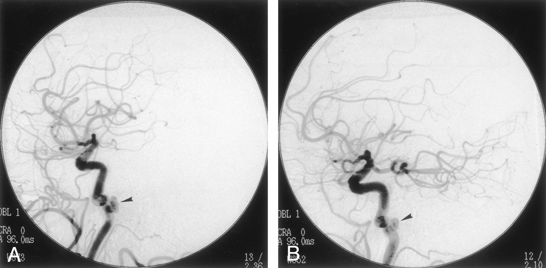
Audiography showed severe to profound sensorineural hearing loss in the right ear with a mild, sloping to severe sensorineural loss on the left. High-resolution (1.25-mm section thickness) coronal and axial CT examinations of the temporal bones were obtained, which revealed erosive lesions involving the petrous bone bilaterally, along the posteromedial wall of the horizontal carotid canals. Lucency was also seen extending from the petrous carotid canal to the cochlea with erosion of the basal and apical turns of the cochlea bilaterally (Figs 1 and 2). The right-side lesion was larger than the left, with more significant erosion of the cochlea. The vestibule, semicircular canals, and internal auditory canals were normal. There was no enlargement of the vestibular aqueduct. MR imaging was performed, revealing questionable flow-related enhancement in these lesions. A diagnostic cerebral angiogram was obtained, which revealed bilateral 5–6-mm aneurysms in the petrous carotid arteries corresponding in location to the CT abnormalities. Subtle changes in diameter were seen in the cervical internal carotid arteries bilaterally, suggesting that these findings represented sequela of remote dissections. An incidental 3-mm right middle meningeal artery aneurysm was also noted.

Fig 1.
  • Download figure
  • Open in new tab
  • Download powerpoint
Fig 1.

Axial (A) and coronal (B) CT images acquired through the temporal bone demonstrate an erosive lesion involving the right petrous carotid canal posteromedially with erosion into the cochlea (black arrowhead). The horizontal petrous carotid canal is indicated by the white arrow. Similar changes were seen on the left.

Fig 2.
  • Download figure
  • Open in new tab
  • Download powerpoint
Fig 2.

Images from right (A) and left (B) common carotid artery injections demonstrate bilateral aneurysms of the horizontal petrous carotid arteries (black arrowhead).

Because of the patient’s age and the presence of bilateral ICA pseudoaneurysms, conservative management was chosen.

Discussion

Vascular lesions involving the petrous segment of the extradural ICA are uncommon. In large part, the rarity of lesions involving the petrous ICA results from the well-protected course of the vessel within the carotid canal, the absence of branches from this portion of the vessel, and the uncommon involvement of the petrous ICA by atheromatous changes. Nevertheless, the anatomic arrangement of the cervico-petrous junction of the ICA may predispose to injury from blunt trauma. As the relatively mobile cervical portion of the ICA enters the carotid canal, the vessel is fixed to the skull base, an arrangement that permits stretching forces to be transmitted to the vessel wall by excessive head movement around the atlanto-occipital joint. Although most often resulting in dissection of the cervical ICA, such forces may also damage the petrous ICA with traumatic pseudoaneurysm formation, the most common cause of petrous aneurysm in most series (3).

A pseudoaneurysm, or false aneurysm, develops when a fibrous tissue capsule forms in response to injury to all layers of an arterial wall (eg, after trauma, surgery, infection, or vasculitis). Such injury may result from a number of mechanisms, including blunt or penetrating trauma, infections, or inflammatory conditions. Although most often resulting from blunt trauma, pseudoaneurysms involving the petrous ICA have been described as rare sequelae of vessel injury from pituitary and sinus surgery as well as from gunshot wounds (1, 4). Mycotic aneurysms of the petrous ICA have also been reported from bacterial and fungal infections (2, 5). The lack of structural integrity of the wall of a pseudoaneurysm often permits rapid expansion and may suggest the cause.

Several reports of rapidly enlarging petrous aneurysms in children have documented involvement of other arterial structures or associated anomalies. Such cases may suggest congenital disorders, including neurofibromatosis (6, 7). Connective tissue disorders such as Marfan syndrome and fibromuscular dysplasia may also be associated with the development of pseudoaneurysms, although involvement of the petrous ICA has not been documented (8).

The presence of a pseudoaneurysm may be demonstrated on CT or MR images. In the case of our patient, the pseudoaneurysm presented as an erosive petrous bone mass on CT images. The differential diagnosis of petrous lesions includes cholesterol granuloma, cholesteatoma, mucocele, chondrosarcoma, granulation tissue, and pseudoaneurysms. The MR imaging appearance of pseudoaneurysm may mimic cholesterol granuloma or mucocele (9). Conventional angiography remains the reference standard for diagnosing vascular lesions such as pseudoaneurysms.

The presenting symptoms of pseudoaneuryms vary depending on the vessels involved and adjacent structures. Symptoms associated with petrous ICA pseudoaneurysms include headache, nasal congestion, and midface pressure and pain (2). A ruptured petrous ICA aneurysm may occur with otorrhagia, epistaxis, and neurologic deficit (10). Sudden sensorineural hearing loss has also been associated with aneurysms (11). Other vascular causes of sensorineural hearing loss are microvascular insufficiency, arteriovenous malformations, vertebrobasilar artery dolichoectasia, thrombosis, and vascular loops (12). The sensorineural hearing loss in these cases is postulated to be a result of cranial nerve VIII compression and dysfunction. Conductive hearing loss in association with a petrous ICA aneurysm has also been reported (13, 14).

Although cranial nerve VIII dysfunction may play a role in our patient’s sensorineural hearing loss, we suspect that it resulted from erosion of the pseudoaneurysm into the cochlea, as seen on CT and MR imaging. Our patient’s sensorineural hearing loss was progressive, reflecting the slow erosion of the cochlea from the pseudoaneurysms. In contrast, the sensorineural hearing loss described in other case reports was usually sudden and associated with recent trauma. In our patient, there is no known history of head trauma; however, in light of the changes in the diameter of cervical internal carotid arteries and an additional aneurysm in the right middle meningeal artery, we suspect that these pseudoaneuryms are the result of remote dissection.

The management of petrous aneurysms is challenging. The primary goals of treatment for petrous aneurysms are to provide symptomatic relief and to minimize the risks of hemodynamic compromise to the cerebrovascular circulation, thromboembolism, and hemorrhage. Treatment of petrous aneurysms was originally limited to rare instances amenable to surgical clip placement or vessel sacrifice versus conservative management (15). More recently, endovascular treatment has become the most often used technique, with sacrifice of the vessel following balloon occlusion testing or endovascular stent placement to preserve the vessel (14, 16, 17). The safety of treatment depends on the anatomy of the aneurysm and segment involved as well as the presence of collateral flow to the brain and the age of the patient. In this case, treatment options were discussed and conservative management was chosen, because there were bilateral ICA aneurysms and the patient was elderly.

Conclusion

Petrous ICA pseudoaneurysms are rare, with only a few case reports in the literature. To our knowledge, this is the first case of bilateral petrous ICA pseudoaneurysms eroding into the cochlea presenting as progressive bilateral sensorineural hearing loss. Thus, in cases of progressive sensorineural hearing loss, a vascular cause such as a pseudoaneurysm should be considered in the differential diagnosis. Imaging studies are integral in evaluating this association.

References

  1. ↵
    Vasama JP, Ramsay H, Markkola A. Petrous internal carotid artery pseudoaneurysm due to gunshot injury. Ann Otol Rhinol Laryngol 2001;110:491–493
    PubMed
  2. ↵
    Tanaka H, Patel U, Shrier DA, Coniglio JU. Pseudoaneurysm of the petrous internal carotid artery after skull base infection and prevertebral abscess drainage. AJNR Am J Neuroradiol 1998;19:502–504
    Abstract
  3. ↵
    Berenstein A, Lasjaunias P. Surgical Neuroangiography. New York: Springer-Verlag;1987
  4. ↵
    Rostomily RC, Newell DW, Grady MS, et al. Gunshot wounds of the internal carotid artery at the skull base: management with vein bypass grafts and a review of the literature. J Trauma 1997;42:123–132
    PubMed
  5. ↵
    Samuel J, Fernandes CM. Mycotic aneurysm of the petrous portion of the internal carotid artery. J Laryngol Otol 1989;103:111–114
    PubMed
  6. ↵
    Frank E, Brown BM, Wilson DF. Asymptomatic fusiform aneurysm of the petrous carotid artery in a patient with von Recklinghausen’s neurofibromatosis. Surg Neurol 1989;32:75–78
    CrossRefPubMed
  7. ↵
    Halbach VV, Higashida RT, Hieshima GB, et al. Aneurysms of the petrous portion of the internal carotid artery: results of treatment with endovascular or surgical occlusion. AJNR Am J Neuroradiol 1990;11:253–257
    Abstract/FREE Full Text
  8. ↵
    Clark ET, Gewertz BL. Pseudoaneurysms. In: Rutherford RB, ed. Vascular Surgery. 4th ed. Philadelphia: WB Saunders;1995 :1153–1161
  9. ↵
    Jackler RK, Parker DA. Radiographic differential diagnosis of petrous apex lesions. Am J Otol 1992;13:561–574
    PubMed
  10. ↵
    Costantino PD, Russell E, Reisch D, Breit RA, Hart C. Ruptured petrous carotid aneurysm presenting with otorrhagia and epistaxis. Am J Otol 1991;12:378–383
    PubMed
  11. ↵
    Colclasure JB, Graham SS. Intracranial aneurysm occurring as sensorineural hearing loss. Otolaryngol Head Neck Surg 1981;89:283–287
    PubMed
  12. ↵
    Murphy TP. Macrovascular sensorineural hearing loss. Am J Otol 1991;12:88–92
    PubMed
  13. ↵
    Reece PH, Higgins N, Hardy DG, Moffat DA. An aneurysm of the petrous internal carotid artery. J Laryngol Otol 1999;113:55–57
    PubMed
  14. ↵
    Coley SC, Clifton A, Britton J. Giant aneurysm of the petrous internal carotid artery: diagnosis and treatment. J Laryngol Otol 1998;112:196–198
    PubMed
  15. ↵
    Lempert TE, Halbach VV, Higashida RT, et al. Endovascular treatment of pseudoaneurysms with electrolytically detachable coils. AJNR Am J Neuroradiol 1998;19:907–911
    Abstract
  16. ↵
    Cheng KM, Chan CM, Cheung YL, et al. Endovascular treatment of radiation-induced petrous internal carotid artery aneurysm presenting with acute haemorrhage: a report of two cases. Acta Neurochir (Wien) 2001;143:351–355; discussion 355–356
  17. ↵
    Alexander MJ, Smith TP, Tucci DL. Treatment of an iatrogenic petrous carotid artery pseudoaneurysm with a Symbiot covered stent: technical case report. Neurosurgery 2002;50:658–662
    CrossRefPubMed
  • Received July 24, 2002.
  • Accepted after revision October 18, 2002.
  • Accepted after revision October 18, 2002.
  • Copyright © American Society of Neuroradiology
View Abstract
PreviousNext
Back to top

In this issue

American Journal of Neuroradiology: 24 (6)
American Journal of Neuroradiology
Vol. 24, Issue 6
1 Jun 2003
  • Table of Contents
  • Index by author
Advertisement
Print
Download PDF
Email Article

Thank you for your interest in spreading the word on American Journal of Neuroradiology.

NOTE: We only request your email address so that the person you are recommending the page to knows that you wanted them to see it, and that it is not junk mail. We do not capture any email address.

Enter multiple addresses on separate lines or separate them with commas.
Bilateral Petrous Internal Carotid Artery Pseudoaneurysms Presenting with Sensorineural Hearing Loss
(Your Name) has sent you a message from American Journal of Neuroradiology
(Your Name) thought you would like to see the American Journal of Neuroradiology web site.
CAPTCHA
This question is for testing whether or not you are a human visitor and to prevent automated spam submissions.
Cite this article
Catherine J. Hwang, Gul Moonis, Robert W. Hurst, Neil Hockstein, Douglas Bigelow
Bilateral Petrous Internal Carotid Artery Pseudoaneurysms Presenting with Sensorineural Hearing Loss
American Journal of Neuroradiology Jun 2003, 24 (6) 1139-1141;

Citation Manager Formats

  • BibTeX
  • Bookends
  • EasyBib
  • EndNote (tagged)
  • EndNote 8 (xml)
  • Medlars
  • Mendeley
  • Papers
  • RefWorks Tagged
  • Ref Manager
  • RIS
  • Zotero
0 Responses
Respond to this article
Share
Bookmark this article
Bilateral Petrous Internal Carotid Artery Pseudoaneurysms Presenting with Sensorineural Hearing Loss
Catherine J. Hwang, Gul Moonis, Robert W. Hurst, Neil Hockstein, Douglas Bigelow
American Journal of Neuroradiology Jun 2003, 24 (6) 1139-1141;
del.icio.us logo Twitter logo Facebook logo Mendeley logo
  • Tweet Widget
  • Facebook Like
  • Google Plus One

Jump to section

  • Article
    • Abstract
    • Case Report
    • Discussion
    • Conclusion
    • References
  • Figures & Data
  • Info & Metrics
  • Responses
  • References
  • PDF

Related Articles

  • No related articles found.
  • PubMed
  • Google Scholar

Cited By...

  • No citing articles found.
  • Crossref
  • Google Scholar

This article has not yet been cited by articles in journals that are participating in Crossref Cited-by Linking.

More in this TOC Section

  • FDG Uptake and Thyroid Cancer Post-BRAF Therapy
  • Normal Facial Nerve Enhancement on VIBE Sequence
  • Contrast Enhanced Pituitary CISS/FIESTA
Show more Head and Neck Imaging

Similar Articles

Advertisement

Indexed Content

  • Current Issue
  • Accepted Manuscripts
  • Article Preview
  • Past Issues
  • Editorials
  • Editor's Choice
  • Fellows' Journal Club
  • Letters to the Editor
  • Video Articles

Cases

  • Case Collection
  • Archive - Case of the Week
  • Archive - Case of the Month
  • Archive - Classic Case

More from AJNR

  • Trainee Corner
  • Imaging Protocols
  • MRI Safety Corner
  • Book Reviews

Multimedia

  • AJNR Podcasts
  • AJNR Scantastics

Resources

  • Turnaround Time
  • Submit a Manuscript
  • Submit a Video Article
  • Submit an eLetter to the Editor/Response
  • Manuscript Submission Guidelines
  • Statistical Tips
  • Fast Publishing of Accepted Manuscripts
  • Graphical Abstract Preparation
  • Imaging Protocol Submission
  • Evidence-Based Medicine Level Guide
  • Publishing Checklists
  • Author Policies
  • Become a Reviewer/Academy of Reviewers
  • News and Updates

About Us

  • About AJNR
  • Editorial Board
  • Editorial Board Alumni
  • Alerts
  • Permissions
  • Not an AJNR Subscriber? Join Now
  • Advertise with Us
  • Librarian Resources
  • Feedback
  • Terms and Conditions
  • AJNR Editorial Board Alumni

American Society of Neuroradiology

  • Not an ASNR Member? Join Now

© 2025 by the American Society of Neuroradiology All rights, including for text and data mining, AI training, and similar technologies, are reserved.
Print ISSN: 0195-6108 Online ISSN: 1936-959X

Powered by HighWire