Advertisement
  • Main menu
  • User menu
  • Search
  • English ▼
    • English
    • Afrikaans
    • Albanian
    • Amharic
    • Arabic
    • Armenian
    • Azerbaijani
    • Basque
    • Belarusian
    • Bengali
    • Bosnian
    • Bulgarian
    • Catalan
    • Cebuano
    • Chichewa
    • Chinese (Simplified)
    • Chinese (Traditional)
    • Corsican
    • Croatian
    • Czech
    • Danish
    • Dutch
    • Esperanto
    • Estonian
    • Filipino
    • Finnish
    • French
    • Frisian
    • Galician
    • Georgian
    • German
    • Greek
    • Gujarati
    • Haitian Creole
    • Hausa
    • Hawaiian
    • Hebrew
    • Hindi
    • Hmong
    • Hungarian
    • Icelandic
    • Igbo
    • Indonesian
    • Irish
    • Italian
    • Japanese
    • Javanese
    • Kannada
    • Kazakh
    • Khmer
    • Korean
    • Kurdish (Kurmanji)
    • Kyrgyz
    • Lao
    • Latin
    • Latvian
    • Lithuanian
    • Luxembourgish
    • Macedonian
    • Malagasy
    • Malay
    • Malayalam
    • Maltese
    • Maori
    • Marathi
    • Mongolian
    • Myanmar (Burmese)
    • Nepali
    • Norwegian
    • Pashto
    • Persian
    • Polish
    • Portuguese
    • Punjabi
    • Romanian
    • Russian
    • Samoan
    • Scottish Gaelic
    • Serbian
    • Sesotho
    • Shona
    • Sindhi
    • Sinhala
    • Slovak
    • Slovenian
    • Somali
    • Spanish
    • Sudanese
    • Swahili
    • Swedish
    • Tajik
    • Tamil
    • Telugu
    • Thai
    • Turkish
    • Ukrainian
    • Urdu
    • Uzbek
    • Vietnamese
    • Welsh
    • Xhosa
    • Yiddish
    • Yoruba
    • Zulu

Main menu

  • Home
  • Content
    • Current Issue
    • Accepted Manuscripts
    • Article Preview
    • Past Issue Archive
    • Video Articles
    • AJNR Case Collection
    • Case of the Week Archive
    • Case of the Month Archive
    • Classic Case Archive
  • Special Collections
    • Advancing NeuroMRI with High-Relaxivity Contrast Agents
    • AJNR Awards
    • Low-Field MRI
    • Alzheimer Disease
    • ASNR Foundation Special Collection
    • Photon-Counting CT
    • View All
  • Multimedia
    • AJNR Podcasts
    • AJNR SCANtastic
    • Trainee Corner
    • MRI Safety Corner
    • Imaging Protocols
  • For Authors
    • Submit a Manuscript
    • Submit a Video Article
    • Submit an eLetter to the Editor/Response
    • Manuscript Submission Guidelines
    • Statistical Tips
    • Fast Publishing of Accepted Manuscripts
    • Graphical Abstract Preparation
    • Imaging Protocol Submission
    • Author Policies
  • About Us
    • About AJNR
    • Editorial Board
    • Editorial Board Alumni
  • More
    • Become a Reviewer/Academy of Reviewers
    • Subscribers
    • Permissions
    • Alerts
    • Feedback
    • Advertisers
    • ASNR Home

User menu

  • Alerts
  • Log in

Search

  • Advanced search
American Journal of Neuroradiology

American Journal of Neuroradiology

ASHNR American Society of Functional Neuroradiology ASHNR American Society of Pediatric Neuroradiology ASSR
  • Alerts
  • Log in

Advanced Search

English ▼
  • English
  • Afrikaans
  • Albanian
  • Amharic
  • Arabic
  • Armenian
  • Azerbaijani
  • Basque
  • Belarusian
  • Bengali
  • Bosnian
  • Bulgarian
  • Catalan
  • Cebuano
  • Chichewa
  • Chinese (Simplified)
  • Chinese (Traditional)
  • Corsican
  • Croatian
  • Czech
  • Danish
  • Dutch
  • Esperanto
  • Estonian
  • Filipino
  • Finnish
  • French
  • Frisian
  • Galician
  • Georgian
  • German
  • Greek
  • Gujarati
  • Haitian Creole
  • Hausa
  • Hawaiian
  • Hebrew
  • Hindi
  • Hmong
  • Hungarian
  • Icelandic
  • Igbo
  • Indonesian
  • Irish
  • Italian
  • Japanese
  • Javanese
  • Kannada
  • Kazakh
  • Khmer
  • Korean
  • Kurdish (Kurmanji)
  • Kyrgyz
  • Lao
  • Latin
  • Latvian
  • Lithuanian
  • Luxembourgish
  • Macedonian
  • Malagasy
  • Malay
  • Malayalam
  • Maltese
  • Maori
  • Marathi
  • Mongolian
  • Myanmar (Burmese)
  • Nepali
  • Norwegian
  • Pashto
  • Persian
  • Polish
  • Portuguese
  • Punjabi
  • Romanian
  • Russian
  • Samoan
  • Scottish Gaelic
  • Serbian
  • Sesotho
  • Shona
  • Sindhi
  • Sinhala
  • Slovak
  • Slovenian
  • Somali
  • Spanish
  • Sudanese
  • Swahili
  • Swedish
  • Tajik
  • Tamil
  • Telugu
  • Thai
  • Turkish
  • Ukrainian
  • Urdu
  • Uzbek
  • Vietnamese
  • Welsh
  • Xhosa
  • Yiddish
  • Yoruba
  • Zulu
  • Home
  • Content
    • Current Issue
    • Accepted Manuscripts
    • Article Preview
    • Past Issue Archive
    • Video Articles
    • AJNR Case Collection
    • Case of the Week Archive
    • Case of the Month Archive
    • Classic Case Archive
  • Special Collections
    • Advancing NeuroMRI with High-Relaxivity Contrast Agents
    • AJNR Awards
    • Low-Field MRI
    • Alzheimer Disease
    • ASNR Foundation Special Collection
    • Photon-Counting CT
    • View All
  • Multimedia
    • AJNR Podcasts
    • AJNR SCANtastic
    • Trainee Corner
    • MRI Safety Corner
    • Imaging Protocols
  • For Authors
    • Submit a Manuscript
    • Submit a Video Article
    • Submit an eLetter to the Editor/Response
    • Manuscript Submission Guidelines
    • Statistical Tips
    • Fast Publishing of Accepted Manuscripts
    • Graphical Abstract Preparation
    • Imaging Protocol Submission
    • Author Policies
  • About Us
    • About AJNR
    • Editorial Board
    • Editorial Board Alumni
  • More
    • Become a Reviewer/Academy of Reviewers
    • Subscribers
    • Permissions
    • Alerts
    • Feedback
    • Advertisers
    • ASNR Home
  • Follow AJNR on Twitter
  • Visit AJNR on Facebook
  • Follow AJNR on Instagram
  • Join AJNR on LinkedIn
  • RSS Feeds

AJNR Awards, New Junior Editors, and more. Read the latest AJNR updates


Improved Turnaround Times | Median time to first decision: 12 days

Research ArticleBrain

MR Imaging in Human Rabies

Jiraporn Laothamatas, Thiravat Hemachudha, Erawady Mitrabhakdi, Pongsak Wannakrairot and Supoch Tulayadaechanont
American Journal of Neuroradiology June 2003, 24 (6) 1102-1109;
Jiraporn Laothamatas
  • Find this author on Google Scholar
  • Find this author on PubMed
  • Search for this author on this site
Thiravat Hemachudha
  • Find this author on Google Scholar
  • Find this author on PubMed
  • Search for this author on this site
Erawady Mitrabhakdi
  • Find this author on Google Scholar
  • Find this author on PubMed
  • Search for this author on this site
Pongsak Wannakrairot
  • Find this author on Google Scholar
  • Find this author on PubMed
  • Search for this author on this site
Supoch Tulayadaechanont
  • Find this author on Google Scholar
  • Find this author on PubMed
  • Search for this author on this site
  • Article
  • Figures & Data
  • Info & Metrics
  • Responses
  • References
  • PDF
Loading

Article Figures & Data

Figures

  • Tables
  • Fig 1.
    • Download figure
    • Open in new tab
    • Download powerpoint
    Fig 1.

    Patient 1. Encephalitic rabies.

    A and B, Coronal fast spin-echo T2-weighted MR images of the brain, and C and D, axial gradient-echo T2-weighted MR images of the cervical cord. A and C were obtained on day 3 and B and D on day 7 after clinical onset. Note slight progression of an ill-defined mild hyperintensity change involving the deep and subcortical white matter (arrow with block in A and double arrows in B), hippocampal gyri (black arrow with circle in A and B), brain stem (arrowhead in A and B), and cervical cord (arrow in C and D).

  • Fig 2.
    • Download figure
    • Open in new tab
    • Download powerpoint
    Fig 2.

    Patient 2. Encephalitic rabies.

    A and B, Axial fast spin-echo T2-weighted MR images of the brain, after the patient received intravenous HRIG, demonstrate ill-defined hyperintensity changes involving the brain stem (arrow in A), both deep and cortical gray matter (arrowheads in B), and deep and subcortical white matter (arrow with block in B).
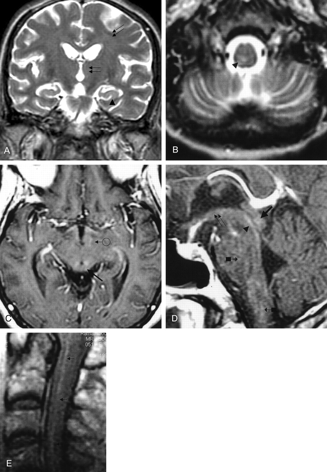
  • Fig 3.
    • Download figure
    • Open in new tab
    • Download powerpoint
    Fig 3.

    Patient 1. Encephalitis rabies.

    A, Coronal and B, axial gadolinium-enhanced T1-weighted fat-suppressed MR images of the brachial plexi, and C, axial gadolinium-enhanced T1-weighted image of the cervical spine reveal ill-defined, mild to moderate enhancement along the left brachial plexus (arrows in A and B) and intradural left dorsal cervical nerve root (arrows in C).

  • Fig 4.
    • Download figure
    • Open in new tab
    • Download powerpoint
    Fig 4.

    Patient 4. Paralytic rabies.

    A, Coronal and B, axial fast spin-echo T2-weighted images of the brain reveal ill-defined mild hyperintensity change at the posterior right paramedian lower medulla and hippocampi (arrowhead in A and B) and deep white matter of the brain (double arrows in A).

    C, Axial and D and E, sagittal gadolinium-enhanced T1-weighted images of the hypothalami, brain stem, and upper cervical cord demonstrate mild to moderate enhancement at the hypothalami (arrow with circle in C and double arrow in D), tectal plate (thick arrow in C and D), periaqueductal gray matter (arrowhead in D), pons and medulla (arrows with block in D), and cervical cord (arrows in E). There is no meningeal enhancement. (Reprinted with permission from reference 3.)

  • Fig 5.
    • Download figure
    • Open in new tab
    • Download powerpoint
    Fig 5.

    Patient 5. Paralytic rabies.

    A, Coronal and B, axial fast spin-echo T2-weighted images of the brain, and C, gadolinium-enhanced axial T1-weighted image of the midcervical cord reveal ill-defined moderate hyperintensity changes of the deep gray matter (double arrows in A), white matter (single arrow in A), and brain stem and dentate nuclei (arrowhead and arrow with block in B). Vivid enhancement of the ventral and dorsal cervical nerve roots (arrows in C) is demonstrated. (Reprinted with permission from reference 3.)

Tables

  • Figures
  • Patient data and study details

    Patient No./Age (y)/SexBite SiteIncubation PeriodSurvival Period (days)Date of MR Study after Clinical OnsetConsciousness LevelMR Studies Performed
    Encephalitic rabies
     1/50/ML wrist7 wk7Day 3Fully consciousBrain, spinal cord, nerve roots, and brachial plexus, with contrast material (Figs 1, 3)
    Day 7ComatoseBrain and spinal cord, without contrast material (Fig 1)
     2/26/FR leg2 mo15Day 2Fully consciousBrain, with contrast material (Fig 2)
    Paralytic rabies
     3/43/FL hand3 mo9Day 4ArousableBrain, with contrast material (no figure)
     4/72/FL leg3 mo13Day 12ComatoseBrain and upper cervical cord, with contrast material (Fig 4)
     5/70/FL face3 wk21Day 20ComatoseBrain, cervical cord, and nerve roots, with contrast material (Fig 5)
PreviousNext
Back to top

In this issue

American Journal of Neuroradiology: 24 (6)
American Journal of Neuroradiology
Vol. 24, Issue 6
1 Jun 2003
  • Table of Contents
  • Index by author
Advertisement
Print
Download PDF
Email Article
Cite this article
0 Responses
Respond to this article
Share
Bookmark this article
MR Imaging in Human Rabies
Jiraporn Laothamatas, Thiravat Hemachudha, Erawady Mitrabhakdi, Pongsak Wannakrairot, Supoch Tulayadaechanont
American Journal of Neuroradiology Jun 2003, 24 (6) 1102-1109;
del.icio.us logo Twitter logo Facebook logo Mendeley logo
  • Tweet Widget

Jump to section

  • Article
    • Abstract
    • Methods
    • Results
    • Discussion
    • Conclusion
    • Acknowledgments
    • Footnotes
    • References
  • Figures & Data
  • Info & Metrics
  • Responses
  • References
  • PDF

Related Articles

  • No related articles found.
  • PubMed
  • Google Scholar

Cited By...

  • The influence of parasite load on transcriptional activity and morphology of a cestode and its ant intermediate host
  • Dual Role of Toll-Like Receptor 7 in the Pathogenesis of Rabies Virus in a Mouse Model
  • Rabies viral encephalitis: clinical determinants in diagnosis with special reference to paralytic form
  • Paralytic rabies after a two week holiday in India
  • Crossref
  • Google Scholar

This article has not yet been cited by articles in journals that are participating in Crossref Cited-by Linking.

More in this TOC Section

  • Progression of Microstructural Damage in Spinocerebellar Ataxia Type 2: A Longitudinal DTI Study
  • SWI or T2*: Which MRI Sequence to Use in the Detection of Cerebral Microbleeds? The Karolinska Imaging Dementia Study
  • Statin Therapy Does Not Affect the Radiographic and Clinical Profile of Patients with TIA and Minor Stroke
Show more Brain

Similar Articles

Advertisement

Indexed Content

  • Current Issue
  • Accepted Manuscripts
  • Article Preview
  • Past Issues
  • Editorials
  • Editor's Choice
  • Fellows' Journal Club
  • Letters to the Editor
  • Video Articles

Cases

  • Case Collection
  • Archive - Case of the Week
  • Archive - Case of the Month
  • Archive - Classic Case

More from AJNR

  • Trainee Corner
  • Imaging Protocols
  • MRI Safety Corner
  • Book Reviews

Multimedia

  • AJNR Podcasts
  • AJNR Scantastics

Resources

  • Turnaround Time
  • Submit a Manuscript
  • Submit a Video Article
  • Submit an eLetter to the Editor/Response
  • Manuscript Submission Guidelines
  • Statistical Tips
  • Fast Publishing of Accepted Manuscripts
  • Graphical Abstract Preparation
  • Imaging Protocol Submission
  • Evidence-Based Medicine Level Guide
  • Publishing Checklists
  • Author Policies
  • Become a Reviewer/Academy of Reviewers
  • News and Updates

About Us

  • About AJNR
  • Editorial Board
  • Editorial Board Alumni
  • Alerts
  • Permissions
  • Not an AJNR Subscriber? Join Now
  • Advertise with Us
  • Librarian Resources
  • Feedback
  • Terms and Conditions
  • AJNR Editorial Board Alumni

American Society of Neuroradiology

  • Not an ASNR Member? Join Now

© 2025 by the American Society of Neuroradiology All rights, including for text and data mining, AI training, and similar technologies, are reserved.
Print ISSN: 0195-6108 Online ISSN: 1936-959X

Powered by HighWire
Email this Article

Thank you for your interest in spreading the word on American Journal of Neuroradiology.

NOTE: We only request your email address so that the person you are recommending the page to knows that you wanted them to see it, and that it is not junk mail. We do not capture any email address.

Enter multiple addresses on separate lines or separate them with commas.
MR Imaging in Human Rabies
(Your Name) has sent you a message from American Journal of Neuroradiology
(Your Name) thought you would like to see the American Journal of Neuroradiology web site.
CAPTCHA
This question is for testing whether or not you are a human visitor and to prevent automated spam submissions.
Cite this article
Jiraporn Laothamatas, Thiravat Hemachudha, Erawady Mitrabhakdi, Pongsak Wannakrairot, Supoch Tulayadaechanont
MR Imaging in Human Rabies
American Journal of Neuroradiology Jun 2003, 24 (6) 1102-1109;

Citation Manager Formats

  • BibTeX
  • Bookends
  • EasyBib
  • EndNote (tagged)
  • EndNote 8 (xml)
  • Medlars
  • Mendeley
  • Papers
  • RefWorks Tagged
  • Ref Manager
  • RIS
  • Zotero

We use cookies on this site to enhance your user experience. By clicking any link on this page you are giving your consent for us to set cookies.