Advertisement

Main menu

  • Home
  • Content
    • Current Issue
    • Accepted Manuscripts
    • Article Preview
    • Past Issue Archive
    • Video Articles
    • AJNR Case Collection
    • Case of the Week Archive
    • Case of the Month Archive
    • Classic Case Archive
  • Special Collections
    • AJNR Awards
    • Low-Field MRI
    • Alzheimer Disease
    • ASNR Foundation Special Collection
    • Photon-Counting CT
    • View All
  • Multimedia
    • AJNR Podcasts
    • AJNR SCANtastic
    • Trainee Corner
    • MRI Safety Corner
    • Imaging Protocols
  • For Authors
    • Submit a Manuscript
    • Submit a Video Article
    • Submit an eLetter to the Editor/Response
    • Manuscript Submission Guidelines
    • Statistical Tips
    • Fast Publishing of Accepted Manuscripts
    • Graphical Abstract Preparation
    • Imaging Protocol Submission
    • Author Policies
  • About Us
    • About AJNR
    • Editorial Board
    • Editorial Board Alumni
  • More
    • Become a Reviewer/Academy of Reviewers
    • Subscribers
    • Permissions
    • Alerts
    • Feedback
    • Advertisers
    • ASNR Home

User menu

  • Alerts
  • Log in

Search

  • Advanced search
American Journal of Neuroradiology

American Journal of Neuroradiology

ASHNR American Society of Functional Neuroradiology ASHNR American Society of Pediatric Neuroradiology ASSR
  • Alerts
  • Log in

Advanced Search

  • Home
  • Content
    • Current Issue
    • Accepted Manuscripts
    • Article Preview
    • Past Issue Archive
    • Video Articles
    • AJNR Case Collection
    • Case of the Week Archive
    • Case of the Month Archive
    • Classic Case Archive
  • Special Collections
    • AJNR Awards
    • Low-Field MRI
    • Alzheimer Disease
    • ASNR Foundation Special Collection
    • Photon-Counting CT
    • View All
  • Multimedia
    • AJNR Podcasts
    • AJNR SCANtastic
    • Trainee Corner
    • MRI Safety Corner
    • Imaging Protocols
  • For Authors
    • Submit a Manuscript
    • Submit a Video Article
    • Submit an eLetter to the Editor/Response
    • Manuscript Submission Guidelines
    • Statistical Tips
    • Fast Publishing of Accepted Manuscripts
    • Graphical Abstract Preparation
    • Imaging Protocol Submission
    • Author Policies
  • About Us
    • About AJNR
    • Editorial Board
    • Editorial Board Alumni
  • More
    • Become a Reviewer/Academy of Reviewers
    • Subscribers
    • Permissions
    • Alerts
    • Feedback
    • Advertisers
    • ASNR Home
  • Follow AJNR on Twitter
  • Visit AJNR on Facebook
  • Follow AJNR on Instagram
  • Join AJNR on LinkedIn
  • RSS Feeds

AJNR Awards, New Junior Editors, and more. Read the latest AJNR updates

Research ArticleBRAIN

Diffusion-Weighted Imaging of Acute Corticospinal Tract Injury Preceding Wallerian Degeneration in the Maturing Human Brain

Avi Mazumdar, Pratik Mukherjee, Jeffrey H. Miller, Hiten Malde and Robert C. McKinstry
American Journal of Neuroradiology June 2003, 24 (6) 1057-1066;
Avi Mazumdar
  • Find this author on Google Scholar
  • Find this author on PubMed
  • Search for this author on this site
Pratik Mukherjee
  • Find this author on Google Scholar
  • Find this author on PubMed
  • Search for this author on this site
Jeffrey H. Miller
  • Find this author on Google Scholar
  • Find this author on PubMed
  • Search for this author on this site
Hiten Malde
  • Find this author on Google Scholar
  • Find this author on PubMed
  • Search for this author on this site
Robert C. McKinstry
  • Find this author on Google Scholar
  • Find this author on PubMed
  • Search for this author on this site
  • Article
  • Figures & Data
  • Info & Metrics
  • Responses
  • References
  • PDF
Loading

Article Figures & Data

Figures

  • Tables
  • Fig 1.
    • Download figure
    • Open in new tab
    • Download powerpoint
    Fig 1.

    Patient 1 was a 4-day-old neonate with a right MCA infarct, which occurred 4 days before MR imaging was performed. At 3- and 6-month clinical follow-up, the patient had clinical progression of the left hemiparesis, consistent with the evolution of Wallerian degeneration.

    Far left, Transverse (top) and coronal (bottom) fast spin-echo T2-weighted images (5000/96 [TR/TE]) show abnormal signal intensity in the right corticospinal tract (arrowhead). Increased signal intensity is present in the region of the right temporal lobe subcortical white matter (black arrow, top), along with areas of decreased signal intensity in the cortical gray matter (black arrows, bottom). A focus of hemorrhage (white arrow) is seen in the corona radiata.

    Middle left, Transverse (top) and coronal (bottom) DW imagess (4000/100) acquired with a spin-echo echo-planar imaging technique show increased signal intensity in the right cerebral peduncle (arrowhead) and subcortical white matter in the territorial infarct (arrow).

    Middle right, Corresponding transverse (top) and coronal (bottom) ADC maps and reduced ADC in the right cerebral peduncle (arrowhead) and subcortical white matter in the territorial infarct (arrow).

    Far right, Transverse DW image (top) and corresponding ADC map (bottom) show involvement of the splenium of the corpus callosum (arrowhead).
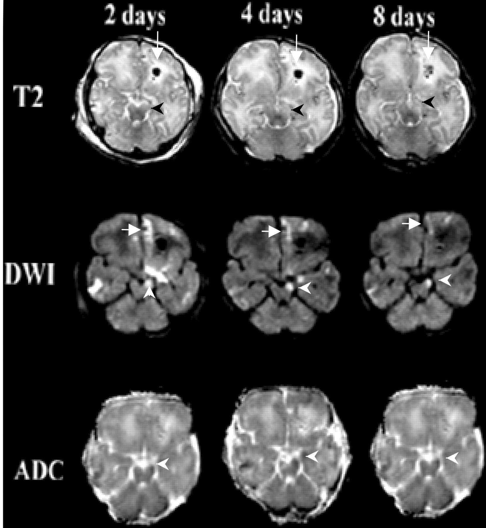
  • Fig 2.
    • Download figure
    • Open in new tab
    • Download powerpoint
    Fig 2.

    Patient 2 was an 8-day-old neonate with a skull fracture after forceps delivery. Transverse images obtained at days 2, 4, and 8.

    Top row, T2-weighted images acquired with a fast spin-echo technique (5000/96) show an area of hemorrhage with surrounding edema in the left frontal lobe (arrow). At 4 and 8 days, progressive development of the increased signal intensity in the left cerebral peduncle is seen (arrowhead).

    Middle row, DW images obtained by using a spin-echo echo-planar imaging sequence (3000/106) show areas of increased signal intensity in the right frontal, right temporal, left frontal lobes, as well as in the left frontopontine tract (arrowhead). Also shown is the area of hemorrhage with surrounding edema in the left frontal lobe (arrow).

    Bottom row, ADC maps confirm truly reduced diffusion in the left cerebral peduncle (arrowhead). Interestingly the diffusion changes in the left frontal lobe subcortical white matter become less apparent on days 4 and 8, consistent with ADC pseudonormalization.
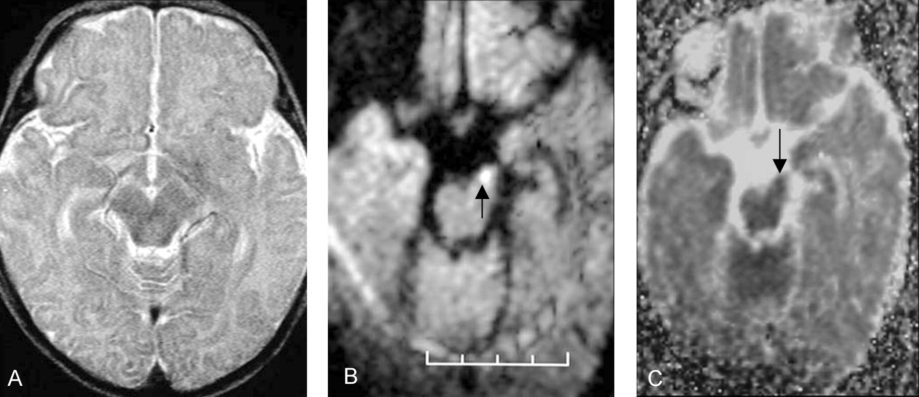
  • Fig 3.
    • Download figure
    • Open in new tab
    • Download powerpoint
    Fig 3.

    Patient 3 was an 8-week-old female infant with a history of seizures that started 3 days before MR imaging was performed. DW imaging had shown a left MCA distribution infarct (not shown). The patient had decreased tone in the right arm at the time of her discharge from the hospital 3 weeks after initial injury; the patient’s initial clinical presentation did not change.

    A, Normal transverse fast spin-echo T2-weighted image (5000/96) shows the left cerebral peduncle.

    B, Transverse DW image acquired with a spin-echo echo-planar imaging technique (4000/100) shows increased signal intensity in the left cerebral peduncle (arrow).

    C, Corresponding ADC map demonstrates that the ADC value in the left cerebral peduncle (arrow) is lower that that in the normal right cerebral peduncle.
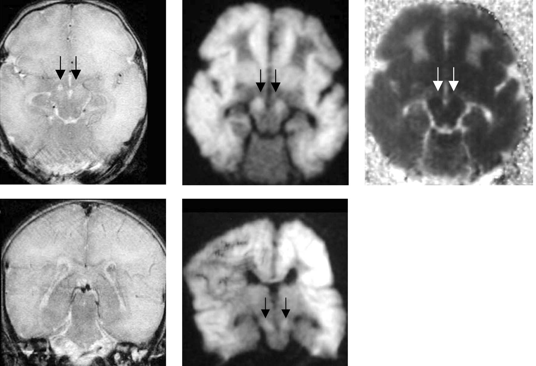
  • Fig 4.
    • Download figure
    • Open in new tab
    • Download powerpoint
    Fig 4.

    Patient 6 was a 5-day-old neonate with hypoxic encephalopathy.

    A, Transverse turbo spin-echo T2-weighted image (5000/96) shows diffusely increased signal intensity in the subcortical white matter in both cerebral hemispheres, with increased signal intensity in both cerebral peduncles (arrows).

    B, Transverse DW image acquired with a spin-echo echo-planar technique (4000/100) shows diffusely increased signal intensity in the subcortical white matter and both cerebral peduncles (arrows).

    C, Transverse ADC map confirms the finding shown in B (arrows).

    D, Coronal T2-weighted image acquired with a turbo spin-echo technique shows no evidence of corticospinal tract injury.

    E, Coronal DW image acquired with a spin-echo echo-planar technique (4000/100) shows bilateral corticospinal tract injury (arrows).

Tables

  • Figures
    • View popup
    TABLE 1:

    Clinical findings in the study group

    Patient/Sex/AgeClinical PresentationInitial Neurologic Findings*Follow-up Findings
    1/M/4 dApnea and seizures after cesarean deliveryL upper-extremity impairmentProgressive L hemiparesis over 6 mo
    2/M/8 dSkull fracture after forceps deliveryR-sided hemiparesisPersistent R hemiparesis at 9-mo follow-up
    3/F/8 wkPresented with seizuresR upper-extremity impairmentNo change over 3 wk
    4/F/19 wkNonaccidental traumaIncreased tone bilateral upper and lower extremitiesNo change over 1 y
    5/M/3 dSkull fracture after failed vacuum extractionMild decreased in tone, bilateral lower extremitiesDelayed motor development, increased tone in all extremities (upper more than lower) at 1-y follow-up
    6/F/5 dSeizures, apneic episodesIncreased tone in bilateral lower extremities, hyperreflexiaNot applicable
    • View popup
    TABLE 2:

    Conventional MR imaging findings

    PatientInfarct Distribution*Projectional White Matter Pathways Involved
    1R MCA territory involving the frontal lobe (including premotor cortex, primary motor cortex), parietal lobe (including somatosensory cortex), and temporal lobeR corticospinal, corticobulbar, corticopontine tracts, genu and splenium of the corpus callosum
    2L and R frontal lobes (prefrontal cortex), R temporal lobeL frontopontine tract, genu of the corpus callosum
    3L MCA territory involving the L frontal lobe (primary motor cortex, premotor and supplementary motor areas, and prefrontal cortex) and parietal lobeL corticospinal tract, L corticobulbar and L corticopontine tracts, genu and splenium of the corpus callosum
    4Bilateral MCA territory involving bilateral frontal, parietal, and temporal lobesL corticospinal, corticobulbar, and corticopontine tracts; genu and splenium of the corpus callosum
    5Bilateral MCA territory, most prominent in the L sensorimotor cortex and in the R parieto-occipital regionBilateral corticospinal, corticobulbar, and corticopontine tracts
    6Bilateral subcortical white matter involving the frontal, parietal, and temporal lobesBilateral corticospinal, corticobulbar, and corticopontine tracts and genu and splenium of the corpus callosum
    • View popup
    TABLE 3:

    DWI findings

    Territory InvolvedInfarct Age (days)Signal IntensityADC, × 10−3 mm2/s
    T2-Weighted ImageDWI
    Patient 1
     L internal capsule4NormalNormal1.03 ± 0.06
     R internal capsule4IncreasedIncreased0.74 ± 0.03
    Patient 2
     L cerebral peduncle2NormalIncreased0.8 ± 0.05
     R cerebral peduncle2NormalNormal1.07 ± 0.05
     L cerebral peduncle4IncreasedIncreased0.9 ± 0.05
     R cerebral peduncle4NormalNormal1.07 ± 0.04
     L cerebral peduncle8IncreasedIncreased0.95 ± 0.06
     R cerebral peduncle8NormalNormal1.08 ± 0.04
    Patient 3
     L cerebral peduncle3NormalIncreased0.79 ± 0.13
     R cerebral peduncle3NormalNormal0.99 ± 0.12
    Patient 4
     L internal capsule8NormalIncreased0.64 ± 0.02
     R internal capsule8NormalNormal0.84 ± 0.04
    Patient 5
     L internal capsule2NormalIncreasedNot applicable
     R internal capsule2NormalIncreasedNot applicable
    Patient 6
     L cerebral peduncle6IncreasedIncreased0.04 ± 0.03
     R cerebral peduncle6IncreasedIncreased0.10 ± 0.02
PreviousNext
Back to top

In this issue

American Journal of Neuroradiology: 24 (6)
American Journal of Neuroradiology
Vol. 24, Issue 6
1 Jun 2003
  • Table of Contents
  • Index by author
Advertisement
Print
Download PDF
Email Article

Thank you for your interest in spreading the word on American Journal of Neuroradiology.

NOTE: We only request your email address so that the person you are recommending the page to knows that you wanted them to see it, and that it is not junk mail. We do not capture any email address.

Enter multiple addresses on separate lines or separate them with commas.
Diffusion-Weighted Imaging of Acute Corticospinal Tract Injury Preceding Wallerian Degeneration in the Maturing Human Brain
(Your Name) has sent you a message from American Journal of Neuroradiology
(Your Name) thought you would like to see the American Journal of Neuroradiology web site.
CAPTCHA
This question is for testing whether or not you are a human visitor and to prevent automated spam submissions.
Cite this article
Avi Mazumdar, Pratik Mukherjee, Jeffrey H. Miller, Hiten Malde, Robert C. McKinstry
Diffusion-Weighted Imaging of Acute Corticospinal Tract Injury Preceding Wallerian Degeneration in the Maturing Human Brain
American Journal of Neuroradiology Jun 2003, 24 (6) 1057-1066;

Citation Manager Formats

  • BibTeX
  • Bookends
  • EasyBib
  • EndNote (tagged)
  • EndNote 8 (xml)
  • Medlars
  • Mendeley
  • Papers
  • RefWorks Tagged
  • Ref Manager
  • RIS
  • Zotero
0 Responses
Respond to this article
Share
Bookmark this article
Diffusion-Weighted Imaging of Acute Corticospinal Tract Injury Preceding Wallerian Degeneration in the Maturing Human Brain
Avi Mazumdar, Pratik Mukherjee, Jeffrey H. Miller, Hiten Malde, Robert C. McKinstry
American Journal of Neuroradiology Jun 2003, 24 (6) 1057-1066;
del.icio.us logo Twitter logo Facebook logo Mendeley logo
  • Tweet Widget
  • Facebook Like
  • Google Plus One

Jump to section

  • Article
    • Abstract
    • Methods
    • Results
    • Discussion
    • Conclusion
    • Acknowledgments
    • References
  • Figures & Data
  • Info & Metrics
  • Responses
  • References
  • PDF

Related Articles

  • No related articles found.
  • PubMed
  • Google Scholar

Cited By...

  • Teaching NeuroImages: Wallerian degeneration in evolving pediatric stroke
  • Injury to the Cerebellum in Term Asphyxiated Newborns Treated with Hypothermia
  • Postoperative Transient Reduced Diffusion in the Ipsilateral Striatum and Thalamus
  • Acute Corticospinal Tract Wallerian Degeneration Is Associated With Stroke Outcome
  • Corticospinal Tract Pre-Wallerian Degeneration: A Novel Outcome Predictor for Pediatric Stroke on Acute MRI
  • Quantified Corticospinal Tract Diffusion Restriction Predicts Neonatal Stroke Outcome
  • Crossref
  • Google Scholar

This article has not yet been cited by articles in journals that are participating in Crossref Cited-by Linking.

More in this TOC Section

  • Enhanced Axonal Metabolism during Early Natalizumab Treatment in Relapsing-Remitting Multiple Sclerosis
  • SWI or T2*: Which MRI Sequence to Use in the Detection of Cerebral Microbleeds? The Karolinska Imaging Dementia Study
  • Progression of Microstructural Damage in Spinocerebellar Ataxia Type 2: A Longitudinal DTI Study
Show more BRAIN

Similar Articles

Advertisement

Indexed Content

  • Current Issue
  • Accepted Manuscripts
  • Article Preview
  • Past Issues
  • Editorials
  • Editor's Choice
  • Fellows' Journal Club
  • Letters to the Editor
  • Video Articles

Cases

  • Case Collection
  • Archive - Case of the Week
  • Archive - Case of the Month
  • Archive - Classic Case

More from AJNR

  • Trainee Corner
  • Imaging Protocols
  • MRI Safety Corner
  • Book Reviews

Multimedia

  • AJNR Podcasts
  • AJNR Scantastics

Resources

  • Turnaround Time
  • Submit a Manuscript
  • Submit a Video Article
  • Submit an eLetter to the Editor/Response
  • Manuscript Submission Guidelines
  • Statistical Tips
  • Fast Publishing of Accepted Manuscripts
  • Graphical Abstract Preparation
  • Imaging Protocol Submission
  • Evidence-Based Medicine Level Guide
  • Publishing Checklists
  • Author Policies
  • Become a Reviewer/Academy of Reviewers
  • News and Updates

About Us

  • About AJNR
  • Editorial Board
  • Editorial Board Alumni
  • Alerts
  • Permissions
  • Not an AJNR Subscriber? Join Now
  • Advertise with Us
  • Librarian Resources
  • Feedback
  • Terms and Conditions
  • AJNR Editorial Board Alumni

American Society of Neuroradiology

  • Not an ASNR Member? Join Now

© 2025 by the American Society of Neuroradiology All rights, including for text and data mining, AI training, and similar technologies, are reserved.
Print ISSN: 0195-6108 Online ISSN: 1936-959X

Powered by HighWire