Advertisement

Main menu

  • Home
  • Content
    • Current Issue
    • Accepted Manuscripts
    • Article Preview
    • Past Issue Archive
    • Video Articles
    • AJNR Case Collection
    • Case of the Week Archive
    • Case of the Month Archive
    • Classic Case Archive
  • Special Collections
    • Advancing NeuroMRI with High-Relaxivity Contrast Agents
    • AJNR Awards
    • Low-Field MRI
    • Alzheimer Disease
    • ASNR Foundation Special Collection
    • Photon-Counting CT
    • View All
  • Multimedia
    • AJNR Podcasts
    • AJNR SCANtastic
    • Trainee Corner
    • MRI Safety Corner
    • Imaging Protocols
  • For Authors
    • Submit a Manuscript
    • Submit a Video Article
    • Submit an eLetter to the Editor/Response
    • Manuscript Submission Guidelines
    • Statistical Tips
    • Fast Publishing of Accepted Manuscripts
    • Graphical Abstract Preparation
    • Imaging Protocol Submission
    • Author Policies
  • About Us
    • About AJNR
    • Editorial Board
    • Editorial Board Alumni
  • More
    • Become a Reviewer/Academy of Reviewers
    • Subscribers
    • Permissions
    • Alerts
    • Feedback
    • Advertisers
    • ASNR Home

User menu

  • Alerts
  • Log in

Search

  • Advanced search
American Journal of Neuroradiology

American Journal of Neuroradiology

ASHNR American Society of Functional Neuroradiology ASHNR American Society of Pediatric Neuroradiology ASSR
  • Alerts
  • Log in

Advanced Search

  • Home
  • Content
    • Current Issue
    • Accepted Manuscripts
    • Article Preview
    • Past Issue Archive
    • Video Articles
    • AJNR Case Collection
    • Case of the Week Archive
    • Case of the Month Archive
    • Classic Case Archive
  • Special Collections
    • Advancing NeuroMRI with High-Relaxivity Contrast Agents
    • AJNR Awards
    • Low-Field MRI
    • Alzheimer Disease
    • ASNR Foundation Special Collection
    • Photon-Counting CT
    • View All
  • Multimedia
    • AJNR Podcasts
    • AJNR SCANtastic
    • Trainee Corner
    • MRI Safety Corner
    • Imaging Protocols
  • For Authors
    • Submit a Manuscript
    • Submit a Video Article
    • Submit an eLetter to the Editor/Response
    • Manuscript Submission Guidelines
    • Statistical Tips
    • Fast Publishing of Accepted Manuscripts
    • Graphical Abstract Preparation
    • Imaging Protocol Submission
    • Author Policies
  • About Us
    • About AJNR
    • Editorial Board
    • Editorial Board Alumni
  • More
    • Become a Reviewer/Academy of Reviewers
    • Subscribers
    • Permissions
    • Alerts
    • Feedback
    • Advertisers
    • ASNR Home
  • Follow AJNR on Twitter
  • Visit AJNR on Facebook
  • Follow AJNR on Instagram
  • Join AJNR on LinkedIn
  • RSS Feeds

AJNR Awards, New Junior Editors, and more. Read the latest AJNR updates


Improved Turnaround Times | Median time to first decision: 12 days

Research ArticleBrain

Diffuse Axonal Injury Associated with Chronic Traumatic Brain Injury: Evidence from T2*-weighted Gradient-echo Imaging at 3 T

Rainer Scheid, Cristoph Preul, Oliver Gruber, Christopher Wiggins and D. Yves von Cramon
American Journal of Neuroradiology June 2003, 24 (6) 1049-1056;
Rainer Scheid
  • Find this author on Google Scholar
  • Find this author on PubMed
  • Search for this author on this site
Cristoph Preul
  • Find this author on Google Scholar
  • Find this author on PubMed
  • Search for this author on this site
Oliver Gruber
  • Find this author on Google Scholar
  • Find this author on PubMed
  • Search for this author on this site
Christopher Wiggins
  • Find this author on Google Scholar
  • Find this author on PubMed
  • Search for this author on this site
D. Yves von Cramon
  • Find this author on Google Scholar
  • Find this author on PubMed
  • Search for this author on this site
  • Article
  • Figures & Data
  • Info & Metrics
  • Responses
  • References
  • PDF
Loading

Article Figures & Data

Figures

  • Tables
  • Fig 1.
    • Download figure
    • Open in new tab
    • Download powerpoint
    Fig 1.

    Frequency and site of traumatic microbleeds according to 10 brain areas. Shown is the total number of traumatic microbleeds in each brain area (front, frontal lobe; temp, temporal lobe; par, parietal lobe; occip, occipital lobe; cereb, cerebellum; bg, basal ganglia; thal, thalamus; cc, corpus callosum; mes, mesencephalon; bs, brain stem).

  • Fig 2.
    • Download figure
    • Open in new tab
    • Download powerpoint
    Fig 2.

    Images of a 20-year-old man who was a passenger in a traffic accident in May 1999; he had not been wearing a seat belt. Multiple traumatic microbleeds are shown in the white matter of the right superior frontal gyrus. Left, T2-weighted image; right: T2*-weighted image. Axial view sections obtained from the identical location. Multiple traumatic microbleeds, which are clearly shown on the T2*-weighted gradient-echo images, are not depicted on the T2-weighted MR images. Note that no T2-hyperintense foci are seen. GCS score, 3; GOS score, 5.
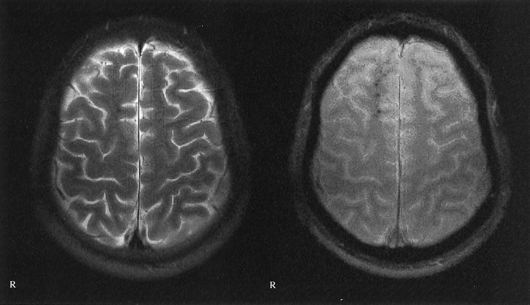
  • Fig 3.
    • Download figure
    • Open in new tab
    • Download powerpoint
    Fig 3.

    Images of a 22-year-old man who was the driver of a car that collided with a truck in June 1999. Multiple traumatic microbleeds are shown at the gray matter-white matter border. Left, T2-weighted image; right, T2*-weighted image. Images were obtained in the same plane. GCS score, 5; GOS score, 4.
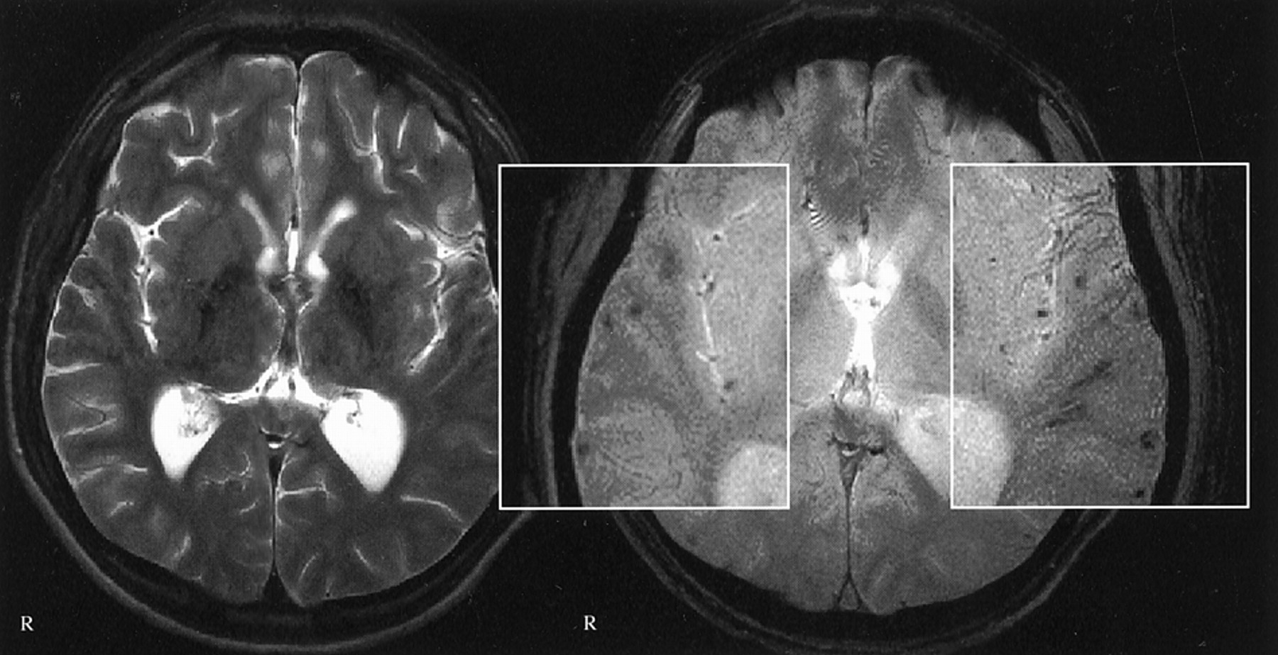
  • Fig 4.
    • Download figure
    • Open in new tab
    • Download powerpoint
    Fig 4.

    Images of a 42-year-old man who was a passenger in a traffic accident in September 2000. Traumatic microbleeds are shown in the posterior corpus callosum. Left, T2-weighted image; right, T2*-weighted image. Note the additional traumatic microbleeds in the left side of the splenium and at the gray matter-white matter border of the frontal lobes, which are not visible on the T2-weighted images. No T2-hyperintense callosal lesions are seen. GCS score, 3; GOS score, 4.

  • Fig 5.
    • Download figure
    • Open in new tab
    • Download powerpoint
    Fig 5.

    Images of a 39-year-old woman who fell off a horse in July 1996. Traumatic microbleeds are shown in the left rostral brain stem (superior cerebellar peduncle), which is a preferential site for diffuse axonal injury. The nearly symmetrical additional larger dark areas are artifacts from the petrous bone. Left, T2-weighted image; right, T2*-weighted image. GCS score, 14; GOS score, 6.

  • Fig 6.
    • Download figure
    • Open in new tab
    • Download powerpoint
    Fig 6.

    Relationships between number and site of traumatic microbleeds and clinical and imaging parameters. Top left, relationship of number of traumatic microbleeds to site of a callosal lesion (P = .0001). Top right, relationship of total number of traumatic microbleeds to the existence of a callosal lesion in general (P = .0000). Bottom left, relationship of total number of traumatic microbleeds to cause of injury (TA, traffic accident; P = .0028). Bottom right, relationship of total number of traumatic microbleeds to existence of inner brain atrophy (P = .0020).

  • Fig 7.
    • Download figure
    • Open in new tab
    • Download powerpoint
    Fig 7.

    Images of a 37-year-old man who was a pedestrian in a traffic accident in January 1992; he was hit by an automobile while under the influence of alcohol. Traumatic microbleed is shown in the left midbrain, adjacent to the red nucleus, 99 months after traumatic brain injury. Left, T2-weighted image; right, T2*-weighted image. GCS score, 4; GOS score, 4.

Tables

  • Figures
    • View popup
    TABLE 1:

    Clinical data

    Number or RangeMedianPercent
    n66
    Age (yr)17–5733
    Sex
     Male5177.3
     Female1522.7
    Time interval
     TBI–MR imaging (mo)3–29223.5
    Type of head injury
     Open1725.8
     Closed4974.2
    Cause of injury
     Traffic accident4568.2
     Fall1624.2
     Blow/assault57.6
    Multiple injuries913.6
    Medical history
     Hypertension11.5
     Diabetes mellitus00
     Lacunar stroke00
     Alcohol abuse34.5
     TBI23
    GCS score (n = 62)3–156
    GOS score3–85
    • Note.—TBI indicates traumatic brain injury; GCS, Glasgow Coma Scale; GOS, Glasgow Outcome Scale.

    • View popup
    TABLE 2:

    Frequency of focal lesions

    Total LesionsRangeMedian + SD
    T13260–312.5 + 6.8*
    T2 hypo168.50–121.0 + 3.6*
    T2 hyper650–100 + 2.25*
    T2*6080–613.75 + 13.9
    • Note.—T1 indicates T1-weighted MR imaging; T2 hypo, T2-weighted MR imaging hypointensities; T2 hyper, T2-weighted MR imaging hyperintensities; T2*, T2*-weighted MR imaging.

    • View popup
    TABLE 3:

    Frequency of specific lesion patterns and sites

    NumberPercent
    MR imaging-negative1015.2
     Concerning TMBs2030.3
    TMBs/DAI
     None or nonisolated5583.3
     Isolated1116.7
    FCC3756
     Isolated1319.7
    Side of lesion(s)
     Right57.6
     Left57.6
     Both3654.5
    Corpus callosum lesion
     None5278.8
     Anterior46.1
     Posterior812.1
     Diffuse23.0
    Brain stem involvement69.1
    Other traumatic lesions (isolated or in combination)
     Traumatic hematomas23
     Gliding contusions69
     SAB710.6
     SDH69
     EDH57.6
     Ischemic lesions34.5
    Residues
     Atrophy/hydrocephalus710.6
    • Note.—TMBs indicates traumatic microbleeds; DAI, diffuse axonal injury; FCC, focal cortical contusion; SAB, subarachnoid hemorrhage; SDH, subdural hematoma; EDH, epidural hematoma.

    • View popup
    TABLE 4:

    Tested inter-relations between the clinical parameters and the neuroradiologic data

    Relationship TestedMedian (Range)RsP
    T2* latency (TBI–MR imaging)−0.2860.020
    T2* type of TBI0.7211
     Closed4.0 (0–61)
     Open2.0 (0–52.5)
    T2* cause of TBI0.0028*
      Traffic accident5.5 (0–61)
     Fall2.5 (0–34)
     Blow/assault0 (0–5)
    T2* multiple injuries0.2481
     No3.5 (0–52.5)
     Yes5.5 (0–61)
    T2* atrophy/hydrocephalus0.0020*
     No3.5 (0–45.5)
     Yes28 (4–61)
    T2* isolated TMBs0.1266
     No3.5 (0–52.5)
     Yes4.5 (0.5–61)
    T2* corpus callosum lesion0.0000*
     No2.0 (0–34)
     Yes26.0 (1.5–61)
    T2* GCS score (n = 62) for total hypo foci−0.4270.000*
    T2* GCS score (n = 62) for hypo foci in corpus callosum−0.4280.000*
    Corpus callosum lesion GCS score (n = 62)0.002*
     No6 (3–15)
     Yes4 (3–14)
    T2* GOS score for total hypo foci−0.0620.618
    T2* GOS score for hypo foci in corpus callosum−0.1030.408
    Corpus callosum lesion GOS score0.3674
     No5 (3–8)
     Yes5 (3–8)
    Isolated TMBs GOS score0.8856
     No5 (3–8)
     Yes5 (4–7)
    Isolated FCC GOS score0.1724
     No5 (3–8)
     Yes4 (3–6)
    • Note.—Rs indicates Spearman rank correlation coefficient; T2*, T2*-weighted MR imaging; TBI, traumatic brain injury; TMBs, traumatic microbleeds; GCS, Glasgow Coma Scale; hypo, hypointense; GOS, Glasgow Outcome Scale; FCC, focal cortical contusion.

PreviousNext
Back to top

In this issue

American Journal of Neuroradiology: 24 (6)
American Journal of Neuroradiology
Vol. 24, Issue 6
1 Jun 2003
  • Table of Contents
  • Index by author
Advertisement
Print
Download PDF
Email Article

Thank you for your interest in spreading the word on American Journal of Neuroradiology.

NOTE: We only request your email address so that the person you are recommending the page to knows that you wanted them to see it, and that it is not junk mail. We do not capture any email address.

Enter multiple addresses on separate lines or separate them with commas.
Diffuse Axonal Injury Associated with Chronic Traumatic Brain Injury: Evidence from T2*-weighted Gradient-echo Imaging at 3 T
(Your Name) has sent you a message from American Journal of Neuroradiology
(Your Name) thought you would like to see the American Journal of Neuroradiology web site.
CAPTCHA
This question is for testing whether or not you are a human visitor and to prevent automated spam submissions.
Cite this article
Rainer Scheid, Cristoph Preul, Oliver Gruber, Christopher Wiggins, D. Yves von Cramon
Diffuse Axonal Injury Associated with Chronic Traumatic Brain Injury: Evidence from T2*-weighted Gradient-echo Imaging at 3 T
American Journal of Neuroradiology Jun 2003, 24 (6) 1049-1056;

Citation Manager Formats

  • BibTeX
  • Bookends
  • EasyBib
  • EndNote (tagged)
  • EndNote 8 (xml)
  • Medlars
  • Mendeley
  • Papers
  • RefWorks Tagged
  • Ref Manager
  • RIS
  • Zotero
0 Responses
Respond to this article
Share
Bookmark this article
Diffuse Axonal Injury Associated with Chronic Traumatic Brain Injury: Evidence from T2*-weighted Gradient-echo Imaging at 3 T
Rainer Scheid, Cristoph Preul, Oliver Gruber, Christopher Wiggins, D. Yves von Cramon
American Journal of Neuroradiology Jun 2003, 24 (6) 1049-1056;
del.icio.us logo Twitter logo Facebook logo Mendeley logo
  • Tweet Widget
  • Facebook Like
  • Google Plus One

Jump to section

  • Article
    • Abstract
    • Methods
    • Results
    • Discussion
    • Conclusion
    • Acknowledgments
    • Footnotes
    • References
  • Figures & Data
  • Info & Metrics
  • Responses
  • References
  • PDF

Related Articles

  • No related articles found.
  • PubMed
  • Google Scholar

Cited By...

  • Cerebral microbleeds: from depiction to interpretation
  • Maximum AmbiGuity Distance for Phase Imaging in Detection of Traumatic Cerebral Microbleeds: An Improvement over Current Imaging Practice
  • Concussion is confusing us all
  • Susceptibility-weighted MRI in mild traumatic brain injury
  • Damage to the Salience Network and Interactions with the Default Mode Network
  • A longitudinal MRI study of traumatic axonal injury in patients with moderate and severe traumatic brain injury
  • Default Mode Network Connectivity Predicts Sustained Attention Deficits after Traumatic Brain Injury
  • Cerebral Microbleeds Are Predictive of Mortality in the Elderly
  • Diffusion Tensor MR Imaging in Children with Pantothenate Kinase-Associated Neurodegeneration with Brain Iron Accumulation and Their Siblings
  • Compensatory cortical activation during performance of an attention task by patients with diffuse axonal injury: a functional magnetic resonance imaging study
  • A review of structural magnetic resonance neuroimaging
  • Crossref
  • Google Scholar

This article has not yet been cited by articles in journals that are participating in Crossref Cited-by Linking.

More in this TOC Section

  • Progression of Microstructural Damage in Spinocerebellar Ataxia Type 2: A Longitudinal DTI Study
  • SWI or T2*: Which MRI Sequence to Use in the Detection of Cerebral Microbleeds? The Karolinska Imaging Dementia Study
  • Statin Therapy Does Not Affect the Radiographic and Clinical Profile of Patients with TIA and Minor Stroke
Show more Brain

Similar Articles

Advertisement

Indexed Content

  • Current Issue
  • Accepted Manuscripts
  • Article Preview
  • Past Issues
  • Editorials
  • Editor's Choice
  • Fellows' Journal Club
  • Letters to the Editor
  • Video Articles

Cases

  • Case Collection
  • Archive - Case of the Week
  • Archive - Case of the Month
  • Archive - Classic Case

More from AJNR

  • Trainee Corner
  • Imaging Protocols
  • MRI Safety Corner
  • Book Reviews

Multimedia

  • AJNR Podcasts
  • AJNR Scantastics

Resources

  • Turnaround Time
  • Submit a Manuscript
  • Submit a Video Article
  • Submit an eLetter to the Editor/Response
  • Manuscript Submission Guidelines
  • Statistical Tips
  • Fast Publishing of Accepted Manuscripts
  • Graphical Abstract Preparation
  • Imaging Protocol Submission
  • Evidence-Based Medicine Level Guide
  • Publishing Checklists
  • Author Policies
  • Become a Reviewer/Academy of Reviewers
  • News and Updates

About Us

  • About AJNR
  • Editorial Board
  • Editorial Board Alumni
  • Alerts
  • Permissions
  • Not an AJNR Subscriber? Join Now
  • Advertise with Us
  • Librarian Resources
  • Feedback
  • Terms and Conditions
  • AJNR Editorial Board Alumni

American Society of Neuroradiology

  • Not an ASNR Member? Join Now

© 2025 by the American Society of Neuroradiology All rights, including for text and data mining, AI training, and similar technologies, are reserved.
Print ISSN: 0195-6108 Online ISSN: 1936-959X

Powered by HighWire