
Article Figures & Data
Figures
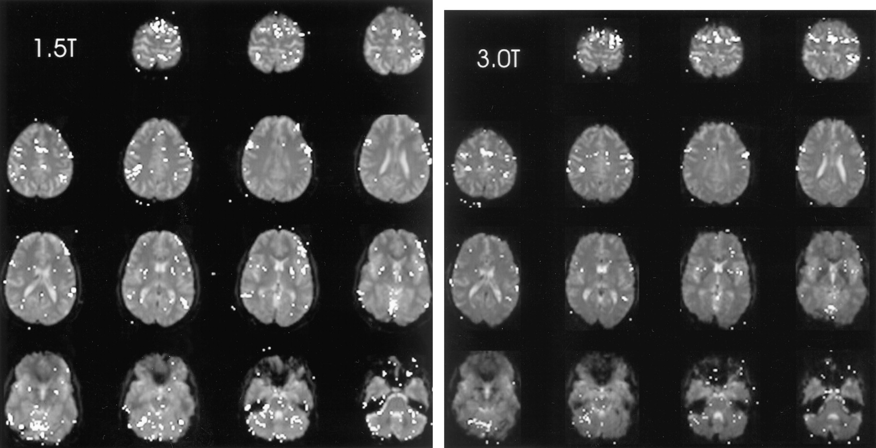
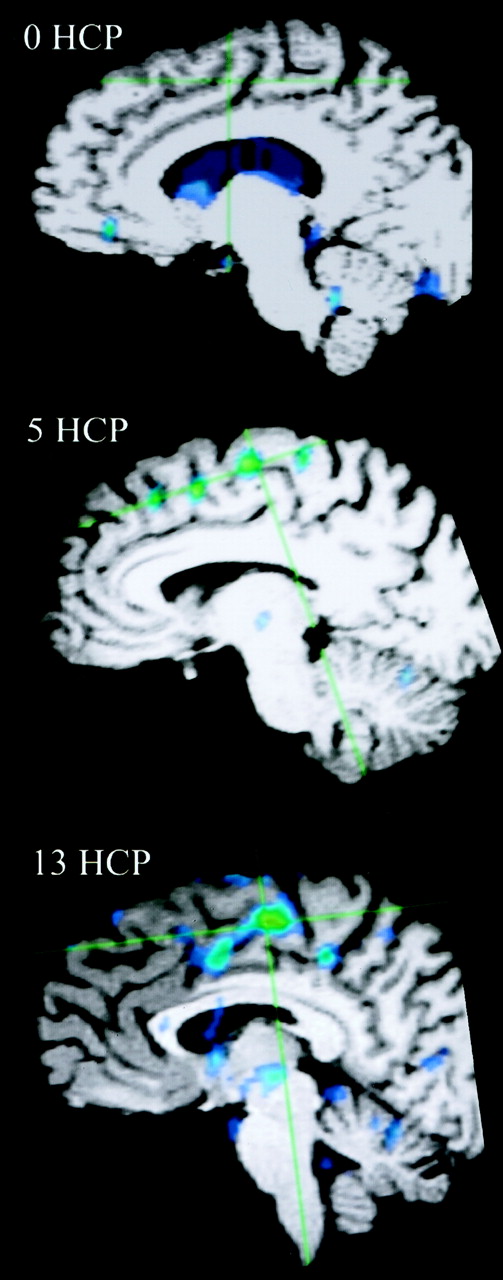
- Fig 1.
Areas of brain activation for the participant with a handicap of 13, rest-versus-golf paradigm for 1.5 T (left) and 3.0 T (right), show a similar pattern for both field strengths. Areas of brain activation are shown overlaid on the axial view images of the brain.
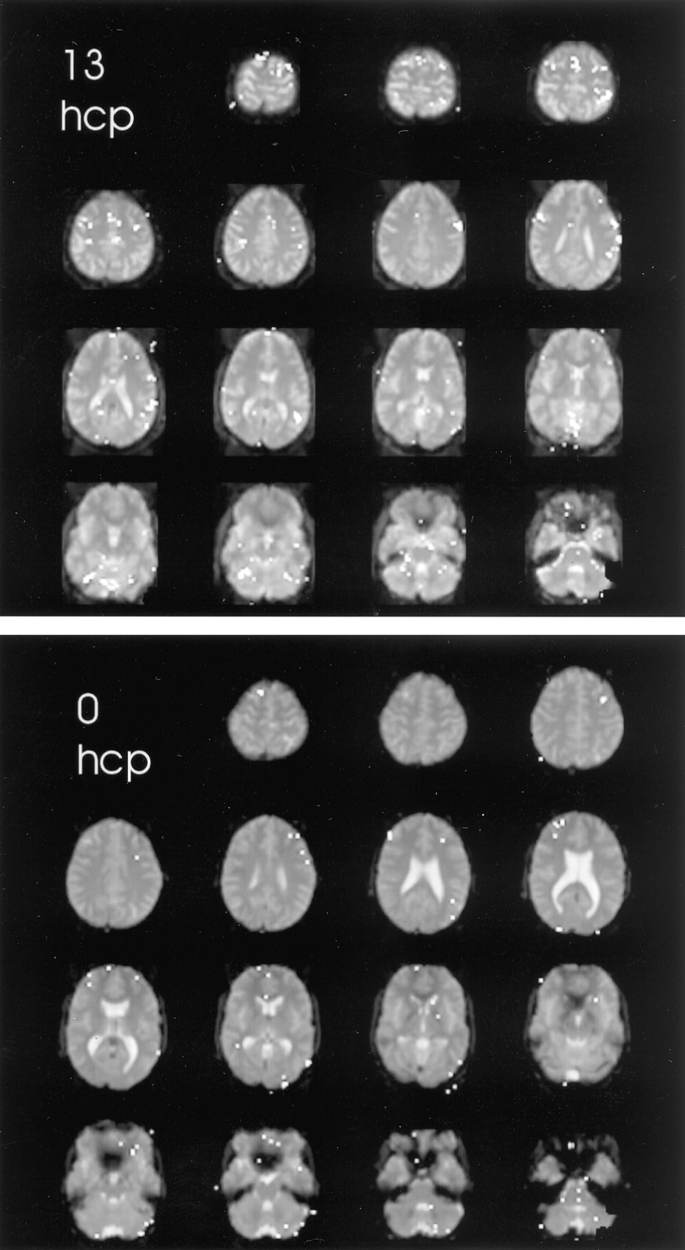
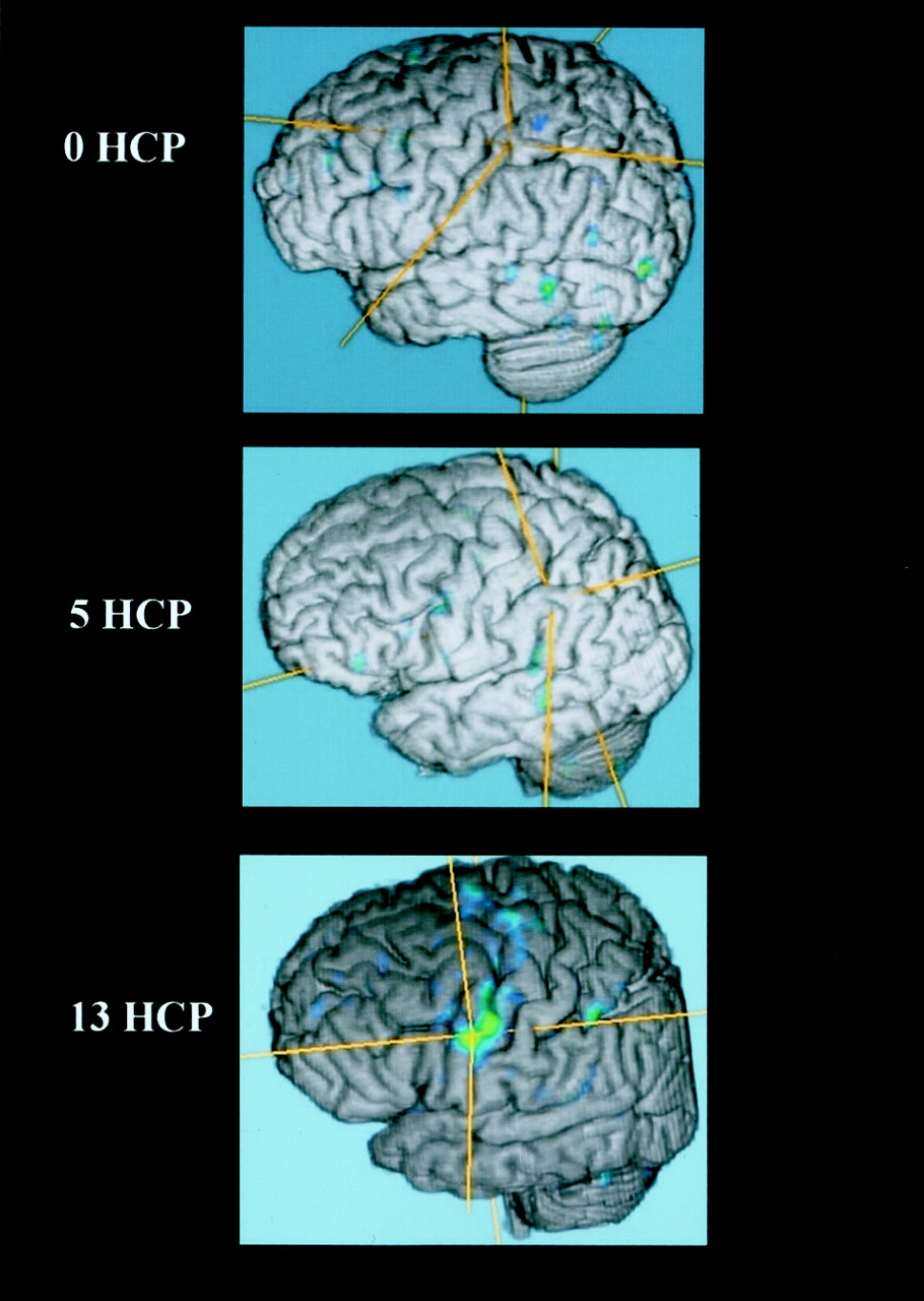
- Fig 2.
Areas of brain activation compared for the participants with handicaps (hcp) of 13 (upper panel) and 0 (lower panel), wall-versus-golf paradigm. Image of the participant with a handicap of 13 can also be compared with the other paradigm shown in Figure 1. The wall-versus-golf paradigm shows overall diminished brain activation, with much less activation in the better player.
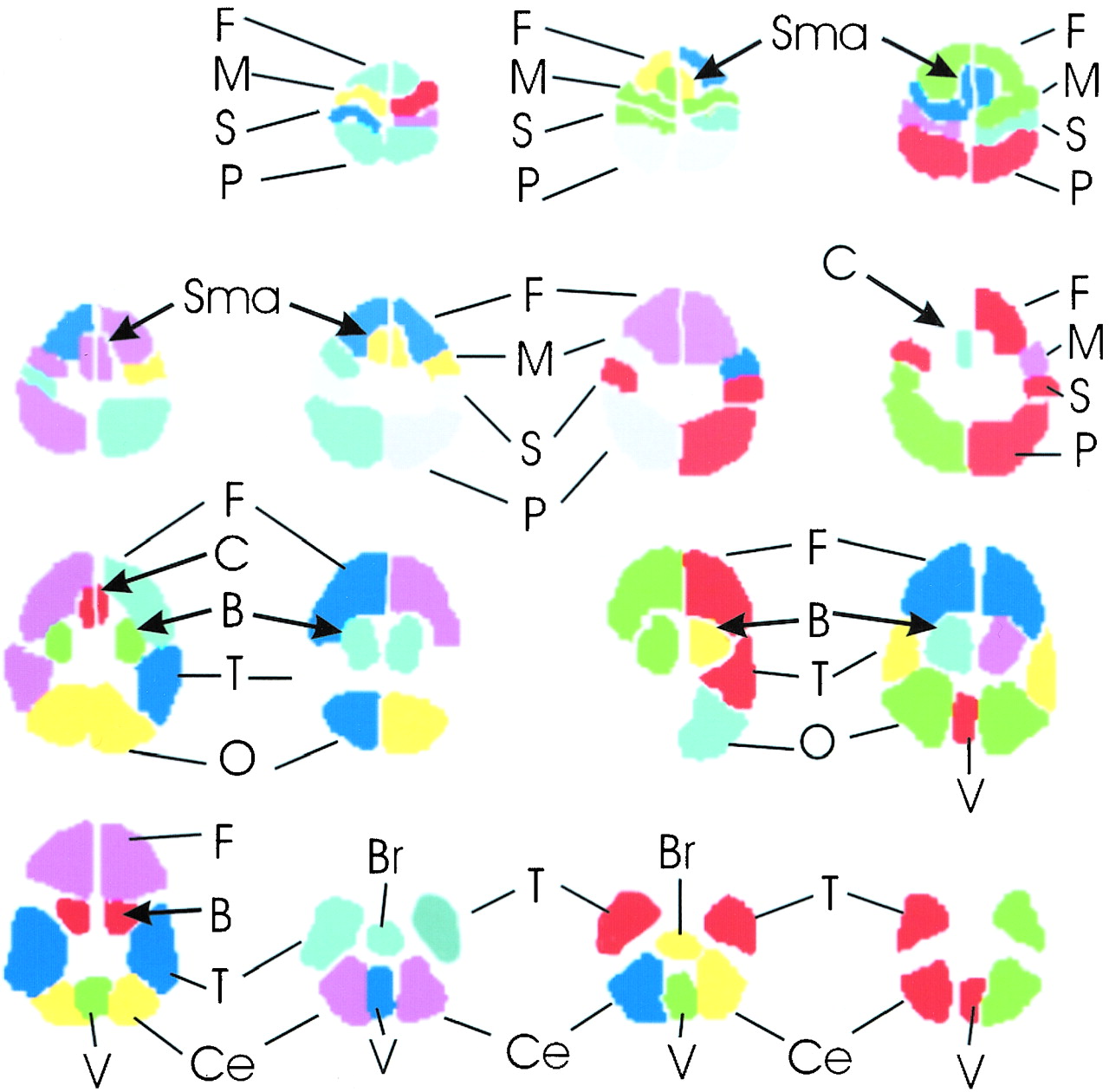
- Fig 3.
Schematic of regions of interest drawn for each participant. The brain regions are shown for each typical section of the functional MR imaging data set, proceeding as in Figures 1 and 2 from the top of the brain at top left to the base of the brain at bottom right. F, frontal lobe; M, motor; S, sensory; P, parietal; Sma, supplementary motor area; C, cingulated; B, basal ganglia; T, temporal lobe; O, occipital lobe; Ce, cerebellar hemisphere; Br, brain stem; V, vermis of cerebellum.
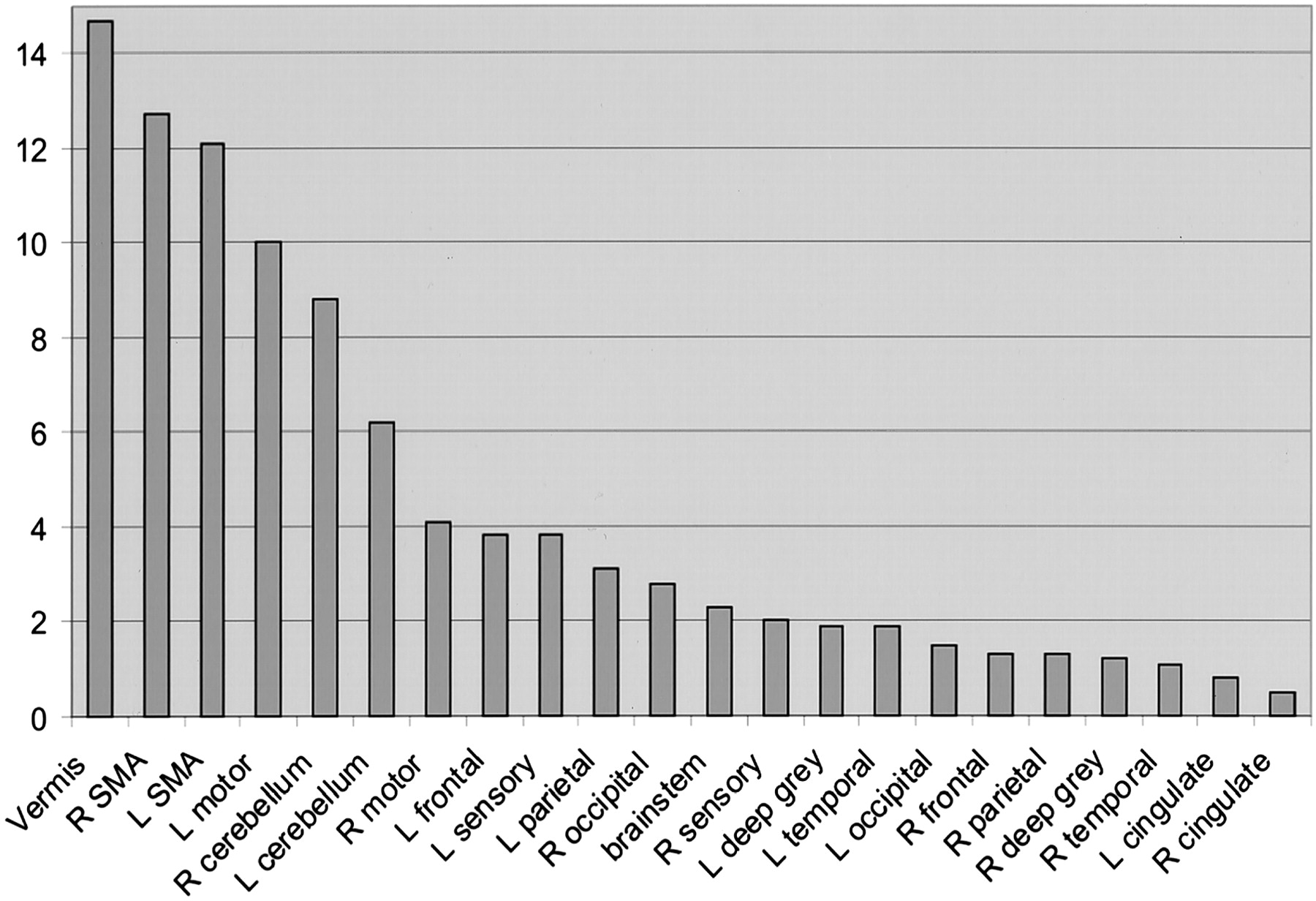
- Fig 4.
Percent area of activation in the participant with a handicap of 13. Data shown represent an average of the percentage area of activation from five separate experiments of the rest-versus-golf paradigm and are sorted from the highest level of activation to the lowest. Vermis of cerebellum, supplementary motor area, motor areas, and cerebellar hemispheres show the greatest activation. R, right; L, left; SMA, supplementary motor area.
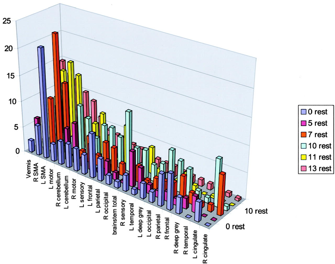
- Fig 5.
All data from the rest-versus-golf paradigm are sorted by areas of activation using the data from the participant with a handicap of 13 from the rest-versus-golf paradigm as a template. R, right; L, left; SMA, supplementary motor area.
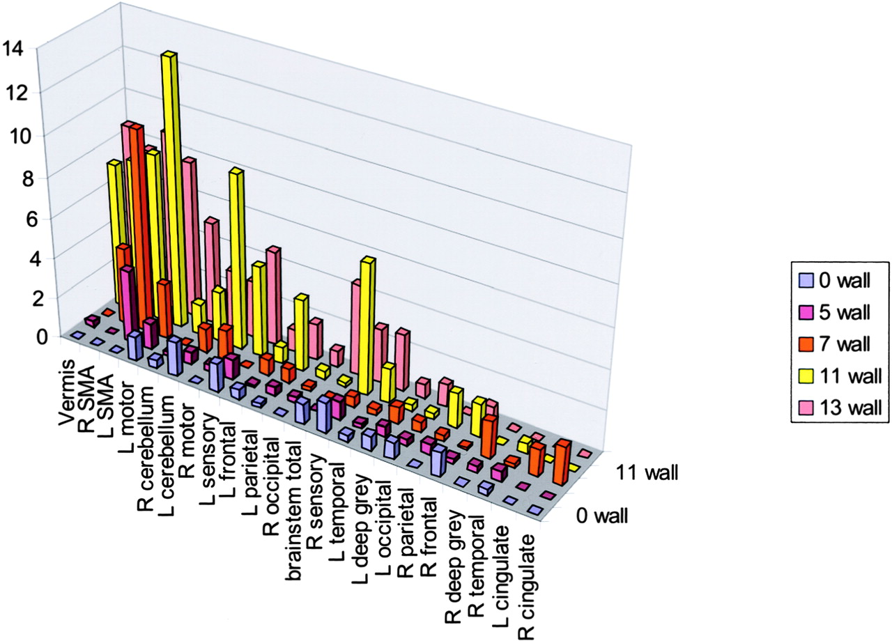
- Fig 6.
Wall-versus-golf paradigm data sorted by areas of activation using the data from the participant with a handicap of 13 as a template, as in Figure 5. R, right; L, left; SMA, supplementary motor area.
- Fig 7.
Activation of wall-versus-golf paradigm of ≥2%. The greatest activation in multiple areas occurred in the participant with the highest handicap. R, right; L, left; SMA, supplementary motor area.
- Fig 8.
Fused functional and 3D T1 gradient-echo data for three participants spanning the handicap (HCP) range show increased activation along the motor cortex in the participant with the highest handicap.
- Fig 9.
Fused functional and 3D T1 gradient-echo data show mesial surface of right hemisphere, with increasing supplementary motor area activation correlated with increasing handicap (HCP).


Tables
Region 0 Rest 0 Wall 5 Rest 5 Wall 7 Rest 7 Wall 10 Rest 11 Rest 11 Wall 13 Rest 13 Wall Vermis 2.3 0 6 0.3 1 0 7.2 8.2 7.2 14.7 8.6 R SMA 5.9 0 0.7 0 9.8 3.8 7.6 13.9 7.7 12.7 7.7 L SMA 21.2 0 0.6 3.6 22.6 10 12.6 16 8.3 12.1 8.9 L motor 3.4 1.2 1.8 1.3 13.8 2.7 2.5 14 13.2 10 7.8 R cerebellum 4.6 0.4 6.3 0.2 0.2 0 9.3 6.1 1.5 8.8 5.1 L cerebellum 4.7 1.7 7.9 0.6 1.7 1.2 7.4 5.6 2.5 6.2 3 R motor 4.4 0 1.7 0.3 5.2 1.5 4.1 8.1 8.7 4.1 2.8 L frontal 8.5 0.5 0.8 0.1 5.2 0.8 7.3 4.4 4.5 3.8 1.1 L sensory 3.8 1.4 5 1 2.1 0 7.2 0.5 0.8 3.8 4.7 L parietal 4.3 0.2 3 0.4 4.8 0.7 6.1 7.5 3.6 3.1 1.8 R occipital 1.8 0 1.4 0.3 2.3 0.2 11.5 1 0.4 2.8 0.8 Brainstem 1.2 1 0.5 0 0 0 5.2 0.7 0.2 2.3 4.5 R sensory 3.1 1.5 6.8 0.9 3.2 0.5 5.4 5.1 6.5 2 2.7 L deep gray 2.9 0.7 1.8 0.5 2.1 0.8 4.1 1.5 1.7 1.9 0.7 L temporal 1.1 0.3 4.3 0.2 1.1 0.3 3.2 0 0.3 1.9 2.8 L occipital 2 0.8 1.3 0.3 2.3 0.5 7.3 0.6 0.3 1.5 1.1 R frontal 6.8 1.2 1.2 0.2 4.6 0.1 5 2.5 1.7 1.3 0.8 R parietal 6.1 0 1.8 0.5 2.7 0.3 5.9 1 1.6 1.3 0 R deep gray 3.1 0 2 0.3 2.1 1.8 1.6 0 0 1.2 0 R temporal 0 0.3 3.1 0.5 0.9 0.2 1 0.4 0.5 1.1 0 L cingulate 3.7 0 0 0 1.3 1.3 8.9 0 0 0.8 0 R cingulate 0 0 0 0 7.6 1.9 0.8 0 0 S0.5 0 Note.—R indicates right; L, left; SMA, supplementary motor area.
* Data are sorted from greatest to least activation, using the average of five series for the participant with a handicap of 13 rest-versus-golf paradigm (boldface type).
Handicap 0 5 7 11 13 Vermis 7.2 8.6 R SMA 3.8 7.7 7.7 L SMA 3.6 10 8.3 8.9 L motor 2.7 13.2 7.8 R cerebellum 5.1 L cerebellum 2.5 3 R motor 8.7 2.8 L frontal L sensory 4.5 4.7 L parietal 3.6 R occipital Brainstem 4.5 R sensory 6.5 2.7 L deep gray L temporal 2.8 L occipital R frontal R parietal R deep gray R temporal L cingulate R cingulate Note.—R indicates right; L, left; SMA, supplementary motor area.


