
Article Figures & Data
Figures
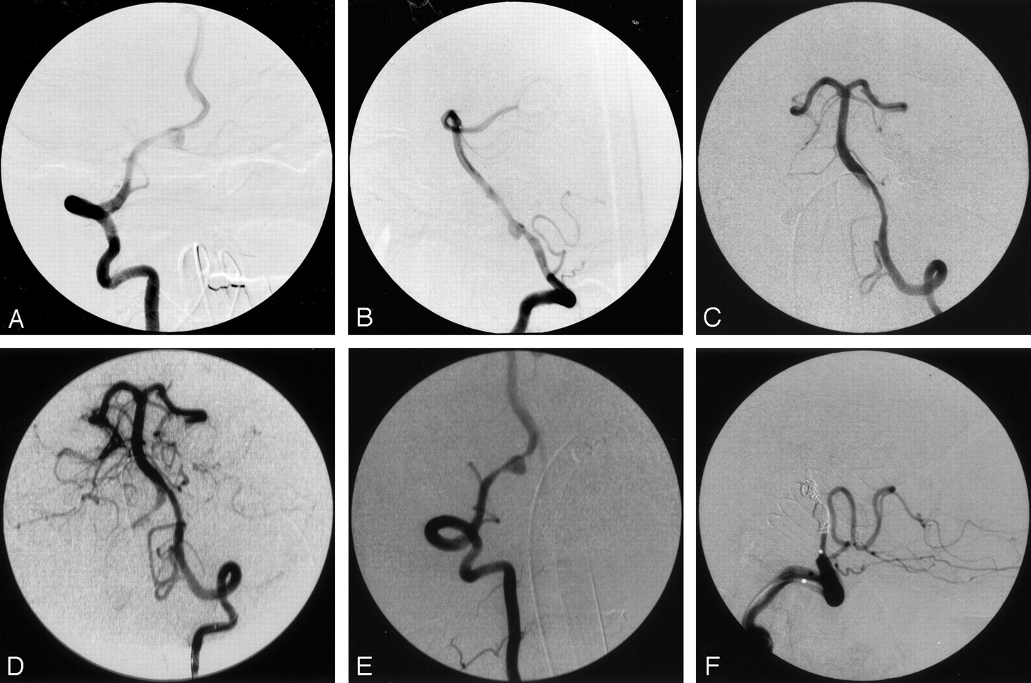
- Fig 1.
Case 1, a 55-year-old woman with acute subarachnoid hemorrhage.
A, Anteroposterior right vertebral angiogram, showing the eccentric location of the aneurysm relative to the vertebral artery.
B, Lateral right vertebral angiogram, showing aneurysm of the V4 segment of the vertebral artery. Tapered narrowing proximal and distal suggests dissection, which is confirmed by visualization of a linear filling defect representing intima.
C, Sixteen hours later, left vertebral anteroposterior angiogram, showing tapered narrowing of the vertebral from propagation of the dissection, with severe narrowing at the vertebrobasilar junction.
D, Left vertebral angiogram after angioplasty, showing markedly improved caliber from vertebral to basilar artery.
E, Anteroposterior right vertebral angiogram, showing pseudoaneurysm between the right posterior inferior cerebellar artery and the vertebrobasilar junction.
F, Lateral right vertebral angiogram after embolization, showing right vertebral occlusion distal to the PICA.
- Fig 2.
Case 2, a 67-year-old man with acute basilar dissection.
A, Anteroposterior right vertebral angiogram (before superselective catheterization), showing occlusion of the right posterior cerebral and left superior cerebellar arteries with small stumps. This was believed to be secondary to thromboembolism. Irregularity of the intradural right vertebral artery, first thought to represent atherosclerosis, may be secondary to dissection. In retrospect, a small intimal flap was visible in the midbasilar segment on the lateral view.
B, After 30 mg of t-PA, there was no improvement; however, following blind catheterization of the right PCA, the right PCA and left SCA show normalized flow.
C, Lateral right vertebral injection, clearly showing a linear filling defect of the basilar artery indicating dissection. This had been present before intervention but was unrecognized until remasking and pixel shifting were performed on the initial runs.
- Fig 3.
Case 2, 6 days later.
Frontal (A) and lateral right vertebral (B) angiograms. There is worsening of the dissection with multiple branch defects and intraluminal clot. Lateral (C) and anteroposterior right vertebral (D) angiograms after second stent placement. The basilar artery lumen is reestablished, and terminal branches now fill. A small residual of the false lumen is seen anterior to the basilar artery.


