Advertisement

Main menu

  • Home
  • Content
    • Current Issue
    • Accepted Manuscripts
    • Article Preview
    • Past Issue Archive
    • Video Articles
    • AJNR Case Collection
    • Case of the Week Archive
    • Case of the Month Archive
    • Classic Case Archive
  • Special Collections
    • AJNR Awards
    • Low-Field MRI
    • Alzheimer Disease
    • ASNR Foundation Special Collection
    • Photon-Counting CT
    • View All
  • Multimedia
    • AJNR Podcasts
    • AJNR SCANtastic
    • Trainee Corner
    • MRI Safety Corner
    • Imaging Protocols
  • For Authors
    • Submit a Manuscript
    • Submit a Video Article
    • Submit an eLetter to the Editor/Response
    • Manuscript Submission Guidelines
    • Statistical Tips
    • Fast Publishing of Accepted Manuscripts
    • Graphical Abstract Preparation
    • Imaging Protocol Submission
    • Author Policies
  • About Us
    • About AJNR
    • Editorial Board
    • Editorial Board Alumni
  • More
    • Become a Reviewer/Academy of Reviewers
    • Subscribers
    • Permissions
    • Alerts
    • Feedback
    • Advertisers
    • ASNR Home

User menu

  • Alerts
  • Log in

Search

  • Advanced search
American Journal of Neuroradiology

American Journal of Neuroradiology

ASHNR American Society of Functional Neuroradiology ASHNR American Society of Pediatric Neuroradiology ASSR
  • Alerts
  • Log in

Advanced Search

  • Home
  • Content
    • Current Issue
    • Accepted Manuscripts
    • Article Preview
    • Past Issue Archive
    • Video Articles
    • AJNR Case Collection
    • Case of the Week Archive
    • Case of the Month Archive
    • Classic Case Archive
  • Special Collections
    • AJNR Awards
    • Low-Field MRI
    • Alzheimer Disease
    • ASNR Foundation Special Collection
    • Photon-Counting CT
    • View All
  • Multimedia
    • AJNR Podcasts
    • AJNR SCANtastic
    • Trainee Corner
    • MRI Safety Corner
    • Imaging Protocols
  • For Authors
    • Submit a Manuscript
    • Submit a Video Article
    • Submit an eLetter to the Editor/Response
    • Manuscript Submission Guidelines
    • Statistical Tips
    • Fast Publishing of Accepted Manuscripts
    • Graphical Abstract Preparation
    • Imaging Protocol Submission
    • Author Policies
  • About Us
    • About AJNR
    • Editorial Board
    • Editorial Board Alumni
  • More
    • Become a Reviewer/Academy of Reviewers
    • Subscribers
    • Permissions
    • Alerts
    • Feedback
    • Advertisers
    • ASNR Home
  • Follow AJNR on Twitter
  • Visit AJNR on Facebook
  • Follow AJNR on Instagram
  • Join AJNR on LinkedIn
  • RSS Feeds

AJNR Awards, New Junior Editors, and more. Read the latest AJNR updates

OtherBRAIN

Proton MR Spectroscopy of Gliomatosis Cerebri: Case Report of Elevated Myoinositol with Normal Choline Levels

Efrat Saraf-Lavi, Brian C. Bowen, Pradip M. Pattany, Evelyn M.L. Sklar, James B. Murdoch and Carol K. Petito
American Journal of Neuroradiology May 2003, 24 (5) 946-951;
Efrat Saraf-Lavi
  • Find this author on Google Scholar
  • Find this author on PubMed
  • Search for this author on this site
Brian C. Bowen
  • Find this author on Google Scholar
  • Find this author on PubMed
  • Search for this author on this site
Pradip M. Pattany
  • Find this author on Google Scholar
  • Find this author on PubMed
  • Search for this author on this site
Evelyn M.L. Sklar
  • Find this author on Google Scholar
  • Find this author on PubMed
  • Search for this author on this site
James B. Murdoch
  • Find this author on Google Scholar
  • Find this author on PubMed
  • Search for this author on this site
Carol K. Petito
  • Find this author on Google Scholar
  • Find this author on PubMed
  • Search for this author on this site
  • Article
  • Figures & Data
  • Info & Metrics
  • Responses
  • References
  • PDF
Loading

Article Figures & Data

Figures

  • Tables
  • Fig 1.
    • Download figure
    • Open in new tab
    • Download powerpoint
    Fig 1.

    Spin-echo MR images. Axial view FLAIR image (left) shows extensive hyperintensity involving the left hemisphere white matter and basal ganglia, bilateral thalami, and splenium of corpus callosum, with mild mass effect. Comparison between unenhanced T1-weighted image (center) and contrast-enhanced T1-weighted image (right) reveals no appreciable enhancement.

  • Fig 2.
    • Download figure
    • Open in new tab
    • Download powerpoint
    Fig 2.

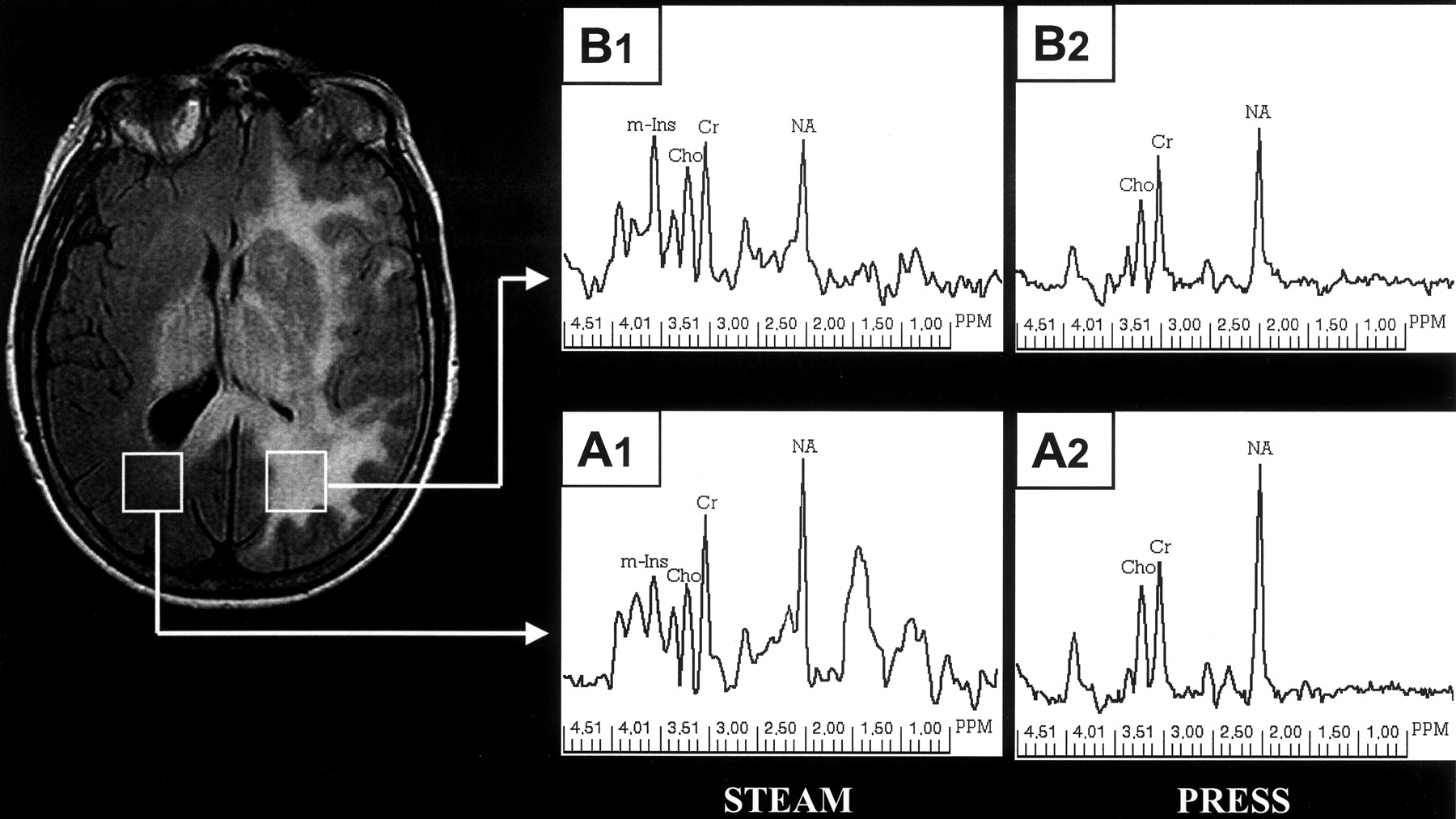
    Single voxel spectroscopy. Localized STEAM (TE 20) spectra were acquired from the normal appearing right periatrial region (A1) and from the hyperintense left periatrial region (B1). PRESS (TE 135) spectra were acquired from the same locations (A2 and B2, respectively). The right and left STEAM spectra are displayed by using the same vertical scale factor, as are the right and left PRESS spectra.

  • Fig 3.
    • Download figure
    • Open in new tab
    • Download powerpoint
    Fig 3.

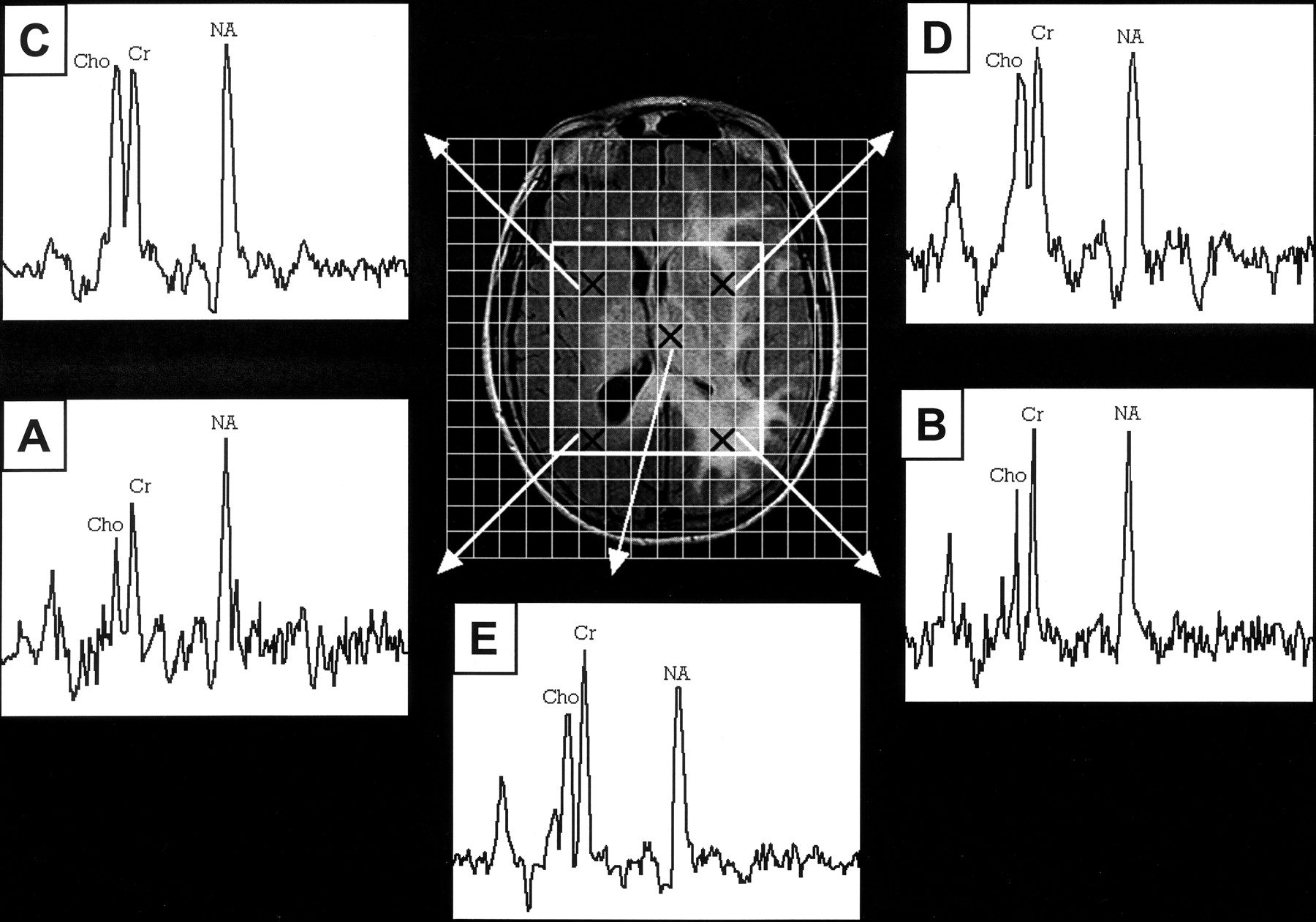
    Spectroscopic imaging. Multi-voxel spectra (TE 135) from five different locations (marked by Xs) indicated on the axial FLAIR localizer. A, Right periatrial region. B, Left periatrial region. C, Right basal ganglia region. D, Left basal ganglia region. E, Left thalamus.

  • Fig 4.
    • Download figure
    • Open in new tab
    • Download powerpoint
    Fig 4.

    Histopathology of left thalamic biopsy specimen. Moderately cellular infiltrating glioma (hematoxylin and eosin; original magnification, ×400).

Tables

  • Figures
    • View popup
    Table 1.

    Metabolite concentration ratios from single-voxel MR spectroscopyα

    RatioSTEAMPRESS
    Left PAβRight PAControlγLeft PARight PAControlγ
    NA/Cr0.810.921.11 ± 0.121.161.451.61 ± 0.17
    Cho/Cr0.230.210.17 ± 0.050.230.250.23 ± 0.03
    m-Ins/Cr0.730.430.46 ± 0.05ndδndnd
    Cho/NA0.280.230.15 ± 0.040.20.180.15 ± 0.02
    m-Ins/NA0.90.470.42 ± 0.04ndndnd
    • α Metabolite concentration ratios must be multiplied by the ratios of protons contributing to the corresponding resonances in order to obtain “peak area ratios.” The ratios for the protons are as follows: NA/Cr = 3/3, Cho/Cr = 9/3, m-Ins/Cr = ∼3.4/3, Cho/NA = 9/3, m-Ins/NA = ∼3.4/3. Thus, the “peak area ratio” for STEAM, left PA Cho/Cr = 0.23 (9/3) = 0.69. The peak area ratios calculated in this way from the concentration ratios yield results that are qualitatively in agreement with the relative peak areas in Figure 2. If the linewidths of the resonances are not excessively broadened or variable, similar qualitative agreement is achieved when relative peak amplitudes, rather than peak areas, are assessed. {Addendum: The estimate of 3.4 protons contributing to the 3.56-ppm myo-Ins resonance was based on two approaches: (1) analysis of synthetic spectra generated with the TE/TM values used in this case, (2) analysis of metabolite peak area ratios (from a standard peak fitting algorithm) relative to concentration ratios (from LCModel) in a database of 124 similarly acquired control spectra (JB Murdoch, unpublished results).}

    • β PA = periatrial region.

    • γ Control data reported as mean ± standard deviation.

    • δ nd = not determined.

PreviousNext
Back to top

In this issue

American Journal of Neuroradiology: 24 (5)
American Journal of Neuroradiology
Vol. 24, Issue 5
1 May 2003
  • Table of Contents
  • Index by author
Advertisement
Print
Download PDF
Email Article

Thank you for your interest in spreading the word on American Journal of Neuroradiology.

NOTE: We only request your email address so that the person you are recommending the page to knows that you wanted them to see it, and that it is not junk mail. We do not capture any email address.

Enter multiple addresses on separate lines or separate them with commas.
Proton MR Spectroscopy of Gliomatosis Cerebri: Case Report of Elevated Myoinositol with Normal Choline Levels
(Your Name) has sent you a message from American Journal of Neuroradiology
(Your Name) thought you would like to see the American Journal of Neuroradiology web site.
CAPTCHA
This question is for testing whether or not you are a human visitor and to prevent automated spam submissions.
Cite this article
Efrat Saraf-Lavi, Brian C. Bowen, Pradip M. Pattany, Evelyn M.L. Sklar, James B. Murdoch, Carol K. Petito
Proton MR Spectroscopy of Gliomatosis Cerebri: Case Report of Elevated Myoinositol with Normal Choline Levels
American Journal of Neuroradiology May 2003, 24 (5) 946-951;

Citation Manager Formats

  • BibTeX
  • Bookends
  • EasyBib
  • EndNote (tagged)
  • EndNote 8 (xml)
  • Medlars
  • Mendeley
  • Papers
  • RefWorks Tagged
  • Ref Manager
  • RIS
  • Zotero
0 Responses
Respond to this article
Share
Bookmark this article
Proton MR Spectroscopy of Gliomatosis Cerebri: Case Report of Elevated Myoinositol with Normal Choline Levels
Efrat Saraf-Lavi, Brian C. Bowen, Pradip M. Pattany, Evelyn M.L. Sklar, James B. Murdoch, Carol K. Petito
American Journal of Neuroradiology May 2003, 24 (5) 946-951;
del.icio.us logo Twitter logo Facebook logo Mendeley logo
  • Tweet Widget
  • Facebook Like
  • Google Plus One

Jump to section

  • Article
    • Abstract
    • Case Report
    • Discussion
    • Conclusion
    • References
  • Figures & Data
  • Info & Metrics
  • Responses
  • References
  • PDF

Related Articles

  • No related articles found.
  • PubMed
  • Google Scholar

Cited By...

  • In Vivo 2-Hydroxyglutarate Monitoring With Edited MR Spectroscopy for the Follow-up of IDH-Mutant Diffuse Gliomas: The IDASPE Prospective Study
  • Gliomatosis Cerebri
  • Crossref
  • Google Scholar

This article has not yet been cited by articles in journals that are participating in Crossref Cited-by Linking.

More in this TOC Section

  • Progression of Microstructural Damage in Spinocerebellar Ataxia Type 2: A Longitudinal DTI Study
  • SWI or T2*: Which MRI Sequence to Use in the Detection of Cerebral Microbleeds? The Karolinska Imaging Dementia Study
  • Statin Therapy Does Not Affect the Radiographic and Clinical Profile of Patients with TIA and Minor Stroke
Show more BRAIN

Similar Articles

Advertisement

Indexed Content

  • Current Issue
  • Accepted Manuscripts
  • Article Preview
  • Past Issues
  • Editorials
  • Editor's Choice
  • Fellows' Journal Club
  • Letters to the Editor
  • Video Articles

Cases

  • Case Collection
  • Archive - Case of the Week
  • Archive - Case of the Month
  • Archive - Classic Case

More from AJNR

  • Trainee Corner
  • Imaging Protocols
  • MRI Safety Corner

Multimedia

  • AJNR Podcasts
  • AJNR Scantastics

Resources

  • Turnaround Time
  • Submit a Manuscript
  • Submit a Video Article
  • Submit an eLetter to the Editor/Response
  • Manuscript Submission Guidelines
  • Statistical Tips
  • Fast Publishing of Accepted Manuscripts
  • Graphical Abstract Preparation
  • Imaging Protocol Submission
  • Evidence-Based Medicine Level Guide
  • Publishing Checklists
  • Author Policies
  • Become a Reviewer/Academy of Reviewers
  • News and Updates

About Us

  • About AJNR
  • Editorial Board
  • Editorial Board Alumni
  • Alerts
  • Permissions
  • Not an AJNR Subscriber? Join Now
  • Advertise with Us
  • Librarian Resources
  • Feedback
  • Terms and Conditions
  • AJNR Editorial Board Alumni

American Society of Neuroradiology

  • Not an ASNR Member? Join Now

© 2025 by the American Society of Neuroradiology All rights, including for text and data mining, AI training, and similar technologies, are reserved.
Print ISSN: 0195-6108 Online ISSN: 1936-959X

Powered by HighWire