Advertisement
  • Main menu
  • User menu
  • Search
  • English ▼
    • English
    • Afrikaans
    • Albanian
    • Amharic
    • Arabic
    • Armenian
    • Azerbaijani
    • Basque
    • Belarusian
    • Bengali
    • Bosnian
    • Bulgarian
    • Catalan
    • Cebuano
    • Chichewa
    • Chinese (Simplified)
    • Chinese (Traditional)
    • Corsican
    • Croatian
    • Czech
    • Danish
    • Dutch
    • Esperanto
    • Estonian
    • Filipino
    • Finnish
    • French
    • Frisian
    • Galician
    • Georgian
    • German
    • Greek
    • Gujarati
    • Haitian Creole
    • Hausa
    • Hawaiian
    • Hebrew
    • Hindi
    • Hmong
    • Hungarian
    • Icelandic
    • Igbo
    • Indonesian
    • Irish
    • Italian
    • Japanese
    • Javanese
    • Kannada
    • Kazakh
    • Khmer
    • Korean
    • Kurdish (Kurmanji)
    • Kyrgyz
    • Lao
    • Latin
    • Latvian
    • Lithuanian
    • Luxembourgish
    • Macedonian
    • Malagasy
    • Malay
    • Malayalam
    • Maltese
    • Maori
    • Marathi
    • Mongolian
    • Myanmar (Burmese)
    • Nepali
    • Norwegian
    • Pashto
    • Persian
    • Polish
    • Portuguese
    • Punjabi
    • Romanian
    • Russian
    • Samoan
    • Scottish Gaelic
    • Serbian
    • Sesotho
    • Shona
    • Sindhi
    • Sinhala
    • Slovak
    • Slovenian
    • Somali
    • Spanish
    • Sudanese
    • Swahili
    • Swedish
    • Tajik
    • Tamil
    • Telugu
    • Thai
    • Turkish
    • Ukrainian
    • Urdu
    • Uzbek
    • Vietnamese
    • Welsh
    • Xhosa
    • Yiddish
    • Yoruba
    • Zulu

Main menu

  • Home
  • Content
    • Current Issue
    • Accepted Manuscripts
    • Article Preview
    • Past Issue Archive
    • Video Articles
    • AJNR Case Collection
    • Case of the Week Archive
    • Case of the Month Archive
    • Classic Case Archive
  • Special Collections
    • Advancing NeuroMRI with High-Relaxivity Contrast Agents
    • AJNR Awards
    • Low-Field MRI
    • Alzheimer Disease
    • ASNR Foundation Special Collection
    • Photon-Counting CT
    • View All
  • Multimedia
    • AJNR Podcasts
    • AJNR SCANtastic
    • Trainee Corner
    • MRI Safety Corner
    • Imaging Protocols
  • For Authors
    • Submit a Manuscript
    • Submit a Video Article
    • Submit an eLetter to the Editor/Response
    • Manuscript Submission Guidelines
    • Statistical Tips
    • Fast Publishing of Accepted Manuscripts
    • Graphical Abstract Preparation
    • Imaging Protocol Submission
    • Author Policies
  • About Us
    • About AJNR
    • Editorial Board
    • Editorial Board Alumni
  • More
    • Become a Reviewer/Academy of Reviewers
    • Subscribers
    • Permissions
    • Alerts
    • Feedback
    • Advertisers
    • ASNR Home

User menu

  • Alerts
  • Log in

Search

  • Advanced search
American Journal of Neuroradiology

American Journal of Neuroradiology

ASHNR American Society of Functional Neuroradiology ASHNR American Society of Pediatric Neuroradiology ASSR
  • Alerts
  • Log in

Advanced Search

English ▼
  • English
  • Afrikaans
  • Albanian
  • Amharic
  • Arabic
  • Armenian
  • Azerbaijani
  • Basque
  • Belarusian
  • Bengali
  • Bosnian
  • Bulgarian
  • Catalan
  • Cebuano
  • Chichewa
  • Chinese (Simplified)
  • Chinese (Traditional)
  • Corsican
  • Croatian
  • Czech
  • Danish
  • Dutch
  • Esperanto
  • Estonian
  • Filipino
  • Finnish
  • French
  • Frisian
  • Galician
  • Georgian
  • German
  • Greek
  • Gujarati
  • Haitian Creole
  • Hausa
  • Hawaiian
  • Hebrew
  • Hindi
  • Hmong
  • Hungarian
  • Icelandic
  • Igbo
  • Indonesian
  • Irish
  • Italian
  • Japanese
  • Javanese
  • Kannada
  • Kazakh
  • Khmer
  • Korean
  • Kurdish (Kurmanji)
  • Kyrgyz
  • Lao
  • Latin
  • Latvian
  • Lithuanian
  • Luxembourgish
  • Macedonian
  • Malagasy
  • Malay
  • Malayalam
  • Maltese
  • Maori
  • Marathi
  • Mongolian
  • Myanmar (Burmese)
  • Nepali
  • Norwegian
  • Pashto
  • Persian
  • Polish
  • Portuguese
  • Punjabi
  • Romanian
  • Russian
  • Samoan
  • Scottish Gaelic
  • Serbian
  • Sesotho
  • Shona
  • Sindhi
  • Sinhala
  • Slovak
  • Slovenian
  • Somali
  • Spanish
  • Sudanese
  • Swahili
  • Swedish
  • Tajik
  • Tamil
  • Telugu
  • Thai
  • Turkish
  • Ukrainian
  • Urdu
  • Uzbek
  • Vietnamese
  • Welsh
  • Xhosa
  • Yiddish
  • Yoruba
  • Zulu
  • Home
  • Content
    • Current Issue
    • Accepted Manuscripts
    • Article Preview
    • Past Issue Archive
    • Video Articles
    • AJNR Case Collection
    • Case of the Week Archive
    • Case of the Month Archive
    • Classic Case Archive
  • Special Collections
    • Advancing NeuroMRI with High-Relaxivity Contrast Agents
    • AJNR Awards
    • Low-Field MRI
    • Alzheimer Disease
    • ASNR Foundation Special Collection
    • Photon-Counting CT
    • View All
  • Multimedia
    • AJNR Podcasts
    • AJNR SCANtastic
    • Trainee Corner
    • MRI Safety Corner
    • Imaging Protocols
  • For Authors
    • Submit a Manuscript
    • Submit a Video Article
    • Submit an eLetter to the Editor/Response
    • Manuscript Submission Guidelines
    • Statistical Tips
    • Fast Publishing of Accepted Manuscripts
    • Graphical Abstract Preparation
    • Imaging Protocol Submission
    • Author Policies
  • About Us
    • About AJNR
    • Editorial Board
    • Editorial Board Alumni
  • More
    • Become a Reviewer/Academy of Reviewers
    • Subscribers
    • Permissions
    • Alerts
    • Feedback
    • Advertisers
    • ASNR Home
  • Follow AJNR on Twitter
  • Visit AJNR on Facebook
  • Follow AJNR on Instagram
  • Join AJNR on LinkedIn
  • RSS Feeds

AJNR Awards, New Junior Editors, and more. Read the latest AJNR updates


Improved Turnaround Times | Median time to first decision: 12 days

Research ArticleBrain

Measurement of Cerebral Blood Flow in Chronic Carotid Occlusive Disease: Comparison of Dynamic Susceptibility Contrast Perfusion MR Imaging with Positron Emission Tomography

Pratik Mukherjee, Hyunseon Christine Kang, Tom O. Videen, Robert C. McKinstry, William J. Powers and Colin P. Derdeyn
American Journal of Neuroradiology May 2003, 24 (5) 862-871;
Pratik Mukherjee
  • Find this author on Google Scholar
  • Find this author on PubMed
  • Search for this author on this site
Hyunseon Christine Kang
  • Find this author on Google Scholar
  • Find this author on PubMed
  • Search for this author on this site
Tom O. Videen
  • Find this author on Google Scholar
  • Find this author on PubMed
  • Search for this author on this site
Robert C. McKinstry
  • Find this author on Google Scholar
  • Find this author on PubMed
  • Search for this author on this site
William J. Powers
  • Find this author on Google Scholar
  • Find this author on PubMed
  • Search for this author on this site
Colin P. Derdeyn
  • Find this author on Google Scholar
  • Find this author on PubMed
  • Search for this author on this site
  • Article
  • Figures & Data
  • Info & Metrics
  • Responses
  • References
  • PDF
Loading

Article Figures & Data

Figures

  • Tables
  • Fig 1.
    • Download figure
    • Open in new tab
    • Download powerpoint
    Fig 1.

    T2*-weighted gradient-echo single shot echo-planar transverse image (800/47; flip angle, 60 degrees), obtained at the level of the sylvian fissures in patient 7 (Table 1), who had left carotid occlusion, shows AIF selection at the M1 and M2 segments of the MCA. Pixels chosen for AIF determination are illustrated in yellow. Ten to 12 pixels were chosen in the MCA, both for an AIF ipsilateral to the occlusion and an AIF contralateral to the occlusion.
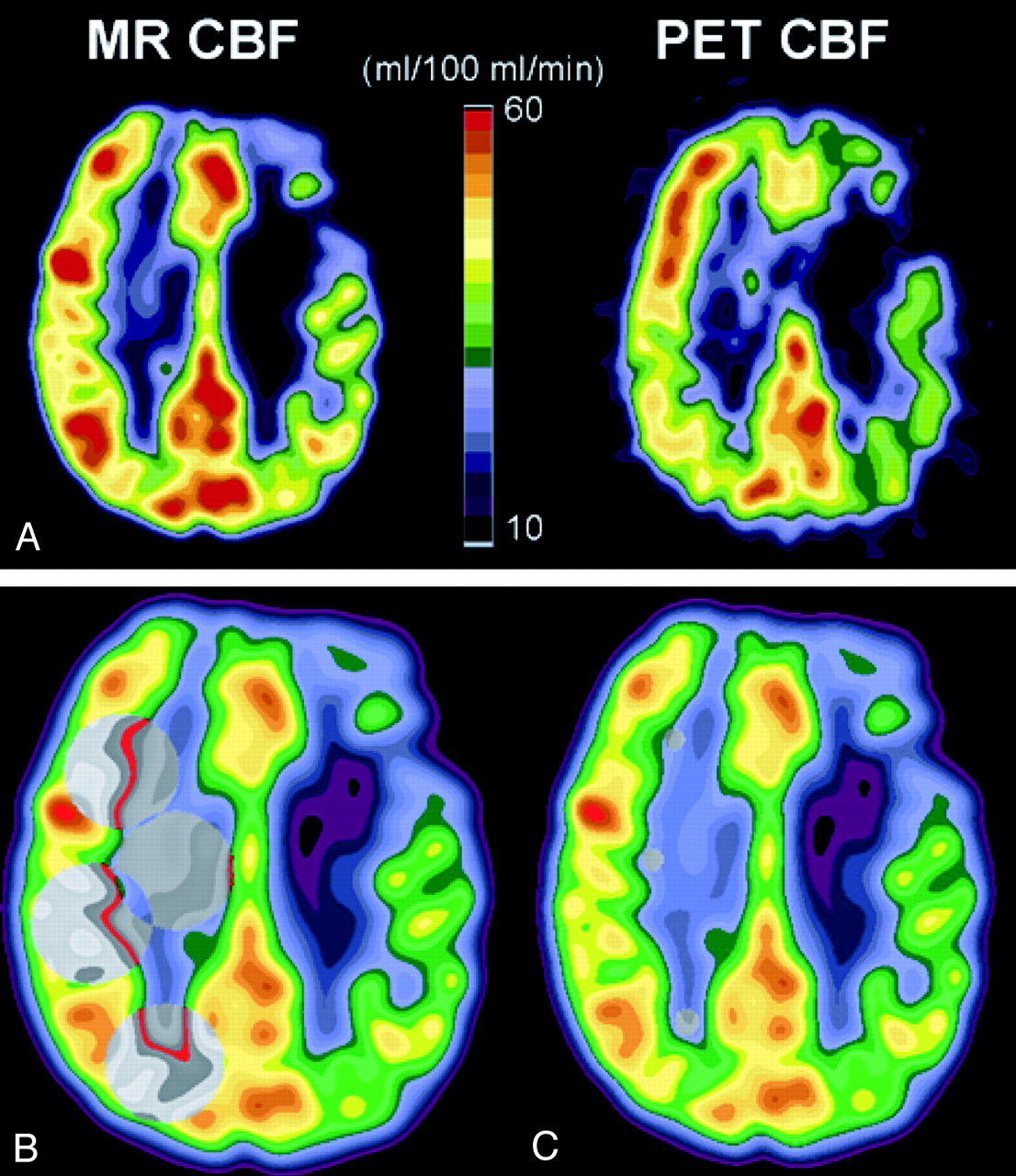
  • Fig 2.
    • Download figure
    • Open in new tab
    • Download powerpoint
    Fig 2.

    CBF in patient 4 (Table 1), who had occlusion of the left internal carotid artery.

    A, CBF measured with dynamic susceptibility contrast-enhanced MR imaging (MR CBF, left) and CBF measured with [15O]-H2O PET (PET CBR, right). Axial view MR image and PET scan are co-registered to the same location, filtered to the same spatial resolution as the PET scan, and displayed with the same color scale. Diffusely decreased CBF in the left hemisphere, ipsilateral to the occluded carotid artery, can be identified on both the MR image and PET scan, with more severely reduced CBF focally within a chronic infarct in the left frontal lobe.

    B, Large regions of interest (gray circles) for CBF measurement are shown on MR image obtained in the hemisphere contralateral to the carotid occlusion. Homologous regions of interest were placed in the hemisphere ipsilateral to the occlusion (see Methods).

    C, Small subcortical regions of interest (gray circles) in normal appearing white matter of the hemisphere contralateral to the carotid occlusion are shown on MR image. These subcortical white matter regions were used to determine normal white matter CBF for the purpose of scaling relative CBF values measured by MR imaging to physically meaningful units of mL/100 mL/min (see Methods).
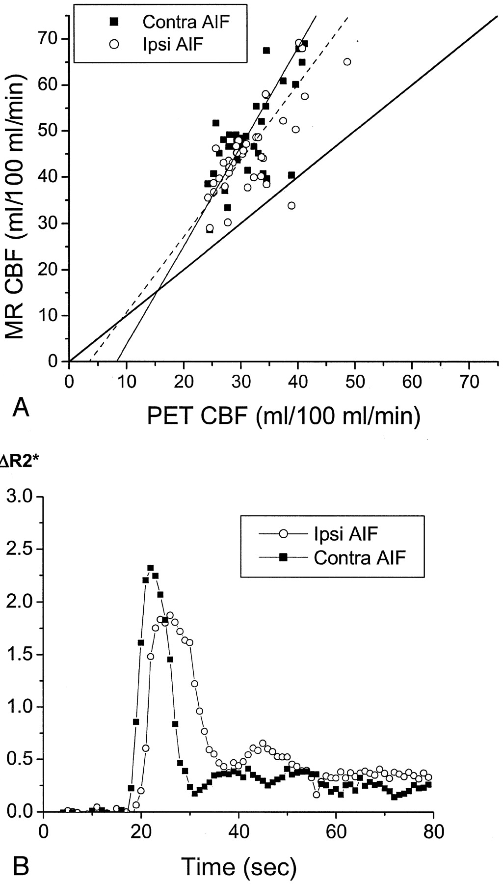
  • Fig 3.
    • Download figure
    • Open in new tab
    • Download powerpoint
    Fig 3.

    CBF in patient 5, a 71-year-old man who had chronic right carotid occlusion.

    A, Graph compares CBF values measured by MR imaging (MR CBF) with those measured by PET (PET CBF). CBF values measured by PET have been corrected for the finite permeability of water through the blood-brain barrier (see Methods). CBF values measured by MR imaging were calculated with AIFs from the proximal MCA, both ipsilateral to the occluded carotid artery (Ipsi AIF, open circles) and contralateral to the occluded carotid artery (Contra AIF, closed squares). The line of identity, with a slope of 1 and a y intercept of 0, is depicted by a solid line. The slopes of the regression line for the data from the ipsilateral AIF (dashed line) and for the data from the contralateral AIF (dotted line) are both close to 1 (Table 2). No statistically significant difference was observed in the slopes or y intercepts of the two regression lines (P > .05). The linear relationship between CBF values measured by MR imaging and those measured by PET is statistically significant for both the ipsilateral AIF (r = 0.88, P < .0001) and the contralateral AIF (r = 0.88, P < .0001). CBF values measured by PET in the subcortical white matter regions used in scaling the CBF values measured by MR imaging was 21.1 mL/100 mL/min, which is close to the assumed value of 22 mL/100 mL/min (see Methods).

    B, Plots of the relaxivity ΔR2* versus time for the AIF in the M1 or M2 segment of the MCA ipsilateral to the carotid occlusion (open circles), and that contralateral to the occlusion (closed squares), show that they are almost identical. ΔR2* was determined as the negative log of susceptibility after baseline normalization. Because susceptibility is a relative measurement and not an absolute quantity, ΔR2* is expressed in arbitrary units. The amplitude of the observed change in ΔR2* is not quantitatively comparable between the ipsilateral and contralateral sides because of differences in factors such as vessel orientation and the extent of partial volume averaging of the blood vessel with surrounding tissue and/or CSF within the selected AIF pixels.

  • Fig 4.
    • Download figure
    • Open in new tab
    • Download powerpoint
    Fig 4.

    Delay and dispersal of the AIF ipsilateral to the occluded carotid artery affects calculated CBF values measured by MR imaging (MR CBF). Conventions used are as described in the legend to Figure 3.

    A, In a 65-year-old man with symptomatic left carotid occlusion (patient 7), CBF values measured by MR imaging systematically overestimated CBF values measured by PET (PET CBF). The linear regression between CBF values measured by MR imaging and those measured by PET has a slope of 1.65 for the ipsilateral AIF (Ipsi AIF, r = 0.79, P < .0001) and 2.14 for the contralateral AIF (Contra AIF, r = 0.81, P < .0001). This difference in slopes is statistically significant (P < .05). CBF value measured by PET in the subcortical white matter regions used in scaling the CBF values measured by MR imaging was 21.9 mL/100 mL/min, which is very close to the assumed value of 22 mL/100 mL/min.

    B, AIFs in patient 7 show a delay in the peak of the ipsilateral AIF (open circles) and a broader peak of the ipsilateral AIF, compared with the contralateral AIF (closed squares).

  • Fig 5.
    • Download figure
    • Open in new tab
    • Download powerpoint
    Fig 5.

    CBF in all seven patients.

    A, Pooled data from all seven patients shows a statistically significant correlation between CBF values measured by MR imaging (MR CBF) and CBF values measured by PET (PET CBF), when CBF values measured by MR imaging are calculated with an AIF ipsilateral to the carotid occlusion (Ipsi AIF, open circles) (r = 0.60, P < .0001) and with an AIF contralateral to the occlusion (Contra AIF, closed squares) (r = 0.54, P < .0001). No significant differences were observed between the correlation coefficients, slopes, or y intercepts of the regression lines for the ipsilateral AIF (dashed line) and the contralateral AIF (dotted line). Both regression lines have slopes significantly <1 (P < .001) and y intercepts significantly >0 (P < .001). Relative MR CBF values were scaled to absolute units by assuming a normal white matter flow rate of 22 mL/100 mL/min (see Methods).

    B, Use of white matter flow rates measured with PET for each patient to scale relative CBF values measured with MR imaging for each patient to absolute units improves the correlation between CBF values measured by MR imaging and those measured by PET, both for the ipsilateral AIF (open circles) (r = 0.85, P < .0001) and the contralateral AIF (closed squares) (r = 0.84, P < .0001). Again, no significant difference was observed between the correlation coefficients, slopes, or y intercepts of the regression lines for the ipsilateral AIF (dashed line) and the contralateral AIF (dotted line). However, both regression lines for PET-scaled CBF values measured by MR imaging versus CBF values measured by PET have slopes significantly >1 (P < .001) and y intercepts significantly <0 (P < .05).

Tables

  • Figures
    • View popup
    TABLE 1:

    Participant clinical data and position emission tomography hemodynamic parameters

    ParticipantAge/Sex/HandednessSide of OcclusionClinical HistoryPET
    CBFCBVMTTOEF
    167/M/RLAsymptomatic0.850.891.051.02
    265/F/RLAsymptomatic0.840.951.131.03
    358/M/RLLeft parietal infarct0.891.301.461.09
    467/M/LLLeft posteriorfrontal infarct0.871.101.260.93
    571/M/RRAsymptomatic0.961.131.170.98
    676/M/RRRight posterior parietal infarct0.981.171.191.04
    765/M/RLLeft frontoparietal infarct0.801.091.371.06
    • All positron emission tomography hemodynamic parameters are ipsilateral-to-contralateral ratios of mean hemispheric values. Cerebral blood volume, mean transit time, and oxygen extraction fraction ratios that exceed the range observed in 18 normal control participants are shown in bold text. The upper limits for normal hemispheric ratios for cerebral blood volume, mean transit time, and oxygen extraction were 1.21, 1.14, and 1.08, respectively. Hemodynamic impairment is clearly shown for participant 3. Participants 4 through 7 may have some degree of autoregulatory vasodilation, with elevated mean transit times.

    • Note.—PET indicates positron emission tomography; CBF, cerebral blood flow; CBV, cerebral blood volume; MTT, mean transit time; OEF, oxygen extraction fraction; M, male; F, female; R, right; L, left.

    • View popup
    TABLE 2:

    Results of linear regression of cerebral blood flow values measured by MR imaging versus cerebral blood flow values measured by positron emission tomography

    ParticipantIpsilateral AIFContralateral AIF
    rSlopey-int.*rSlopey-int.*
    10.650.489.300.660.479.11
    20.750.702.630.720.674.42
    30.500.4220.80.620.5515.9
    40.881.045.200.850.889.10
    50.880.940.380.880.970.27
    60.801.05−9.440.821.17−14.7
    70.791.65−5.740.812.14−17.7
    Pooled†0.600.55 ± 0.0516.6 ± 1.990.540.54 ± 0.0517.6 ± 2.26
    • For each participant and for the pooled data, the linear correlations between cerebral blood flow values measured by MR imaging and cerebral blood flow values measured by by positron emission tomography, both for ipsilateral and contralateral arterial input functions, are statistically significant (P < .001).

    • * y-intercepts of regression lines are expressed in mL/100 mL/min.

    • † Mean linear regression parameters of pooled data from all seven participants (Fig 5A) and standard errors of the mean for slopes and y-intercepts of regression lines.

    • Note.—AIF indicates arterial input function; y-int., y intercepts of the regression lines. The slopes represent cerebral blood flow values measured by MR imaging versus cerebral blood flow values measured by positron emission tomography and therefore are dimensionless.

PreviousNext
Back to top

In this issue

American Journal of Neuroradiology: 24 (5)
American Journal of Neuroradiology
Vol. 24, Issue 5
1 May 2003
  • Table of Contents
  • Index by author
Advertisement
Print
Download PDF
Email Article
Cite this article
0 Responses
Respond to this article
Share
Bookmark this article
Measurement of Cerebral Blood Flow in Chronic Carotid Occlusive Disease: Comparison of Dynamic Susceptibility Contrast Perfusion MR Imaging with Positron Emission Tomography
Pratik Mukherjee, Hyunseon Christine Kang, Tom O. Videen, Robert C. McKinstry, William J. Powers, Colin P. Derdeyn
American Journal of Neuroradiology May 2003, 24 (5) 862-871;
del.icio.us logo Twitter logo Facebook logo Mendeley logo
  • Tweet Widget

Jump to section

  • Article
    • Abstract
    • Methods
    • Results
    • Discussion
    • Conclusion
    • Acknowledgments
    • Footnotes
    • References
  • Figures & Data
  • Info & Metrics
  • Responses
  • References
  • PDF

Related Articles

  • No related articles found.
  • PubMed
  • Google Scholar

Cited By...

  • Augmentation of perfusion with simultaneous vasodilator and inotropic agents in experimental acute middle cerebral artery occlusion: a pilot study
  • Bayesian Estimation of CBF Measured by DSC-MRI in Patients with Moyamoya Disease: Comparison with 15O-Gas PET and Singular Value Decomposition
  • Evaluation of 4D Vascular Flow and Tissue Perfusion in Cerebral Arteriovenous Malformations: Influence of Spetzler-Martin Grade, Clinical Presentation, and AVM Risk Factors
  • Influence of the Arterial Input Function on Absolute and Relative Perfusion-Weighted Imaging Penumbral Flow Detection: A Validation With 15O-Water Positron Emission Tomography
  • Recommendations for Imaging of Acute Ischemic Stroke: A Scientific Statement From the American Heart Association
  • The Performance of MRI-Based Cerebral Blood Flow Measurements in Acute and Subacute Stroke Compared With 15O-Water Positron Emission Tomography: Identification of Penumbral Flow
  • The Acetazolamide Challenge: Techniques and Applications in the Evaluation of Chronic Cerebral Ischemia
  • Influence of Arterial Input Function on Hypoperfusion Volumes Measured With Perfusion-Weighted Imaging
  • Crossref
  • Google Scholar

This article has not yet been cited by articles in journals that are participating in Crossref Cited-by Linking.

More in this TOC Section

  • Multimodal CT Provides Improved Performance for Lacunar Infarct Detection
  • Optimal MRI Sequence for Identifying Occlusion Location in Acute Stroke: Which Value of Time-Resolved Contrast-Enhanced MRA?
  • Evaluating the Effects of White Matter Multiple Sclerosis Lesions on the Volume Estimation of 6 Brain Tissue Segmentation Methods
Show more Brain

Similar Articles

  • Acetazolamide-Loaded QSM and Hyperperfusion
  • Using Data-Driven Methods to Improve Brain Blood Flow Measurements in Cerebrovascular Disease with Dynamic Imaging
  • Acetazolamide-Loaded Dynamic 7T MR Quantitative Susceptibility Mapping in Major Cerebral Artery Steno-Occlusive Disease: Comparison with PET
  • Improved White Matter Cerebrovascular Reactivity after Revascularization in Patients with Steno-Occlusive Disease
  • Chronic Middle Cerebral Artery Occlusion: A Hemodynamic and Metabolic Study with Positron-Emission Tomography
See more
Advertisement

Indexed Content

  • Current Issue
  • Accepted Manuscripts
  • Article Preview
  • Past Issues
  • Editorials
  • Editor's Choice
  • Fellows' Journal Club
  • Letters to the Editor
  • Video Articles

Cases

  • Case Collection
  • Archive - Case of the Week
  • Archive - Case of the Month
  • Archive - Classic Case

More from AJNR

  • Trainee Corner
  • Imaging Protocols
  • MRI Safety Corner
  • Book Reviews

Multimedia

  • AJNR Podcasts
  • AJNR Scantastics

Resources

  • Turnaround Time
  • Submit a Manuscript
  • Submit a Video Article
  • Submit an eLetter to the Editor/Response
  • Manuscript Submission Guidelines
  • Statistical Tips
  • Fast Publishing of Accepted Manuscripts
  • Graphical Abstract Preparation
  • Imaging Protocol Submission
  • Evidence-Based Medicine Level Guide
  • Publishing Checklists
  • Author Policies
  • Become a Reviewer/Academy of Reviewers
  • News and Updates

About Us

  • About AJNR
  • Editorial Board
  • Editorial Board Alumni
  • Alerts
  • Permissions
  • Not an AJNR Subscriber? Join Now
  • Advertise with Us
  • Librarian Resources
  • Feedback
  • Terms and Conditions
  • AJNR Editorial Board Alumni

American Society of Neuroradiology

  • Not an ASNR Member? Join Now

© 2025 by the American Society of Neuroradiology All rights, including for text and data mining, AI training, and similar technologies, are reserved.
Print ISSN: 0195-6108 Online ISSN: 1936-959X

Powered by HighWire
Email this Article

Thank you for your interest in spreading the word on American Journal of Neuroradiology.

NOTE: We only request your email address so that the person you are recommending the page to knows that you wanted them to see it, and that it is not junk mail. We do not capture any email address.

Enter multiple addresses on separate lines or separate them with commas.
Measurement of Cerebral Blood Flow in Chronic Carotid Occlusive Disease: Comparison of Dynamic Susceptibility Contrast Perfusion MR Imaging with Positron Emission Tomography
(Your Name) has sent you a message from American Journal of Neuroradiology
(Your Name) thought you would like to see the American Journal of Neuroradiology web site.
CAPTCHA
This question is for testing whether or not you are a human visitor and to prevent automated spam submissions.
Cite this article
Pratik Mukherjee, Hyunseon Christine Kang, Tom O. Videen, Robert C. McKinstry, William J. Powers, Colin P. Derdeyn
Measurement of Cerebral Blood Flow in Chronic Carotid Occlusive Disease: Comparison of Dynamic Susceptibility Contrast Perfusion MR Imaging with Positron Emission Tomography
American Journal of Neuroradiology May 2003, 24 (5) 862-871;

Citation Manager Formats

  • BibTeX
  • Bookends
  • EasyBib
  • EndNote (tagged)
  • EndNote 8 (xml)
  • Medlars
  • Mendeley
  • Papers
  • RefWorks Tagged
  • Ref Manager
  • RIS
  • Zotero

We use cookies on this site to enhance your user experience. By clicking any link on this page you are giving your consent for us to set cookies.