
Article Figures & Data
Figures
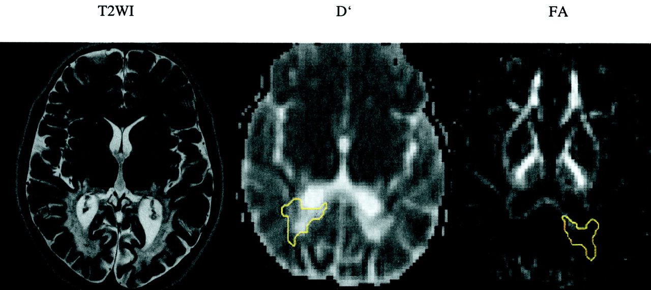
- Fig 1.
T2-weighed image (T2WI) and D′ and FA maps of the blind patient (patient 2) shows confluent and symmetric abnormalities in the deep white matter of both occipital lobes. Regions of interest are shown on D′ (−r) and FA (−l) maps (yellow outlines).
- Fig 2.
T2-weighted images (T2WI) and D′ and FA maps of patient 1.
A, No signal intensity alteration is shown in the parietal white matter.
B, No signal intensity alteration is shown in the occipital deep white matter. Regions of interest are shown on D′ (−r) and FA (−l) maps (yellow outlines).
- Fig 3.
T2-weighted image (T2WI) and D′ and FA maps of patient 3. No signal intensity alteration is shown in the centrum semiovale. Regions of interest are shown on D′ (−r) and FA (−l) maps (yellow outlines).


Tables
D′ Patient 1 Patient 2 Patient 3 Control Participants Mean +1 SD Mean +2 SD Mean SD ROI T2 Mean SD ROI T2 Mean SD ROI T2 Mean SD PLIC-l 0.65 0.02 9 0 0.70 0.08 9 1 0.70 0.05 9 1 0.65 0.02 0.67 0.68 PLIC-r 0.67 0.03 8 0 0.69 0.06 9 1 0.68 0.06 9 1 CSO-l 0.66 0.03 29 0 0.78 0.08 36 1 0.70 0.03 51 0 0.66 0.02 0.68 0.70 CSO-r 0.66 0.02 25 0 0.77 0.10 38 1 0.71 0.03 52 0 FWM-l 0.77 0.04 62 0 0.74 0.04 80 0 0.75 0.05 100 0 0.74 0.02 0.76 0.78 FWM-r 0.77 0.04 80 0 0.74 0.05 103 0 0.73 0.06 105 0 PWM-l 0.79 0.08 85 0 1.10 0.36 160 1 0.66 0.09 112 0 0.71 0.02 0.73 0.76 PWM-r 0.82 0.08 75 0 1.08 0.42 206 1 0.71 0.07 158 0 TWM-l 0.82 0.08 93 0 1.13 0.30 91 1 0.81 0.08 100 0 0.77 0.02 0.79 0.80 TWM-r 0.81 0.08 113 0 1.32 0.36 94 1 0.79 0.10 130 0 OWM-l 0.81 0.15 90 0 1.82 0.54 77 1 0.72 0.21 69 0 0.70 0.04 0.73 0.77 OWM-r 0.78 0.17 71 0 1.72 0.53 85 1 0.71 0.12 96 0 GCC-l 0.84 0.30 24 0 0.81 0.12 22 0 0.72 0.20 21 0 0.81 0.09 0.90 1.00 GCC-r 0.83 0.20 18 0 0.80 0.16 20 0 0.68 0.09 26 0 SCC-l 0.78 0.18 28 0 1.92 0.35 16 1 0.63 0.25 27 0 0.73 0.06 0.78 0.84 SCC-r 0.79 0.13 20 0 2.13 0.38 18 1 0.61 0.10 19 0 ALIC-l 0.72 0.06 11 0 0.77 0.04 13 0 0.86 0.10 16 1 0.73 0.05 0.79 0.84 ALIC-r 0.75 0.09 9 0 0.73 0.06 8 0 0.82 0.11 12 1 Note.—D′ indicates isotropic diffusion; ROI, region of interest; T2, T2 value on T2-weighted MR images; -l, on the left side; -r, on the right side; PLIC, posterior limb of the internal capsule; CSO, centrum semiovale; FWM, frontal white matter; PWM, parietal white matter; TWM, temporal white matter; OWM, occipital white matter; GCC, genu corporis callosi; SCC, splenium corporis callosi; ALIC, anterior limb of the internal capsule. Mean diffusivity values for three patients with X-linked adrenoleukodystrophy, indicated as mean values and intra-region of interest SDs (inter-pixel SDs), are compared with those of seven age-matched normal control participants, indicated as mean values and inter-region of interest SDs. Values shown on light gray background are between 1 and 2 SDs of the mean, and values shown on dark gray background are >2 SDs of the mean, compared with values for normal control participants. T2 values are either 0 for T2-weighted images without evidence of demyelination in the specific region or 1 for T2-weighted images with evidence of demyelination.
FA Patient 1 Patient 2 Patient 3 Control Participant Mean −1 SD Mean −2 SD Mean SD ROI T2 Mean SD ROI T2 Mean SD ROI T2 Mean SD PLIC-l 0.76 0.05 9 0 0.68 0.06 9 1 0.73 0.10 9 1 0.76 0.02 0.75 0.73 PLIC-r 0.72 0.06 8 0 0.65 0.06 9 1 0.75 0.04 9 1 CSO-l 0.56 0.08 29 0 0.32 0.11 36 1 0.50 0.06 51 0 0.54 0.05 0.48 0.43 CSO-r 0.57 0.07 25 0 0.32 0.13 38 1 0.48 0.07 52 0 FWM-l 0.36 0.10 62 0 0.37 0.10 80 0 0.33 0.09 100 0 0.38 0.04 0.34 0.31 FWM-r 0.37 0.10 80 0 0.38 0.09 103 0 0.38 0.11 105 0 PWM-l 0.33 0.09 85 0 0.11 0.06 160 1 0.39 0.07 112 0 0.40 0.02 0.38 0.35 PWM-r 0.31 0.11 75 0 0.11 0.05 206 1 0.40 0.08 158 0 TWM-l 0.44 0.08 93 0 0.13 0.04 91 1 0.40 0.07 100 0 0.47 0.03 0.43 0.40 TWM-r 0.42 0.10 113 0 0.12 0.03 94 1 0.41 0.08 130 0 OWM-l 0.47 0.10 90 0 0.11 0.07 77 1 0.46 0.10 69 0 0.46 0.04 0.42 0.38 OWM-r 0.44 0.11 71 0 0.10 0.05 85 1 0.44 0.13 96 0 GCC-l 0.66 0.13 24 0 0.68 0.12 22 0 0.69 0.12 21 0 0.72 0.07 0.66 0.59 GCC-r 0.75 0.10 18 0 0.64 0.13 20 0 0.67 0.10 26 0 SCC-l 0.72 0.10 28 0 0.21 0.03 16 1 0.79 0.15 27 0 0.78 0.05 0.73 0.68 SCC-r 0.76 0.06 20 0 0.20 0.02 18 1 0.81 0.08 19 0 ALIC-l 0.53 0.13 11 0 0.49 0.09 13 0 0.38 0.06 16 1 0.54 0.06 0.48 0.43 ALIC-r 0.62 0.08 9 0 0.60 0.12 8 0 0.43 0.04 12 1 Note.—FA indicates fractional anisotropy; ROI, region of interest; T2, T2 value on T2-weighted MR images; -l, on the left side; -r, on the right side; PLIC, posterior limb of the internal capsule; CSO, centrum semiovale; FWM, frontal white matter; PWM, parietal white matter; TWM, temporal white matter; OWM, occipital white matter; GCC, genu corporis callosi; SCC, splenium corporis callosi; ALIC, anterior limb of the internal capsule. Fractional anisotropy values for three patients with X-linked adrenoleukodystrophy, indicated as mean values and intra-region of interest SDs (inter-pixel SDs), are compared with those of seven age-matched normal control participants, indicated as mean values and inter-region of interest SDs. Values shown on light gray background are between 1 and 2 SDs of the mean, and values shown on dark gray background are >2 SDs of the mean, compared with values for normal control participants. T2 values are either 0 for T2-weighted images without evidence of demyelination in the specific region or 1 for T2-weighted images with evidence of demyelination.


