
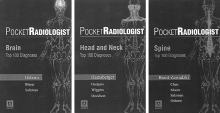
Anne G. Osborn. Philadelphia, PA: W.B. Saunders Company; 2002. 303 pages. Book, $59.95; CD-ROM software, $79.95.
H. Ric Harnsberger. Philadelphia, PA: W.B. Saunders Company; 2002. 305 pages. Book, $59.95; CD-ROM software, $79.95.
Michael Brant-Zawadzki. Philadelphia, PA: W.B. Saunders Company; 2002. 301 pages. Book, $59.95; CD-ROM software, $79.95.
It is rare for a revolutionary idea to come to fruition in the medical publishing world. With the appearance of the new series of (5 × 8“) soft cover volumes, PocketRadiologist, and the accompanying Personal Digital Assistant (PDA) software for use on Palm OS-based PDA, a new and forward thinking method of carrying and dispersing imaging information is now at every radiologist’s disposal.
Although this review centers on the three volumes that deal with neuroradiology, there are now 10 such volumes available: abdominal, brain, cardiac, chest, head & neck, interventional, musculoskeletal, pediatrics, spine, and vascular. If the intent is to have a portable source of information, then the PDA version makes most sense, because 10 of these books, even though they are relatively small in size, would not fit in one’s pocket. Each book and its PDA version can be purchased separately ($59.95 for the book, $79.95 for the PDA) or together, and the publisher plans to offer the entire set of 10 books/PDAs at a discounted price.
The authors of the neuroradiology series— Osborn for Brain, Harnsberger for Head & Neck, and Brant-Zawadski for Spine—have all used the same format. They identify what they consider to be the top 100 diagnoses in each area. Next, they present the appropriate images, key facts, imaging findings, associated findings, and differential diagnoses. Pathologic findings and clinic issues are presented next in an outline (bullet-like) format. One could quibble about what should or should not be included in this list (everyone will have one or two “favorites” that did not make the top 100), but basically, the authors have identified those conditions that are most likely to be encountered.
The neuroradiology books/PDAs have many positive features, including high quality images, interspersed beautiful color drawings, succinct, accurate, important facts regarding a disease (with many “gems” thrown in), and very useful clinical information. Space precludes detailing all the areas that struck me positively, but briefly, these include color illustrations that are magnificent and are the best I’ve ever seen to highlight lesions throughout the nervous system (eg, cochlea dysplasias, otosclerosis, endolymphatic sac anomalies, spinal dysplasias, spinal cord/canal masses, vascular disease of the neck vessels and intracranial vessels, traumatic lesions of the brain), clinical staging of tumors, Hunt and Hess classification of subarachnoid hemorrhage, and nodal classification levels.
Unlike many volumes on the market, this set is not intended to “quiz” the user regarding the proper diagnosis. You must know or have a high suspicion of the diagnosis and can then avail yourself of the key facts concerning the abnormality.
The drawbacks of these volumes are very few, in my opinion. Considering the premise under which these were produced (ie, quick, easily accessible information), the reader does come away with pieces of information, not in-depth analyses. One gets an overall picture of what is going on but does not acquire a great depth of knowledge. This is not meant as a criticism but rather to point out that a purchaser of these books may want to have a more comprehensive textbook for cross-referencing. Nonetheless, it is important to realize that these authors have put at our disposal a warehouse of information in a very useable format.
For future versions, I recommend integration of more technical information, particularly regarding MR imaging studies. A ready reference to and recommendations for MR angiography, CT angiography, diffusion-weighted imaging, perfusion, and spectroscopy would be helpful. This whole area of techniques could constitute another volume in this series.
In addition, the index could be improved. Although it is understood that the index contains just the list of the “top 100 diagnoses,” some consideration should have been given to cite the pages in which other diagnoses are mentioned. The top 100 could have been in printed in a boldface font, for example, and the others could have also been presented in alphabetical order but in a lightface font. If the reader wants to look up a certain fact, the current index is of no help; it should be expanded.
I would have liked to have seen more state-of-the-art imaging; for example, the brain abscess description could have included single voxel spectra, the cerebral infarction could have included a perfusion MR image with a regional cerebral blood volume map, and the higher grade gliomas should have included an example of multi-voxel spectra. Again, these are minor criticisms considering the overall value. I can visualize the books/PDAs being used at film review sessions and conferences at which questions about pathology, staging, and clinical issues are raised.
In summary, the authors have produced valuable, high quality books/PDAs, which have “broken the mold” in radiology publications. I cannot think of a practicing neuroradiologist who would not find these to be useful or a resident or fellow who would not benefit from having this information “at the ready.” I recommend these PocketRadiologist books and CDs in the highest possible terms. Now, if the American Board of Radiology would just let candidates for ABR certification bring their PDAs into the oral examinations…
- Copyright © American Society of Neuroradiology
In this issue


Jump to section
Related Articles
Cited By...
- No citing articles found.