
Article Figures & Data
Figures
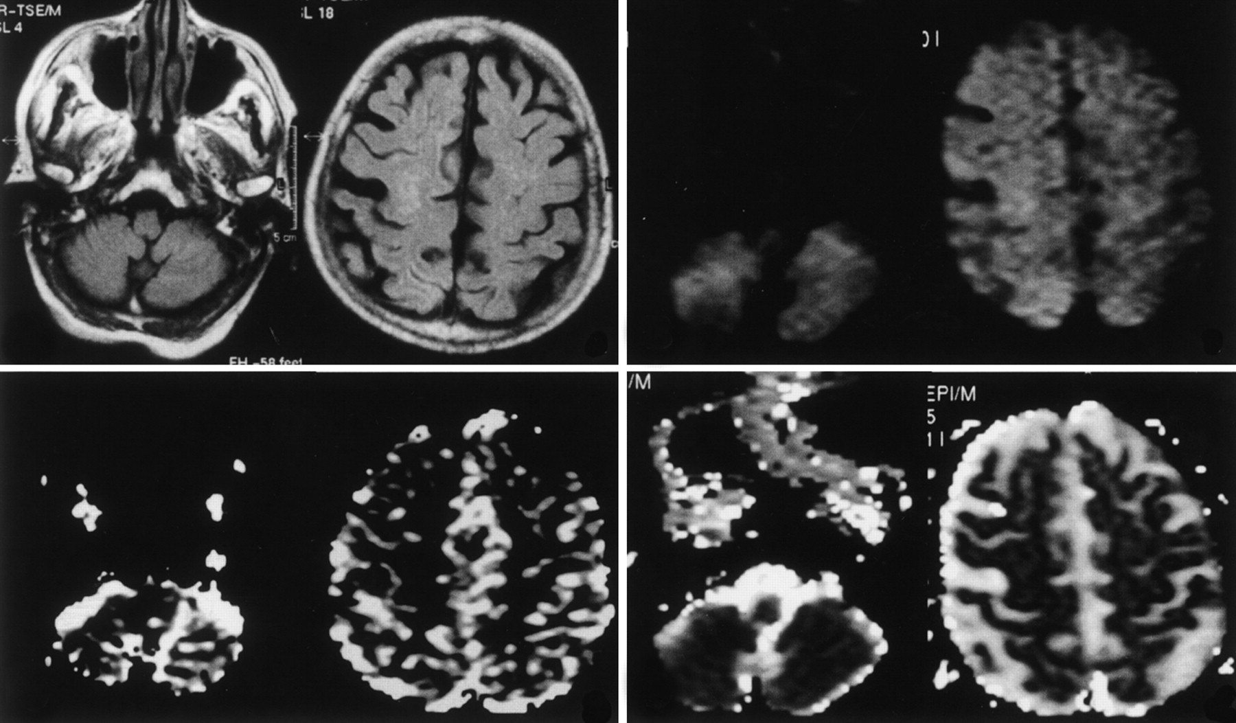
- Fig 1.
Images obtained during the ictal phase on day 1 in an 83-year-old woman with EPC. Top left, T2-weighted FLAIR MR images show diffuse and slight hyperintensity in the right frontotemporal cortex. Bottom left, rCBF maps show hyperperfusion of the right cerebral hemisphere and the contralateral cerebellum (arrows). Top right, On DW images, the same regions show marked hyperintensity. Bottom right, Corresponding decreased ADC is shown.
- Fig 2.
Postictal studies obtained on day 15 after the onset of seizures. Top left, FLAIR MR images demonstrate near-complete resolution of the cortical hyperintensity. rCBF maps (bottom left), DW images (top right), and ADC maps (bottom right) show near-complete resolution of the previous perturbations, both in the cerebral cortex and in the contralateral cerebellar hemisphere.


