
Article Figures & Data
Figures
- Fig 1.
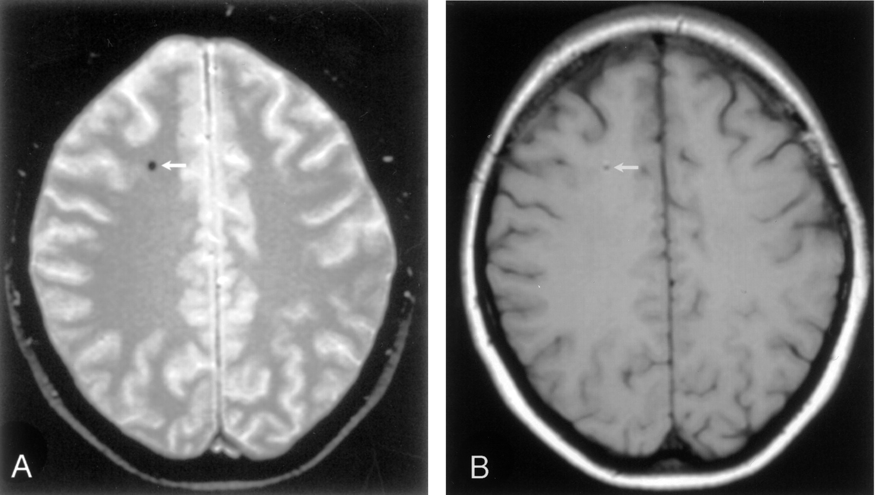
Patient 2.
A, Axial view T2*-weighted gradient-echo image (710/27.6/2 [TR/TE/number of excitations]) shows a black hole (arrow). Repeat images obtained after 1 year appeared unchanged, with no increase in number or size of black holes (not shown).
B, Corresponding axial view T2-weighted spin-echo image (1380/60/2) shows the same black hole with a typical white halo (arrow).
- Fig 2.
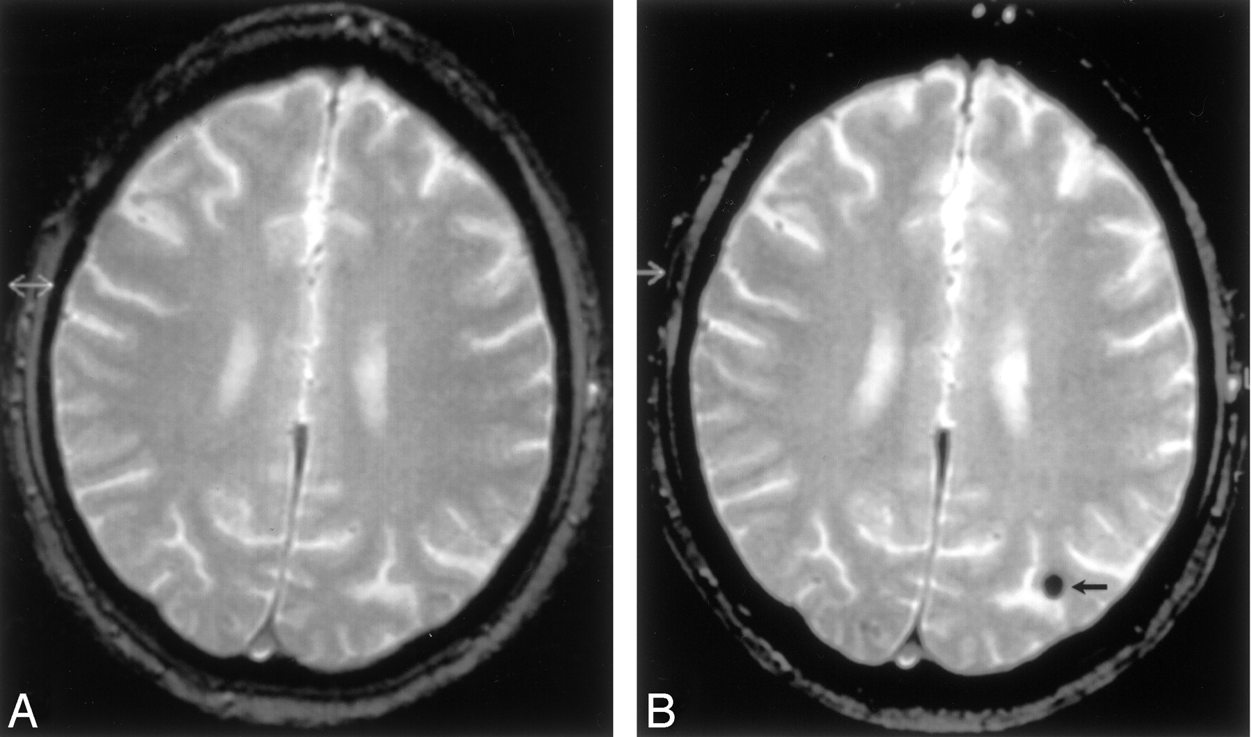
Patient 3.
A, Axial view T2*-weighted gradient-echo image (710/27.6/2), obtained 1 week before double valve replacement, shows no apparent abnormalities.
B, Axial view T2*-weighted gradient-echo image (710/27.6/2), obtained 5 months after successful double valve replacement, shows one of the two large black holes (arrow)


In this issue


Jump to section
Related Articles
Cited By...
- No citing articles found.