
Article Figures & Data
Figures
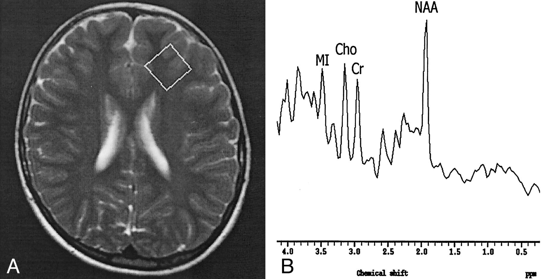
- Fig 1.
Findings in a 6-year-old girl with stage II SSPE.
A, Axial T2-weighted localizer image (2875/120) shows a 2 × 2 × 2-cm voxel placed in the left FSWM.
B, MR spectrum (single voxel, PRESS; 2000/31/256) shows a normal NAA/Cr ratio and increased Cho/Cr and MI/Cr ratios.
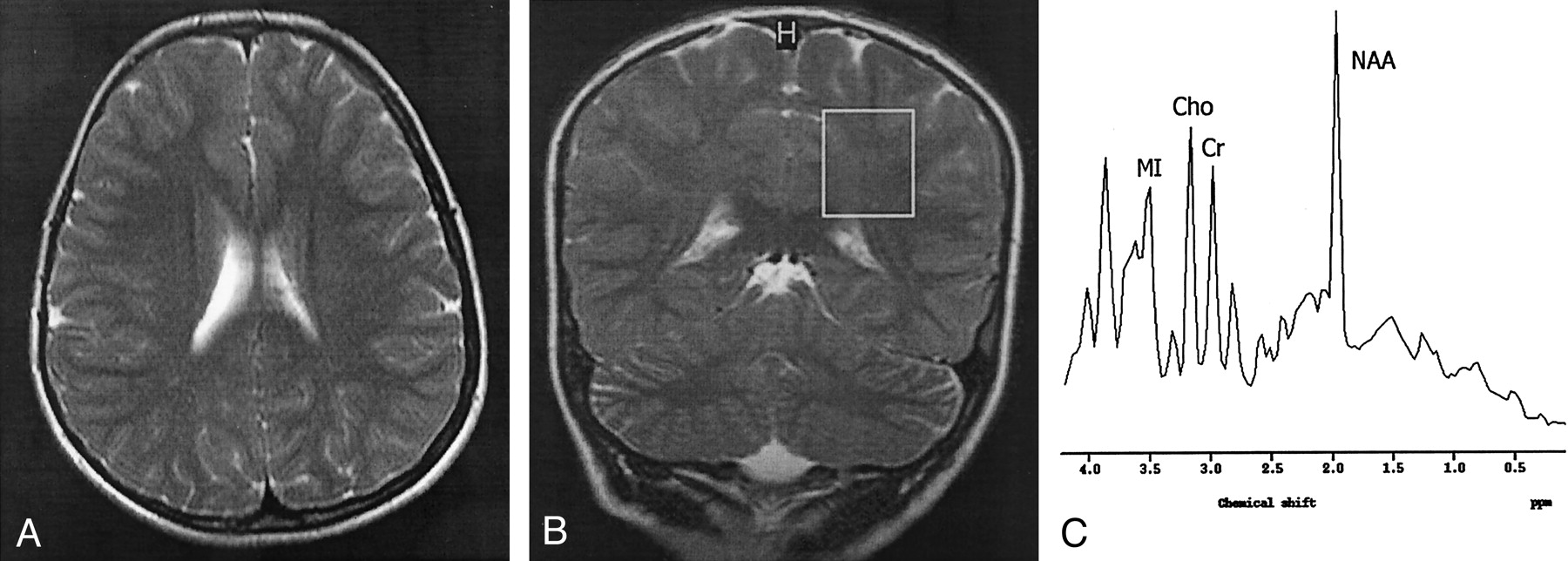
- Fig 2.
Findings in an 8-year-old girl with stage II SSPE.
A, Axial T2-weighted image (4530/100) shows normal brain parenchymal findings.
B, Coronal T2-weighted localizer image (2875/120) shows a 2 × 2 × 2-cm voxel placed in the left POWM.
C, MR spectrum (single voxel, PRESS; 2000/31/256) shows a normal NAA/Cr ratio and increased Cho/Cr and MI/Cr ratios.
- Fig 3.
Findings in an 8-year-old boy with stage III SSPE.
A, Coronal T2-weighted image (4530/100) shows periventricular hyperintensities and cerebral and brain stem atrophy.
B, Axial T2-weighted localizer image (2875/120) shows a 2 × 2 × 2-cm voxel placed in the left POWM.
C, MR spectrum (single voxel, PRESS; 2000/31/256) shows a substantially decreased NAA/Cr ratio, increased Cho/Cr, Ins/Cr ratios, and lipid and lactate peaks.
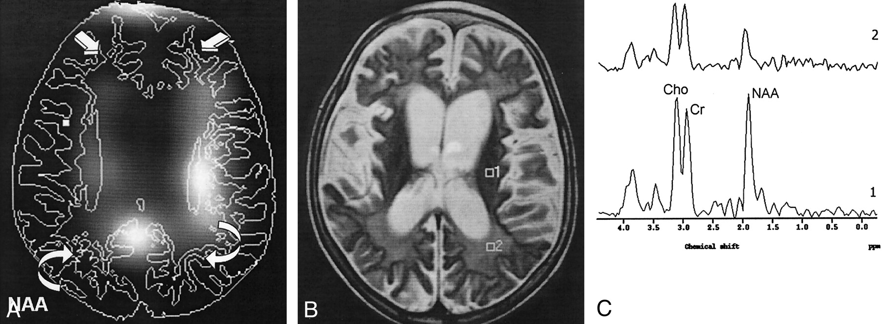
- Fig 4.
Findings in a 7-year-old girl with stage III SSPE.
A, CSI map of NAA shows decreased NAA signal intensity in the FSWM (straight arrows) and POWM (curved arrows).
B, Axial T2-weighted localizer image (2875/120) for MVS shows periventricular hyperintensities and cerebral atrophy.
C, MR spectrum (1500/136) (1) shows a normal NAA/Cr ratio and increased Cho/Cr ratios in left periventricular white matter. MR spectrum (2) shows substantially decreased NAA/Cr and increased Cho/Cr ratios in the left POWM.
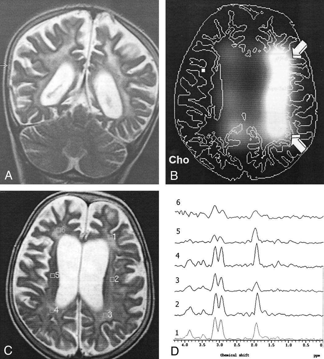
- Fig 5.
Findings in an 8-year-old boy with stage III SSPE.
A, Coronal T2-weighted image (4530/100) shows periventricular hyperintensities and cerebral atrophy.
B, CSI map of Cho shows increased Cho signal intensity in the left periventricular white matter (arrows).
C, Axial T2-weighted localizer image (2875/120) for MVS shows diffuse periventricular hyperintensities and cerebral atrophy.
D, MR spectrum (1500/136) (3) shows significantly decreased NAA/Cr and increased Cho/Cr ratios in the left POWM.


Tables
Patient/Age, y/Sex Vaccination Age at Measles Infection Time after Diagnosis Stage at Diagnosis Clinical and Neurologic Findings MRI Findings Stage II at MRS 1/8/F No 7 mo 1 y II Choreoathetosis, myoclonus, difficulty speaking, tonic convulsion, mental deterioration Normal 2/8/M Yes NA* 6 mo II Myoclonus, ataxia, spasticity, tonic-clonic seizures, difficulty speaking Normal 3/6/F Yes 12 mo 9 mo II Myoclonus, ataxia, mental deterioration, walking with assistance Normal Stage III at MRS 1/8/M Yes 11 mo 4 y II No speech, decerebrate rigidity, bedridden, no responsiveness to any stimulus Cerebral, cerebellar, and brainstem atrophy, periventricular hyperintensities 2/8/M No 6 mo 3 y II No speech, opisthotonos, decerebrate and decorticate rigidity, no responsiveness to any stimulus, bedridden Cerebral and cerebellar atrophy, periventricular hyperintensities 3/7/F Yes 16 mo 2 y II Decerebrate rigidity, bedridden, no speech Cerebral and cerebellar atrophy, periventricular hyperintensities * Not applicable.
Subjects NAA/Cr Cho/Cr MI/Cr NAA/Cho FSWM POWM FSWM POWM FSWM POWM FSWM POWM Stage II SSPE 1.62 ± 0.03 1.56 ± 0.05 1.05 ± 0.01* 1.15 ± 0.07* 1.42 ± 0.15* 1.34 ± 0.01* 1.55 ± 0.16* 1.36 ± 0.13* Stage III SSPE 1.08 ± 0.22* 0.91 ± 0.17* 1.43 ± 0.19* 1.33 ± 0.23* 2.35 ± 0.09* 2.66 ± 0.10* 0.75 ± 0.18* 0.68 ± 0.04* Control 1.74 ± 0.12 1.69 ± 0.19 0.64 ± 0.11 0.66 ± 0.12 0.66 ± 0.11 0.63 ± 0.12 2.78 ± 0.50 2.59 ± 0.50 Note.—Data are the mean ± SDs.
* P < .01 versus control group.


