
Article Figures & Data
Figures
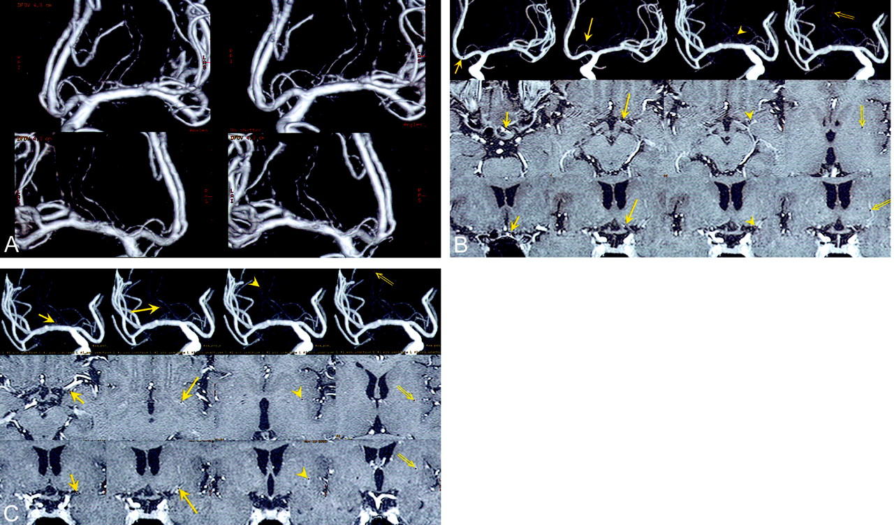
- Fig 1.
Images in a patient with no pathology (contralateral side of a right ICA aneurysm, which was treated with endovascular obliteration).
A, 3D DSA (volume rendering) image, which can be seen stereoscopically, clearly shows one left RAH and one left LSA (upper, anteroposterior view; lower, posteroanterior view).
B, Common cursor image, which simultaneously displays 3D DSA (MIP), axial, and coronal contrast-enhanced MR images, clearly shows the passing course of the left RAH (short arrow, origin of the RAH; long arrow, top of the cranial loop; arrowhead, immediately before entering the anterior perforating substance; double arrow, location at the putamen). The corresponding points are indicated as small red dots.
C, Common cursor image clearly shows the passing course of the left LSA (short arrow, location in the sylvian fissure; long arrow, location at the anterior perforating substance; arrowhead and double arrow, locations at the putamen). The corresponding points are indicated as small red dots.
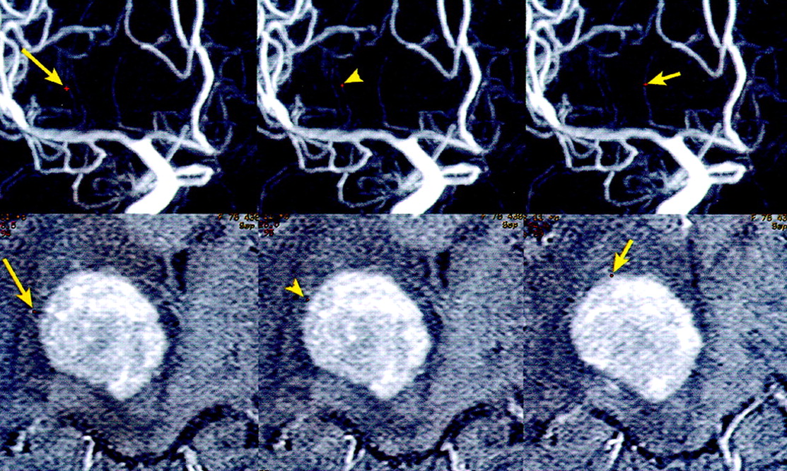
- Fig 2.
Common cursor image in a patient with a right thalamic malignant lymphoma. The image, which simultaneously displays 3D DSA (MIP) and axial contrast-enhanced MR images, clearly shows the relation between the tumor and two LSAs (long arrow and arrowhead) or one RAH (short arrow). The corresponding points are indicated as small red dots.


In this issue


Jump to section
Related Articles
Cited By...
- Diagnostic accuracy of three-dimensional-rotational angiography and heavily T2-weighted volumetric magnetic resonance fusion imaging for the diagnosis of spinal arteriovenous shunts
- Evaluation of CT angiography for visualisation of the lenticulostriate artery: difference between normotensive and hypertensive patients