
Article Figures & Data
Figures
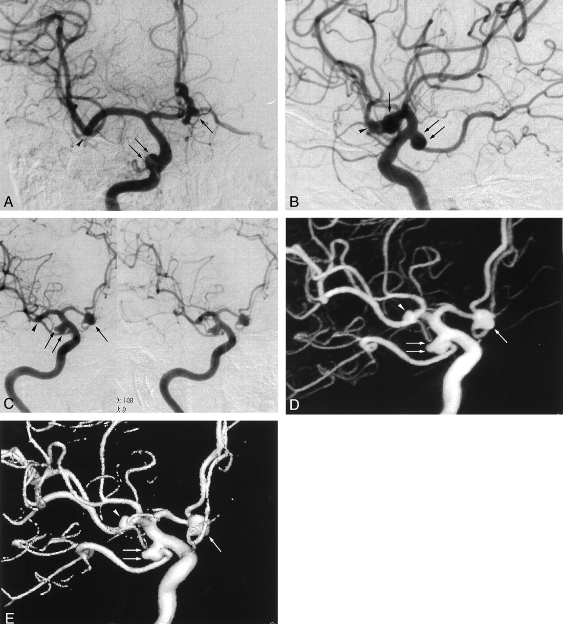
- Fig 1.
Images from the case of a 64-year-old female patient with multiple aneurysms.
A, Anteroposterior standard 2D DSA shows right anterior (arrow) and posterior communicating (double arrows) artery aneurysms, but the right middle cerebral artery aneurysm cannot be visualized (arrowhead).
B, Lateral standard 2D DSA shows right anterior (arrow) and posterior communicating (double arrows) artery aneurysms, but the right middle cerebral artery aneurysm cannot be visualized (arrowhead).
C, Rotational DSA image, which can be seen stereoscopically, clearly shows the relationship of the right anterior (arrow) and posterior communicating (double arrows) artery aneurysms to the neighboring vessels and to the aneurysmal necks. The right middle cerebral artery aneurysm (arrowhead) can be seen, but the relationship to the neighboring vessels and the aneurysmal neck are obscured by the superimposition of the surrounding arteries. Note that minimal misregistrations are observed.
D, MIP image clearly shows the relationship of the right anterior (arrow) and posterior communicating (double arrows) artery aneurysms to the neighboring vessels and to the aneurysmal necks. The right middle cerebral artery aneurysm (arrowhead) can be seen, but the relationship to the neighboring vessels and the aneurysmal neck are obscured by the superimposition of the surrounding arteries. Minimal misregistrations do not create artifacts.
E, SSD image clearly shows the relationship of the right anterior (arrow) and posterior communicating (double arrows) artery aneurysms to the neighboring vessels and to the aneurysmal necks. The right middle cerebral artery aneurysm (arrowhead) is seen, and the relationship to the neighboring vessels and aneurysmal neck are easily recognized. Minimal misregistrations do not create artifacts.
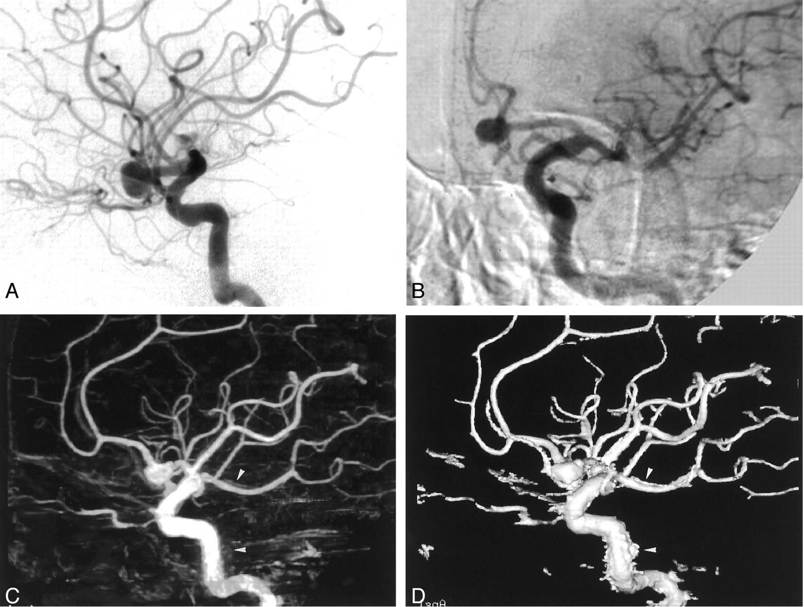
- Fig 2.
Images from the case of a 67-year-old female patient with a right middle cerebral artery aneurysm.
A, Anteroposterior standard 2D DSA image. The aneurysm can be identified (arrow), but the identification of the presence of aneurysmal lobulation and the relationship to neighboring arteries is difficult.
B, Lateral standard 2D DSA image. The aneurysm can be identified (arrow), but the presence of aneurysmal lobulation and the relationship to neighboring arteries is difficult to discern.
C, Rotational DSA image, which can be seen stereoscopically, clearly shows the aneurysm (arrow), but the superimposition of many neighboring arteries makes it difficult to evaluate the presence of aneurysmal lobulation and the relationship to neighboring arteries.
D, MIP image clearly shows the aneurysmal lobulation and relationship to neighboring arteries (arrow).
E, SSD image is especially clear in showing the aneurysmal lobulation and relationship to neighboring arteries.
- Fig 3.
Images from the case of a 61-year-old male patient with a left anterior communicating artery aneurysm.
A, Lateral standard 2D DSA image. Few image artifacts are noted.
B, Rotational DSA image. Image artifacts are severe.
C, MIP image. Image artifacts create blurring (arrowheads).
D, SSD image. Image artifacts create abnormal irregular structures (arrowheads).


Tables
Location Aneurysms (n) Anterior communicating artery 11 Posterior communicating artery 10 Middle cerebral artery 9 Posterior inferior cerebellar artery 3 Ophthalmic artery 2 Basilar artery 2 Internal cerebral artery 1 Anterior choroidal artery 1 Pericarosal artery 1 Total 40 Presence ofAneurysm Presence of Lobulation Visualization of Aneurysmal Neck Relationship to Neighboring Artery Overall Image Quality DSA 4.50 + 0.92 3.27 + 1.27 2.50 + 0.88 2.37 + 0.94 3.90 + 0.31†‡§ Rotation 4.83 + 0.45 3.83 + 1.23 3.60 + 0.65* 3.57 + 0.70* 3.56 + 0.54§ MIP 4.93 + 0.27* 3.94 + 1.08* 3.86 + 0.39* 3.86 + 0.43*† 3.44 + 0.54§ SSD 5.00 + 0.00* 4.54 + 0.99*†‡ 4.00 + 0.00*† 4.00 + 0.00*† 3.06 + 0.25 Note.—DSA indicates digital subtraction angiography; MIP, maximum intensity projection; SSD, surface shaded display.
* Value is significantly greater than that of DSA.
† Value is significantly greater than that of rotational DSA.
‡ Value is significantly greater than that of MIP.
§ Value is significantly greater than that of SSD.
In this issue


Jump to section
Related Articles
Cited By...
- Improving visualization of three-dimensional aneurysm features via segmentation with upsampled resolution and gradient enhancement (SURGE)
- Retrograde 3D rotational venography (3DRV) for venous sinus stent placement in idiopathic intracranial hypertension
- Technology developments in endovascular treatment of intracranial aneurysms
- Guidelines for the Management of Patients With Unruptured Intracranial Aneurysms: A Guideline for Healthcare Professionals From the American Heart Association/American Stroke Association
- Diagnostic quality and accuracy of low dose 3D-DSA protocols in the evaluation of intracranial aneurysms
- Reducing radiation dose while maintaining diagnostic image quality of cerebral three-dimensional digital subtraction angiography: an in vivo study in swine
- MRA Versus DSA for Follow-Up of Coiled Intracranial Aneurysms: A Meta-Analysis
- Standard of practice: embolization of ruptured and unruptured intracranial aneurysms
- Angiographic CT with Intravenous Contrast Injection Compared with Conventional Rotational Angiography in the Diagnostic Work-Up of Cerebral Aneurysms
- Patient-Specific Computational Hemodynamics of Intracranial Aneurysms from 3D Rotational Angiography and CT Angiography: An In Vivo Reproducibility Study
- Large-Cohort Comparison Between Three-Dimensional Time-of-Flight Magnetic Resonance and Rotational Digital Subtraction Angiographies in Intracranial Aneurysm Detection
- Comparison of 2D Digital Subtraction Angiography and 3D Rotational Angiography in the Evaluation of Dome-to-Neck Ratio
- Efficacy of DynaCT Digital Angiography in the Detection of the Fistulous Point of Dural Arteriovenous Fistulas
- 3D Digital Subtraction Angiography of Intracranial Aneurysms: Comparison of Flat Panel Detector with Conventional Image Intensifier TV System Using a Vascular Phantom
- Safety of Cerebral Digital Subtraction Angiography in Children: Complication Rate Analysis in 241 Consecutive Diagnostic Angiograms