Advertisement

Main menu

  • Home
  • Content
    • Current Issue
    • Accepted Manuscripts
    • Article Preview
    • Past Issue Archive
    • Video Articles
    • AJNR Case Collection
    • Case of the Week Archive
    • Case of the Month Archive
    • Classic Case Archive
  • Special Collections
    • AJNR Awards
    • Low-Field MRI
    • Alzheimer Disease
    • ASNR Foundation Special Collection
    • Photon-Counting CT
    • View All
  • Multimedia
    • AJNR Podcasts
    • AJNR SCANtastic
    • Trainee Corner
    • MRI Safety Corner
    • Imaging Protocols
  • For Authors
    • Submit a Manuscript
    • Submit a Video Article
    • Submit an eLetter to the Editor/Response
    • Manuscript Submission Guidelines
    • Statistical Tips
    • Fast Publishing of Accepted Manuscripts
    • Graphical Abstract Preparation
    • Imaging Protocol Submission
    • Author Policies
  • About Us
    • About AJNR
    • Editorial Board
    • Editorial Board Alumni
  • More
    • Become a Reviewer/Academy of Reviewers
    • Subscribers
    • Permissions
    • Alerts
    • Feedback
    • Advertisers
    • ASNR Home

User menu

  • Alerts
  • Log in

Search

  • Advanced search
American Journal of Neuroradiology

American Journal of Neuroradiology

ASHNR American Society of Functional Neuroradiology ASHNR American Society of Pediatric Neuroradiology ASSR
  • Alerts
  • Log in

Advanced Search

  • Home
  • Content
    • Current Issue
    • Accepted Manuscripts
    • Article Preview
    • Past Issue Archive
    • Video Articles
    • AJNR Case Collection
    • Case of the Week Archive
    • Case of the Month Archive
    • Classic Case Archive
  • Special Collections
    • AJNR Awards
    • Low-Field MRI
    • Alzheimer Disease
    • ASNR Foundation Special Collection
    • Photon-Counting CT
    • View All
  • Multimedia
    • AJNR Podcasts
    • AJNR SCANtastic
    • Trainee Corner
    • MRI Safety Corner
    • Imaging Protocols
  • For Authors
    • Submit a Manuscript
    • Submit a Video Article
    • Submit an eLetter to the Editor/Response
    • Manuscript Submission Guidelines
    • Statistical Tips
    • Fast Publishing of Accepted Manuscripts
    • Graphical Abstract Preparation
    • Imaging Protocol Submission
    • Author Policies
  • About Us
    • About AJNR
    • Editorial Board
    • Editorial Board Alumni
  • More
    • Become a Reviewer/Academy of Reviewers
    • Subscribers
    • Permissions
    • Alerts
    • Feedback
    • Advertisers
    • ASNR Home
  • Follow AJNR on Twitter
  • Visit AJNR on Facebook
  • Follow AJNR on Instagram
  • Join AJNR on LinkedIn
  • RSS Feeds

AJNR Awards, New Junior Editors, and more. Read the latest AJNR updates

Research ArticleBrain

Reproducibility of Primary Motor Cortex Somatotopy Under Controlled Conditions

Hatem Alkadhi, Gerard R. Crelier, Sabina Hotz Boendermaker, Xavier Golay, Marie-Claude Hepp-Reymond and Spyros S. Kollias
American Journal of Neuroradiology October 2002, 23 (9) 1524-1532;
Hatem Alkadhi
  • Find this author on Google Scholar
  • Find this author on PubMed
  • Search for this author on this site
Gerard R. Crelier
  • Find this author on Google Scholar
  • Find this author on PubMed
  • Search for this author on this site
Sabina Hotz Boendermaker
  • Find this author on Google Scholar
  • Find this author on PubMed
  • Search for this author on this site
Xavier Golay
  • Find this author on Google Scholar
  • Find this author on PubMed
  • Search for this author on this site
Marie-Claude Hepp-Reymond
  • Find this author on Google Scholar
  • Find this author on PubMed
  • Search for this author on this site
Spyros S. Kollias
  • Find this author on Google Scholar
  • Find this author on PubMed
  • Search for this author on this site
  • Article
  • Figures & Data
  • Info & Metrics
  • Responses
  • References
  • PDF
Loading

Article Figures & Data

Figures

  • Tables
  • Fig 1.
    • Download figure
    • Open in new tab
    • Download powerpoint
    Fig 1.

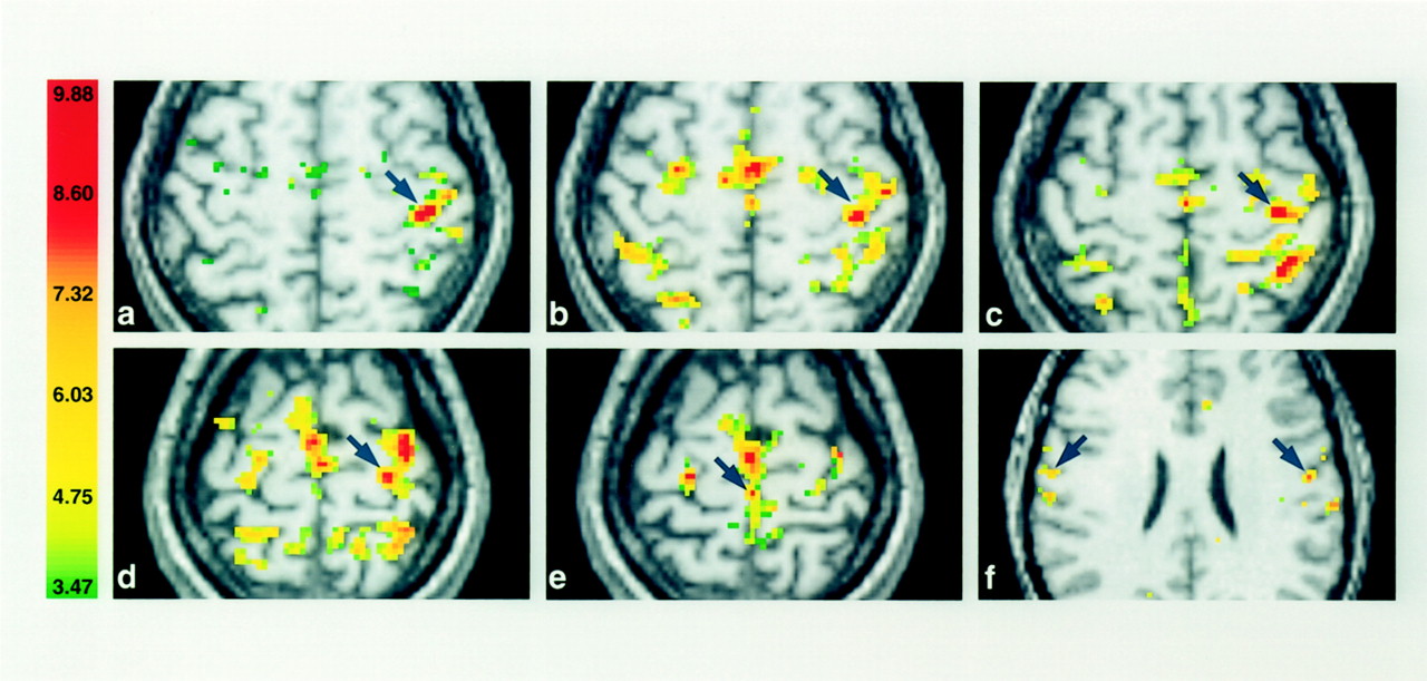
    Activation in contralateral M1 (arrows) displayed in axial sections for one subject in the first session. The right side of the sections corresponds to the left hemisphere, and the numbers in the color bar correspond to t values.

    A and B, The fingers (A) and hand (B) are in almost identical locations (z plane, +58).

    C and D, The wrist (C) and elbow (D) representations are located more medially, superior and posterior along the course of M1 (z plane, +59 and +61, respectively).

    E and F, Note the considerable overlap of activated volumes within the arm and the clear separation of the foot (E) and tongue (F) (z plane, +66 and +28, respectively).

  • Fig 2.
    • Download figure
    • Open in new tab
    • Download powerpoint
    Fig 2.

    Activation of contralateral M1 in the second session in the same subject as in Figure 1. A comparison with the images in Figure 1 reveals a high degree of reproducibility in the somatotopy despite variations in the activated volumes.

    A and B, Fingers (A) and hand (B).

    C and D, Wrist (C) and elbow (D).

    E and F, Foot (E) and tongue (F).

  • Fig 3.
    • Download figure
    • Open in new tab
    • Download powerpoint
    Fig 3.

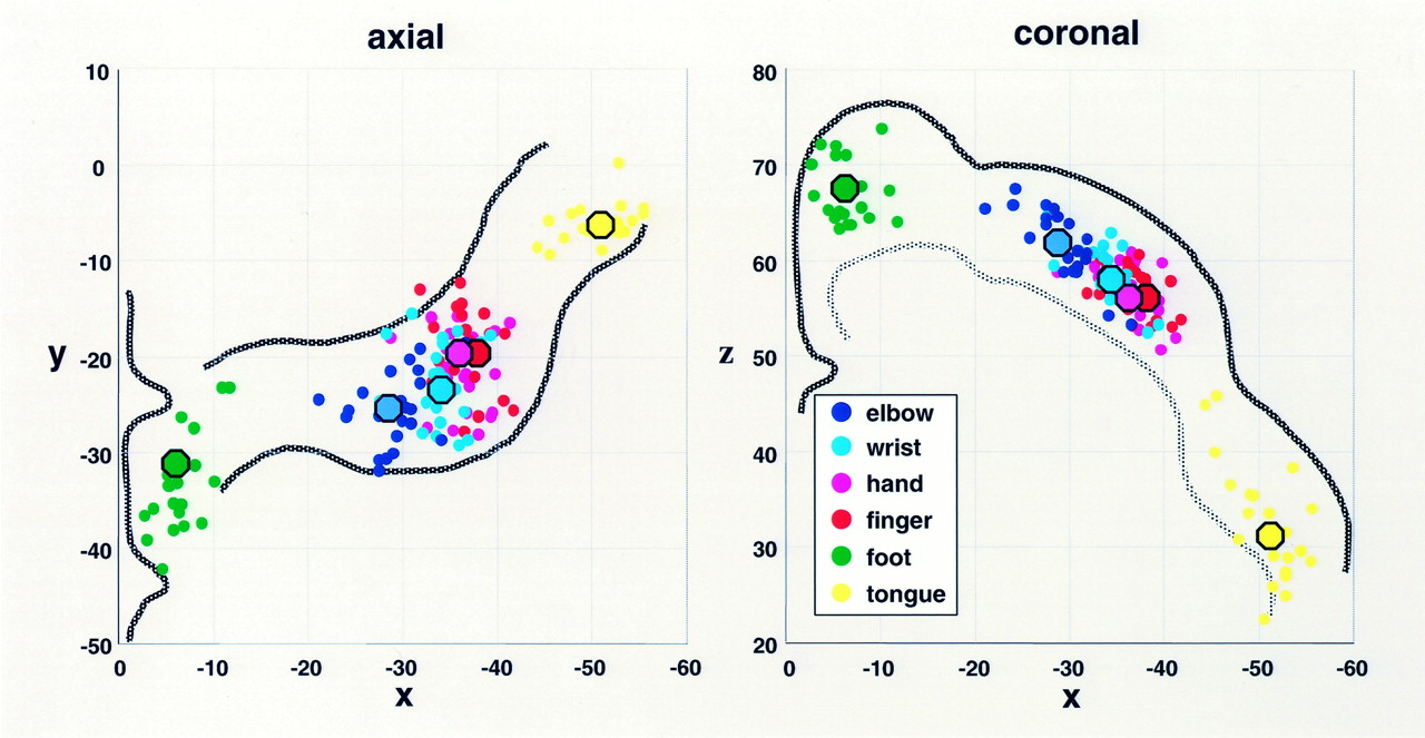
    Two-dimensional scatter plots of the COGs in the 12 subjects (two sessions per subject) in the contralateral M1. Small dots represent individual COGs, and large dots indicate the mean COGs. Note the separate subdivisions for the foot, arm, and tongue and the clear somatotopic gradients within the arm representations in both the axial and coronal planes. The x, y, and z coordinates corresponding to those in Talairach space (21). Left, Axial plane with approximate contour of the precentral gyrus. Right, Coronal plane with the cortical surface and limited to the white matter.

  • Fig 4.
    • Download figure
    • Open in new tab
    • Download powerpoint
    Fig 4.

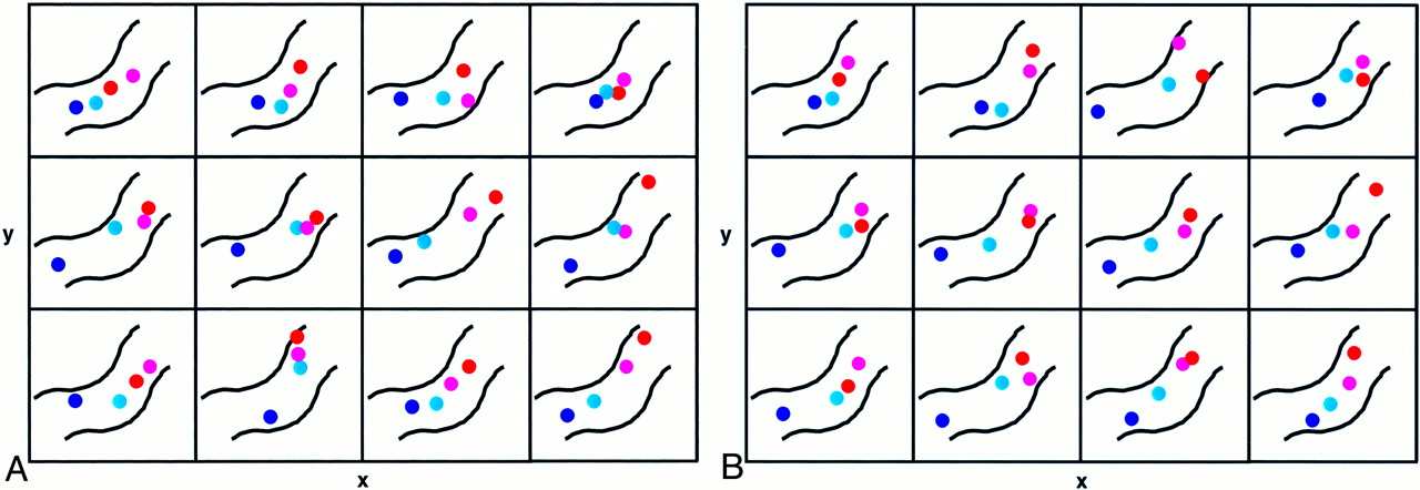
    Two-dimensional scatter plots of within-forearm COGs in contralateral M1 in the 12 subjects. COGs plane for the fingers, hand, wrist, and elbow are displayed in the axial plane.

    A, First experimental session. The approximate contour of the precentral gyrus is outlined (as in Fig 3 left image, with the same colors as in Fig 3). The width x of each rectangle is 20 mm and the height y is 18 mm in Talairach space. Note the preserved somatotopic gradient in all 12 individual hand and forearm representations.

    B, Second experimental session. Note the highly similar distribution and preserved somatotopic gradient of within-forearm COGs in almost all subjects compared with those of first session.

Tables

  • Figures
    • View popup
    TABLE 1:

    Activation in contralateral M1 areas of body parts

    ParadigmVolume of Contralateral M1, mm3Maximum t Value*COG
    Mean ± SDRangexyz
    Hand5566 ± 24732976–96009.7 ± 2.0−36 ± 3−22 ± 458 ± 3
    Fingers2972 ± 12111264–51298.4 ± 2.6−37 ± 2−20 ± 558 ± 2
    Wrist4409 ± 20911520–83209.4 ± 2.4−34 ± 3−23 ± 459 ± 3
    Elbow2267 ± 11581200–78248.2 ± 2.3−29 ± 4−25 ± 461 ± 5
    Foot1457 ± 986256–38885.9 ± 1.2−6 ± 3−33 ± 566 ± 5
    Tongue†
     Left3079 ± 20341088–71527.9 ± 1.5−52 ± 3−5 ± 329 ± 6
     Right3042 ± 164512–62727.3 ± 1.756 ± 2−6 ± 328 ± 5
    • Note.—Data are from the single-subject analysis. Numbers of subjects were as follows: 24 for the hand, fingers, wrist, and elbow experiments and 22 for the tongue and foot experiments.

    • * Data are the mean ± SD.

    • † For the tongue movements, the bilateral representations in the left and right hemispheres are shown.

    • View popup
    TABLE 2:

    Statistical comparisons

    ComparisonP Value from t TestMean Overlapping Volume, %*
    xyz
    Hand versus wrist<.001<.07<.00149% ± 14
    Hand versus elbow<.001<.001<.00128% ± 16
    Finger versus hand>.05>.05>.0586% ± 10
    Finger versus wrist<.001<.001<.00134% ± 14
    Finger versus elbow<.001<.001<.00114% ± 10
    Wrist versus elbow<.001<.001<.00132% ± 14
    • Note.—Comparisons of the COGs and the percentage of overlapping volumes for the finger, hand, wrist, and elbow movements in contralateral M1. Data are from the single-subject analysis. Numbers of subjects were as follows: 24 for the hand, fingers, wrist, and elbow experiments and 22 for the tongue and foot experiments.

    • * Data are the mean ± SD.

    • View popup
    TABLE 3:

    Reproducibility of the COGs and activated volumes in contralateral M1

    Experiment and SessionMean Volumexyz
    Fingers
     First2972 ± 1211−37 ± 2−20 ± 558 ± 2
     Second3023 ± 1263−37 ± 3−19 ± 457 ± 3
    Wrist
     First4409 ± 2091−34 ± 3−23 ± 459 ± 3
     Second3519 ± 1760−34 ± 3−22 ± 559 ± 3
    Elbow
     First2267 ± 1158−29 ± 4−25 ± 461 ± 5
     Second1984 ± 453−29 ± 2−26 ± 362 ± 3
    Hand
     First5566 ± 2473−36 ± 3−22 ± 458 ± 3
     Second5002 ± 1309−37 ± 2−20 ± 457 ± 3
    Foot
     First1457 ± 986−6 ± 3−33 ± 566 ± 5
     Second1436 ± 955−6 ± 3−33 ± 568 ± 4
    Tongue, Left
     First3079 ± 2034−52 ± 3−5 ± 329 ± 6
     Second3907 ± 2185−51 ± 4−6 ± 133 ± 6
    Tongue, Right
     First3042 ± 164056 ± 2−6 ± 328 ± 5
     Second2589 ± 212156 ± 4−7 ± 431 ± 6
    • Note.—COGs are the Talairach coordinates. Data are from the single-subject analysis. Numbers of subjects were as follows: 12 for the hand, fingers, wrist, and elbow experiments and 11 for the tongue and foot experiments.

PreviousNext
Back to top

In this issue

American Journal of Neuroradiology: 23 (9)
American Journal of Neuroradiology
Vol. 23, Issue 9
1 Oct 2002
  • Table of Contents
  • Index by author
Advertisement
Print
Download PDF
Email Article

Thank you for your interest in spreading the word on American Journal of Neuroradiology.

NOTE: We only request your email address so that the person you are recommending the page to knows that you wanted them to see it, and that it is not junk mail. We do not capture any email address.

Enter multiple addresses on separate lines or separate them with commas.
Reproducibility of Primary Motor Cortex Somatotopy Under Controlled Conditions
(Your Name) has sent you a message from American Journal of Neuroradiology
(Your Name) thought you would like to see the American Journal of Neuroradiology web site.
CAPTCHA
This question is for testing whether or not you are a human visitor and to prevent automated spam submissions.
Cite this article
Hatem Alkadhi, Gerard R. Crelier, Sabina Hotz Boendermaker, Xavier Golay, Marie-Claude Hepp-Reymond, Spyros S. Kollias
Reproducibility of Primary Motor Cortex Somatotopy Under Controlled Conditions
American Journal of Neuroradiology Oct 2002, 23 (9) 1524-1532;

Citation Manager Formats

  • BibTeX
  • Bookends
  • EasyBib
  • EndNote (tagged)
  • EndNote 8 (xml)
  • Medlars
  • Mendeley
  • Papers
  • RefWorks Tagged
  • Ref Manager
  • RIS
  • Zotero
0 Responses
Respond to this article
Share
Bookmark this article
Reproducibility of Primary Motor Cortex Somatotopy Under Controlled Conditions
Hatem Alkadhi, Gerard R. Crelier, Sabina Hotz Boendermaker, Xavier Golay, Marie-Claude Hepp-Reymond, Spyros S. Kollias
American Journal of Neuroradiology Oct 2002, 23 (9) 1524-1532;
del.icio.us logo Twitter logo Facebook logo Mendeley logo
  • Tweet Widget
  • Facebook Like
  • Google Plus One

Jump to section

  • Article
    • Abstract
    • Methods
    • Results
    • Discussion
    • Conclusion
    • Footnotes
    • References
  • Figures & Data
  • Info & Metrics
  • Responses
  • References
  • PDF

Related Articles

  • No related articles found.
  • PubMed
  • Google Scholar

Cited By...

  • A mosaic of whole-body representations in human motor cortex
  • "Characteristics and stability of sensorimotor activity driven by isolated-muscle group activation in a human with tetraplegia"
  • Neural Representations of Contextual Guidance in Visual Search of Real-World Scenes
  • Feature-Independent Neural Coding of Target Detection during Search of Natural Scenes
  • Connectivity-Based Parcellation of Human Cingulate Cortex and Its Relation to Functional Specialization
  • Dynamic Changes in Brain Activity during Prism Adaptation
  • Diffusion-Weighted Imaging Tractography-Based Parcellation of the Human Lateral Premotor Cortex Identifies Dorsal and Ventral Subregions with Anatomical and Functional Specializations
  • Positive Emotions Preferentially Engage an Auditory-Motor "Mirror" System
  • Crossref
  • Google Scholar

This article has not yet been cited by articles in journals that are participating in Crossref Cited-by Linking.

More in this TOC Section

  • Predictors of Reperfusion in Patients with Acute Ischemic Stroke
  • Enhanced Axonal Metabolism during Early Natalizumab Treatment in Relapsing-Remitting Multiple Sclerosis
  • SWI or T2*: Which MRI Sequence to Use in the Detection of Cerebral Microbleeds? The Karolinska Imaging Dementia Study
Show more Brain

Similar Articles

Advertisement

Indexed Content

  • Current Issue
  • Accepted Manuscripts
  • Article Preview
  • Past Issues
  • Editorials
  • Editor's Choice
  • Fellows' Journal Club
  • Letters to the Editor
  • Video Articles

Cases

  • Case Collection
  • Archive - Case of the Week
  • Archive - Case of the Month
  • Archive - Classic Case

More from AJNR

  • Trainee Corner
  • Imaging Protocols
  • MRI Safety Corner
  • Book Reviews

Multimedia

  • AJNR Podcasts
  • AJNR Scantastics

Resources

  • Turnaround Time
  • Submit a Manuscript
  • Submit a Video Article
  • Submit an eLetter to the Editor/Response
  • Manuscript Submission Guidelines
  • Statistical Tips
  • Fast Publishing of Accepted Manuscripts
  • Graphical Abstract Preparation
  • Imaging Protocol Submission
  • Evidence-Based Medicine Level Guide
  • Publishing Checklists
  • Author Policies
  • Become a Reviewer/Academy of Reviewers
  • News and Updates

About Us

  • About AJNR
  • Editorial Board
  • Editorial Board Alumni
  • Alerts
  • Permissions
  • Not an AJNR Subscriber? Join Now
  • Advertise with Us
  • Librarian Resources
  • Feedback
  • Terms and Conditions
  • AJNR Editorial Board Alumni

American Society of Neuroradiology

  • Not an ASNR Member? Join Now

© 2025 by the American Society of Neuroradiology All rights, including for text and data mining, AI training, and similar technologies, are reserved.
Print ISSN: 0195-6108 Online ISSN: 1936-959X

Powered by HighWire