
Article Figures & Data
Figures
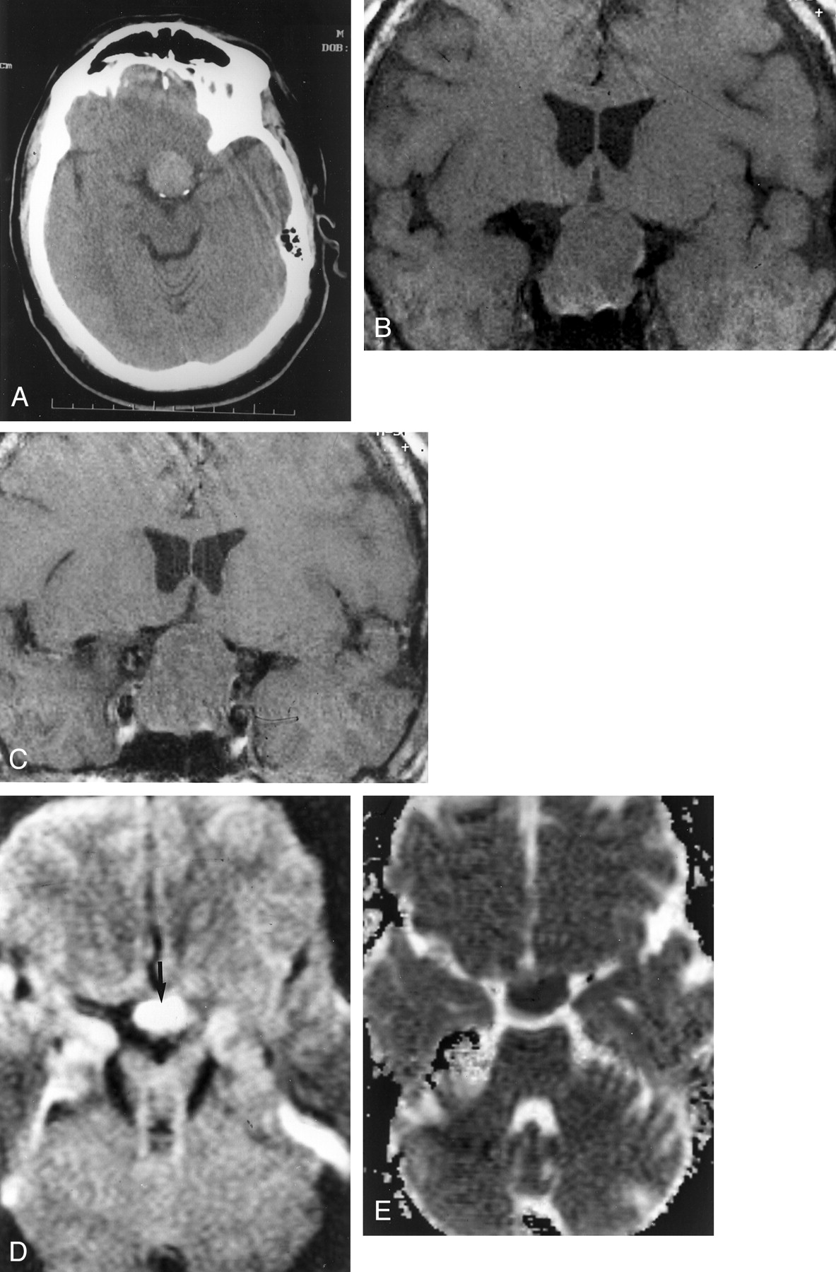
- Fig 1.
The case of a 57-year-old man who presented with decreased vision in his right eye.
A, Nonenhanced CT scan shows a homogeneous, nonhemorrhagic, hyperattenuated intrasellar mass.
B, Coronal spin-echo T1-weighted MR image (500/20 [TR/TE]) shows a large homogeneous intrasellar mass that compresses the optic chiasm, consistent with a nonhemorrhagic macroadenoma.
C, Coronal spin-echo T1-weighted MR image (500/20) shows no change after the administration of contrast medium (0.05 mmol/kg).
D, Tensor diffusion-weighted MR image (b = 1000 mm2/s) shows markedly increased signal intensity (arrow) within the pituitary mass, compared with that in normal gray and white matter.
E, ADC map shows markedly decreased signal intensity within the pituitary mass; mean ADC was 0.49 (10–3 mm2/s).
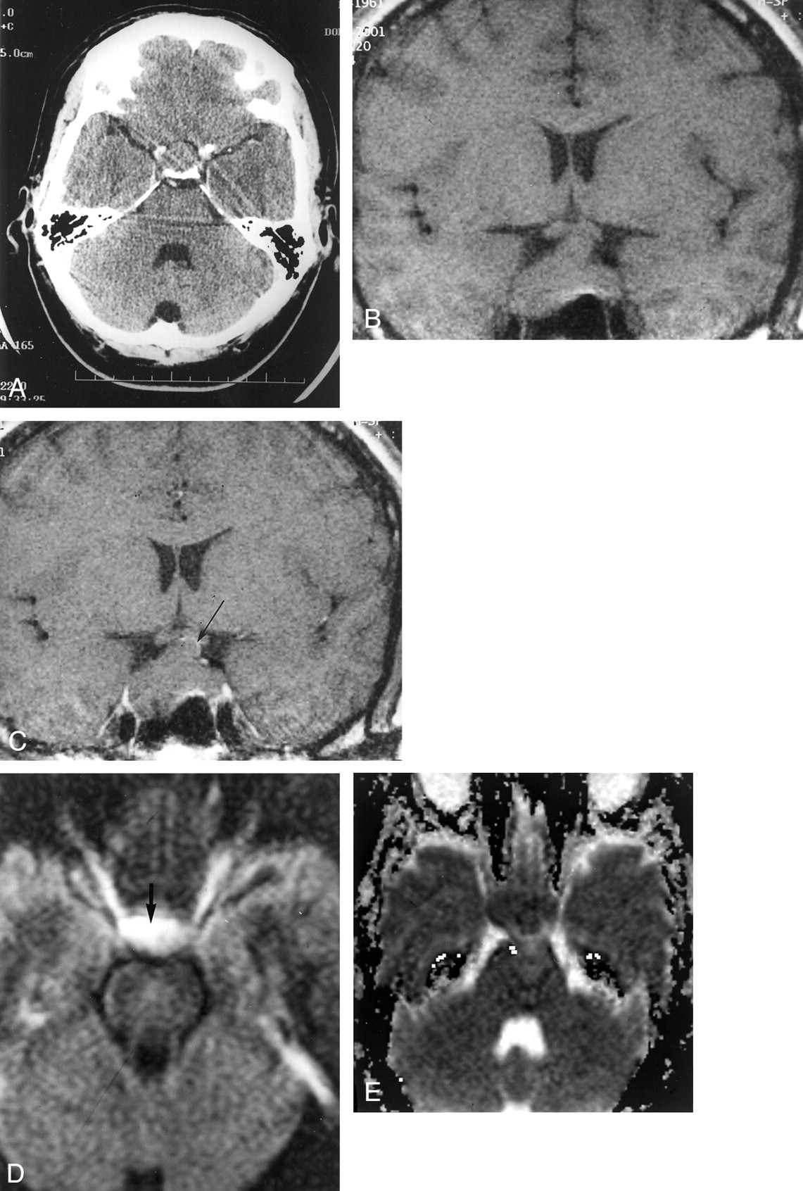
- Fig 2.
The case of a 40-year-old man who awoke with severe bilateral retro-orbital pain.
A, Axial contrast-enhanced CT scan shows a nonenhancing homogeneous intrasellar mass. No hemorrhage is seen.
B, Coronal spin-echo T1-weighted MR image (500/20 [TR/TE]) shows a nonhemorrhagic intrasellar mass with extension to the optic chiasm.
C, Coronal spin-echo T1-weighted MR image (500/20) shows minimal curvilinear marginal enhancement (arrow) after the administration of contrast medium (0.05 mmol/kg).
D, Tensor diffusion-weighted MR image (b = 1000 mm2/s) shows increased signal intensity (arrow) within the pituitary mass, compared with that in normal gray and white matter.
E, ADC map shows decreased signal intensity within the pituitary mass; mean ADC was 0.63 (10–3 mm2/s).
- Fig 3.
The case of a 64-year-old man with histologically confirmed pituitary adenoma without evidence of hemorrhage or infarction.
A, Coronal spin-echo T1-weighted MR image (500/20 [TR/TE]) shows a large homogeneous intrasellar mass compressing the optic chasm, consistent with a nonhemorrhagic macroadenoma.
B, Coronal spin-echo T1-weighted MR image (500/20) shows diffuse uniform enhancement after the administration of contrast medium (0.05 mmol/kg).
C and D, Tensor diffusion-weighted MR images (b = 1000 mm2/s) show isointensity within the pituitary mass (arrows) compared with that in normal brain.
E and F, ADC maps show isointensity within the pituitary mass (arrows) compared with that in normal brain.
- Fig 4.
The case of a 69-year-old woman with histologically confirmed pituitary adenoma without evidence of hemorrhage or infarction.
A, Coronal spin-echo T1-weighted MR image (500/20 [TR/TE]) shows a homogeneous intrasellar mass extending inferiorly, with remodeling of the sellar floor.
B, Tensor diffusion-weighted MR image (b = 1000 mm2/s) shows isointensity within the pituitary mass (arrows) compared with that in normal brain.
C, ADC map shows isointensity within the pituitary mass (arrow) compared with that in normal brain; mean ADC was 0.90 (10–3 mm2/s).


In this issue


Jump to section
Related Articles
Cited By...
- Pituitary tumour apoplexy due to pituitary adenoma in a dog: clinical, 3T MRI and CT features
- Intravoxel Incoherent Motion in Normal Pituitary Gland: Initial Study with Turbo Spin-Echo Diffusion-Weighted Imaging
- Evaluation of Diffusivity in the Anterior Lobe of the Pituitary Gland: 3D Turbo Field Echo with Diffusion-Sensitized Driven-Equilibrium Preparation