Advertisement

Main menu

  • Home
  • Content
    • Current Issue
    • Accepted Manuscripts
    • Article Preview
    • Past Issue Archive
    • Video Articles
    • AJNR Case Collection
    • Case of the Week Archive
    • Case of the Month Archive
    • Classic Case Archive
  • Special Collections
    • AJNR Awards
    • Low-Field MRI
    • Alzheimer Disease
    • ASNR Foundation Special Collection
    • Photon-Counting CT
    • View All
  • Multimedia
    • AJNR Podcasts
    • AJNR SCANtastic
    • Trainee Corner
    • MRI Safety Corner
    • Imaging Protocols
  • For Authors
    • Submit a Manuscript
    • Submit a Video Article
    • Submit an eLetter to the Editor/Response
    • Manuscript Submission Guidelines
    • Statistical Tips
    • Fast Publishing of Accepted Manuscripts
    • Graphical Abstract Preparation
    • Imaging Protocol Submission
    • Author Policies
  • About Us
    • About AJNR
    • Editorial Board
    • Editorial Board Alumni
  • More
    • Become a Reviewer/Academy of Reviewers
    • Subscribers
    • Permissions
    • Alerts
    • Feedback
    • Advertisers
    • ASNR Home

User menu

  • Alerts
  • Log in

Search

  • Advanced search
American Journal of Neuroradiology

American Journal of Neuroradiology

ASHNR American Society of Functional Neuroradiology ASHNR American Society of Pediatric Neuroradiology ASSR
  • Alerts
  • Log in

Advanced Search

  • Home
  • Content
    • Current Issue
    • Accepted Manuscripts
    • Article Preview
    • Past Issue Archive
    • Video Articles
    • AJNR Case Collection
    • Case of the Week Archive
    • Case of the Month Archive
    • Classic Case Archive
  • Special Collections
    • AJNR Awards
    • Low-Field MRI
    • Alzheimer Disease
    • ASNR Foundation Special Collection
    • Photon-Counting CT
    • View All
  • Multimedia
    • AJNR Podcasts
    • AJNR SCANtastic
    • Trainee Corner
    • MRI Safety Corner
    • Imaging Protocols
  • For Authors
    • Submit a Manuscript
    • Submit a Video Article
    • Submit an eLetter to the Editor/Response
    • Manuscript Submission Guidelines
    • Statistical Tips
    • Fast Publishing of Accepted Manuscripts
    • Graphical Abstract Preparation
    • Imaging Protocol Submission
    • Author Policies
  • About Us
    • About AJNR
    • Editorial Board
    • Editorial Board Alumni
  • More
    • Become a Reviewer/Academy of Reviewers
    • Subscribers
    • Permissions
    • Alerts
    • Feedback
    • Advertisers
    • ASNR Home
  • Follow AJNR on Twitter
  • Visit AJNR on Facebook
  • Follow AJNR on Instagram
  • Join AJNR on LinkedIn
  • RSS Feeds

AJNR Awards, New Junior Editors, and more. Read the latest AJNR updates

Research ArticleBRAIN

Turbulence and Circulating Cerebral Emboli Detectable at Doppler Ultrasonography: A Differentiation Study in a Stenotic Middle Cerebral Artery Model

Yi Yang, Donald G. Grosset, Qiu Li, Ashfaq Shuaib and Kennedy R. Lees
American Journal of Neuroradiology August 2002, 23 (7) 1229-1236;
Yi Yang
  • Find this author on Google Scholar
  • Find this author on PubMed
  • Search for this author on this site
Donald G. Grosset
  • Find this author on Google Scholar
  • Find this author on PubMed
  • Search for this author on this site
Qiu Li
  • Find this author on Google Scholar
  • Find this author on PubMed
  • Search for this author on this site
Ashfaq Shuaib
  • Find this author on Google Scholar
  • Find this author on PubMed
  • Search for this author on this site
Kennedy R. Lees
  • Find this author on Google Scholar
  • Find this author on PubMed
  • Search for this author on this site
  • Article
  • Figures & Data
  • Info & Metrics
  • Responses
  • References
  • PDF
Loading

Article Figures & Data

Figures

  • Tables
  • Fig 1.
    • Download figure
    • Open in new tab
    • Download powerpoint
    Fig 1.

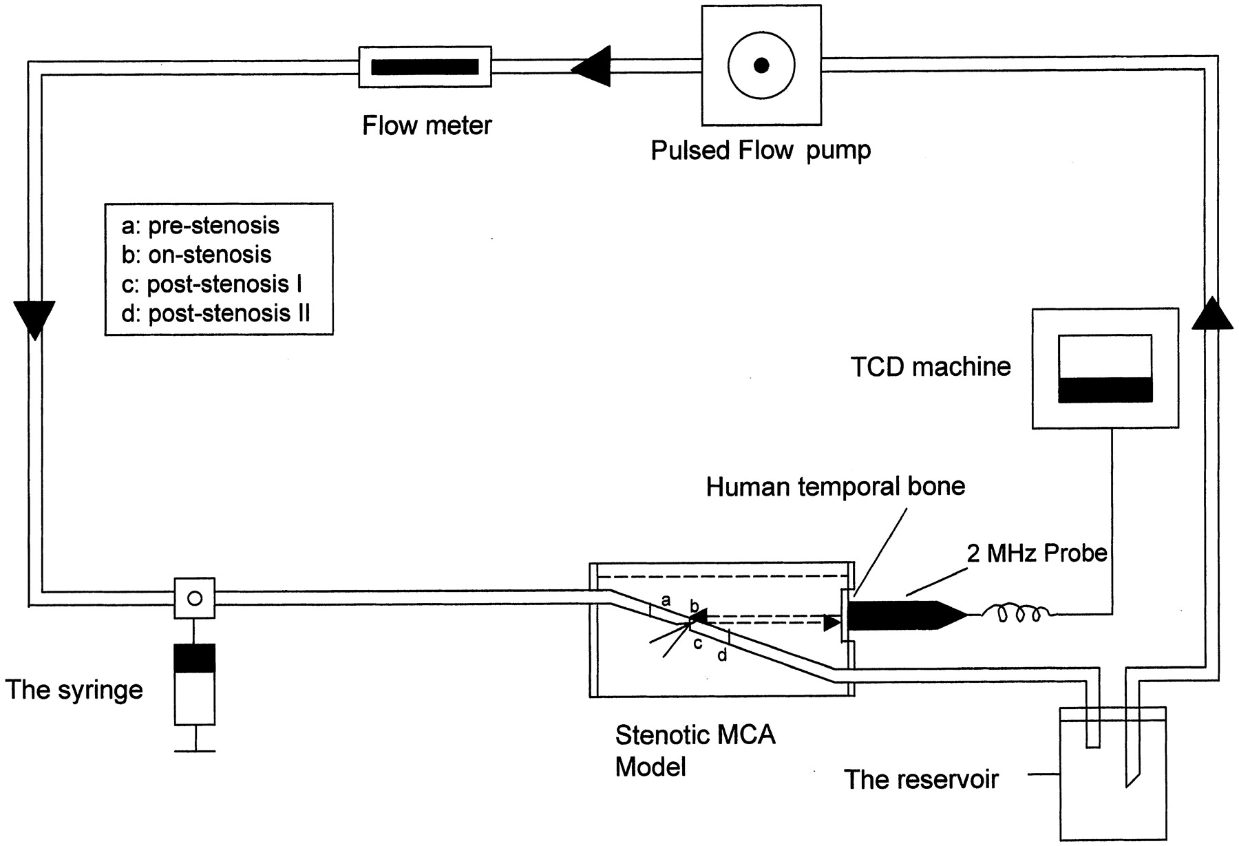
    Schematic illustration of the stenotic MCA model and circuit. Diluted RBC suspension was circulated in a pulsed fashion by a pulsed pump, and the embolic materials were injected into the circuit via the side port. Turbulence and embolic signals as well as flow velocities were continuously recorded from a TCD probe, which was coupled with the outside of temporal bone.

  • Fig 2.
    • Download figure
    • Open in new tab
    • Download powerpoint
    Fig 2.

    Characterization of Doppler waveform at different zones along the stenotic artery (65% reduced area). Flow of RBC suspension displayed a normal Doppler velocity profile at the prestenosis zone. However, at the stenosis zone, the flow showed a sharp increase in systolic velocity and the signal intensity started to distribute unevenly; at poststenosis I zone, the velocity tended to decrease, but a high intensity turbulent signal in the central waveform and apparent reversal flow became notable. The flow profile at poststenosis zone II returned to normal but had a lower systolic velocity.

  • Fig 3.
    • Download figure
    • Open in new tab
    • Download powerpoint
    Fig 3.

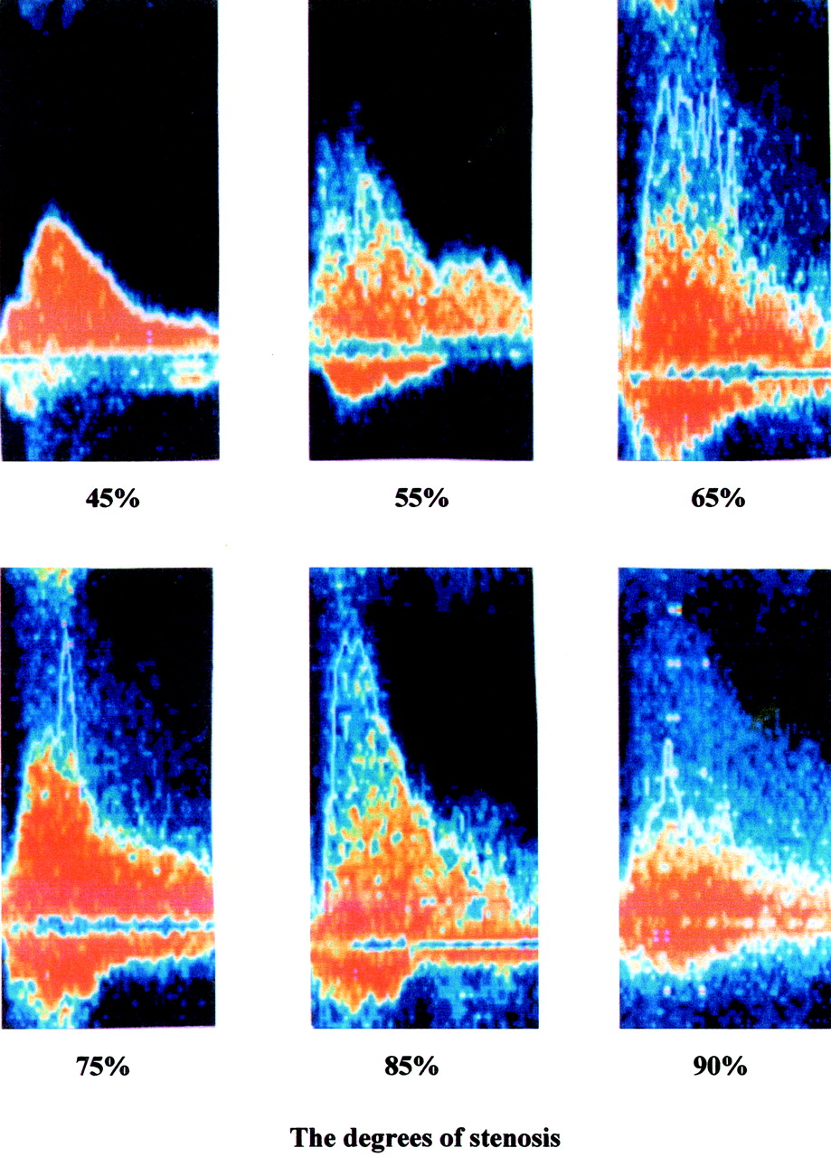
    Dynamic profile of flow turbulence associated with different degrees of arterial stenosis. The transition of flow turbulence did not occur until the degree of stenosis of the artery increased to 55%. At stenoses of 66% and 75%, turbulence became most typical and was accompanied by reversed flow. Turbulence remained but decreased when degree of stenosis increased to 85% or 90%. Both systolic and diastolic velocities increased in association with the rising degree of stenosis, but systolic velocities changed more predominately.

  • Fig 4.
    • Download figure
    • Open in new tab
    • Download powerpoint
    Fig 4.

    Correlation between degree of stenosis and average signal intensity of flow turbulence sampled at poststenosis zone I. Association between average Doppler signal intensities sampled at poststenosis zone I and degrees of stenosis of the artery looks like a tea spoon. Poor correlation existed between the two indices (r = 0.72, P = .07).

  • Fig 5.
    • Download figure
    • Open in new tab
    • Download powerpoint
    Fig 5.

    Correlation between degree of stenosis and Doppler mean flow velocity at poststenosis zone I. Mean velocities calculated at poststenosis zone I showed a linear relationship, with an increase in degree of stenosis after logarithmic transformation of mean flow velocity.

  • Fig 6.
    • Download figure
    • Open in new tab
    • Download powerpoint
    Fig 6.

    Visual differentiation between turbulence signals and embolic signals. Turbulence signals showed a high intensity area in the center of the systolic phase of the Doppler flow waveform and were usually accompanied by jet flow velocity, harsh noise, and reversed flow. However, embolic signals were featured as high intensity points that randomly superimposed on the flow waveform of either the systolic or diastolic period, accompanied by harmonic chirps.

Tables

  • Figures
  • Conversion of cross-sectional area and diameter reductions

    Reduction (%)
    Reduction of cross-sectional area4555657590
    Reduction of diameter2633415069
PreviousNext
Back to top

In this issue

American Journal of Neuroradiology: 23 (7)
American Journal of Neuroradiology
Vol. 23, Issue 7
1 Aug 2002
  • Table of Contents
  • Index by author
Advertisement
Print
Download PDF
Email Article

Thank you for your interest in spreading the word on American Journal of Neuroradiology.

NOTE: We only request your email address so that the person you are recommending the page to knows that you wanted them to see it, and that it is not junk mail. We do not capture any email address.

Enter multiple addresses on separate lines or separate them with commas.
Turbulence and Circulating Cerebral Emboli Detectable at Doppler Ultrasonography: A Differentiation Study in a Stenotic Middle Cerebral Artery Model
(Your Name) has sent you a message from American Journal of Neuroradiology
(Your Name) thought you would like to see the American Journal of Neuroradiology web site.
CAPTCHA
This question is for testing whether or not you are a human visitor and to prevent automated spam submissions.
Cite this article
Yi Yang, Donald G. Grosset, Qiu Li, Ashfaq Shuaib, Kennedy R. Lees
Turbulence and Circulating Cerebral Emboli Detectable at Doppler Ultrasonography: A Differentiation Study in a Stenotic Middle Cerebral Artery Model
American Journal of Neuroradiology Aug 2002, 23 (7) 1229-1236;

Citation Manager Formats

  • BibTeX
  • Bookends
  • EasyBib
  • EndNote (tagged)
  • EndNote 8 (xml)
  • Medlars
  • Mendeley
  • Papers
  • RefWorks Tagged
  • Ref Manager
  • RIS
  • Zotero
0 Responses
Respond to this article
Share
Bookmark this article
Turbulence and Circulating Cerebral Emboli Detectable at Doppler Ultrasonography: A Differentiation Study in a Stenotic Middle Cerebral Artery Model
Yi Yang, Donald G. Grosset, Qiu Li, Ashfaq Shuaib, Kennedy R. Lees
American Journal of Neuroradiology Aug 2002, 23 (7) 1229-1236;
del.icio.us logo Twitter logo Facebook logo Mendeley logo
  • Tweet Widget
  • Facebook Like
  • Google Plus One

Jump to section

  • Article
    • Abstract
    • Methods
    • Results
    • Discussion
    • Conclusion
    • Acknowledgments
    • Footnotes
    • References
  • Figures & Data
  • Info & Metrics
  • Responses
  • References
  • PDF

Related Articles

  • No related articles found.
  • PubMed
  • Google Scholar

Cited By...

  • No citing articles found.
  • Crossref
  • Google Scholar

This article has not yet been cited by articles in journals that are participating in Crossref Cited-by Linking.

More in this TOC Section

  • Enhanced Axonal Metabolism during Early Natalizumab Treatment in Relapsing-Remitting Multiple Sclerosis
  • Progression of Microstructural Damage in Spinocerebellar Ataxia Type 2: A Longitudinal DTI Study
  • SWI or T2*: Which MRI Sequence to Use in the Detection of Cerebral Microbleeds? The Karolinska Imaging Dementia Study
Show more BRAIN

Similar Articles

Advertisement

Indexed Content

  • Current Issue
  • Accepted Manuscripts
  • Article Preview
  • Past Issues
  • Editorials
  • Editor's Choice
  • Fellows' Journal Club
  • Letters to the Editor
  • Video Articles

Cases

  • Case Collection
  • Archive - Case of the Week
  • Archive - Case of the Month
  • Archive - Classic Case

More from AJNR

  • Trainee Corner
  • Imaging Protocols
  • MRI Safety Corner
  • Book Reviews

Multimedia

  • AJNR Podcasts
  • AJNR Scantastics

Resources

  • Turnaround Time
  • Submit a Manuscript
  • Submit a Video Article
  • Submit an eLetter to the Editor/Response
  • Manuscript Submission Guidelines
  • Statistical Tips
  • Fast Publishing of Accepted Manuscripts
  • Graphical Abstract Preparation
  • Imaging Protocol Submission
  • Evidence-Based Medicine Level Guide
  • Publishing Checklists
  • Author Policies
  • Become a Reviewer/Academy of Reviewers
  • News and Updates

About Us

  • About AJNR
  • Editorial Board
  • Editorial Board Alumni
  • Alerts
  • Permissions
  • Not an AJNR Subscriber? Join Now
  • Advertise with Us
  • Librarian Resources
  • Feedback
  • Terms and Conditions
  • AJNR Editorial Board Alumni

American Society of Neuroradiology

  • Not an ASNR Member? Join Now

© 2025 by the American Society of Neuroradiology All rights, including for text and data mining, AI training, and similar technologies, are reserved.
Print ISSN: 0195-6108 Online ISSN: 1936-959X

Powered by HighWire