
Article Figures & Data
Figures
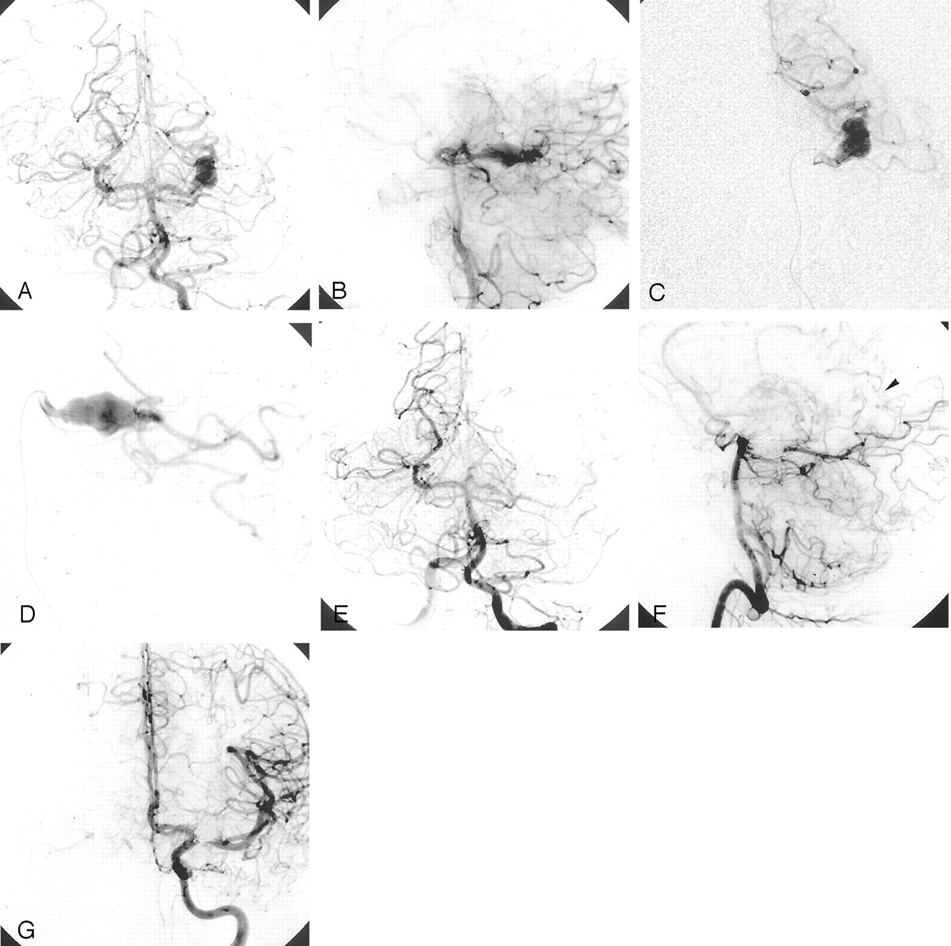
- Fig 1.
Case 3. Large fusiform aneurysm of the left P2 segment.
A and B, Left vertebral artery injection, frontal (A) and lateral (B) views.
C and D, Selective PCA injection, frontal (C) and lateral (D) views.
E–G, After embolization with GDC coils, frontal (E) and oblique (F) views obtained with a vertebral artery injection and frontal view (G) obtained with a left carotid injection shows occlusion of the P2 segment aneurysm and PCA, with distal perfusion via leptomeningeal anastomoses (arrowhead in F).
- Fig 2.
Case 7. Large saccular partially thrombosed aneurysm of the left P2 segment.
A and B, Vertebral artery injection, frontal (A) and lateral (B) projections.
C and D, After embolization, oblique view obtained with a vertebral artery injection (C) and lateral view obtained with a left carotid injection (D) show occlusion of the aneurysm and P2 segment, with distal perfusion of the PCA territory via a leptomeningeal supply. (arrowheads in D).
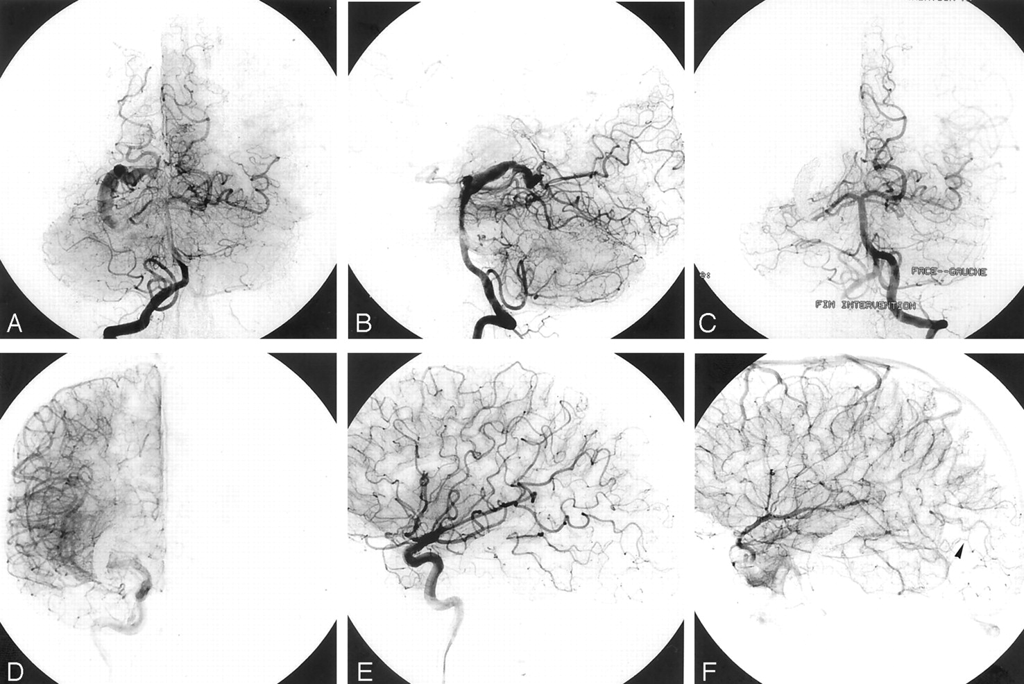
- Fig 3.
Case 9. Giant, partially thrombosed serpentine aneurysm of the right P2 segment.
A and B, Right vertebral artery injection, frontal (A) and lateral (B) views.
C, After embolization, frontal view obtained with a left vertebral artery injection shows complete occlusion of the P2 segment and aneurysm.
D–F, After embolization, frontal (D) and lateral early (E) and late (F) views obtained with a right carotid artery injection show the leptomeningeal supply to the right PCA territory (arrowhead in F).
- Fig 4.
Schematic drawing of the midbrain surrounded by the PCA. The PCA is divided into four segments. The P2 segment begins at the PCoA-PCA junction and courses through the distal peduncular and ambient cisterns to the posterior part of the midbrain.


Tables
Summary of data in 10 patients with P2 segment aneurysms
Patient No./Sex/Age (y) Aneurysm Type, Side, and Size Presenting Symptom Test Treatment Type and Year Clinical Outcome and Follow-Up Findings 1/43/M Saccular, R, giant thrombosis Headaches, phosphenes Balloon occlusion Balloon, 1990 Excellent, no headaches, phosphenes gone; at 11-mo angiography, no recanalization; MR imaging at 6 y, two-thirds reduction in aneurysmal mass 2/53/F Saccular, R, giant thrombosis Headaches, LHH Balloon occlusion, clinical examination Balloon, 1994 LHH, no headaches; at 6-mo angiography, no recanalization 3/20/M Fusiform, L, large Headaches Angiography GDC, 1996 Excellent, no headaches; at 1-y angiography, no recanalization 4/60/M Fusiform, L, large Headaches Balloon occlusion GDC, 1998 Excellent, no headaches; at 1-y angiography, no recanalization 5/49/F Fusiform, L, giant thrombosis Gertsmann syndrome Angiography GDC, 1998 Excellent, no deficit; at 1-y angiography, no recanalization 6/18/F Saccular, L, large SAH, hematoma Angiography Histoacryl, 1998 Excellent, no deficit; at 1-y angiography, no recanalization 7/49/F Saccular, L, large Headaches Balloon occlusion GDC, 1999 Excellent, no headaches; at 1-y angiography, no recanalization 8/47/M Fusiform, L, giant thrombosis SAH, diplopia Balloon occlusion GDC, 1999 Excellent, no deficit; at 1-y angiography, no recanalization 9/26/M Serpentine, R, giant thrombosis Headaches Angiography GDC, 1999 Excellent, no headaches; at 1-y angiography, no recanalization 10/70/F Saccular, R, large Headaches None None Well 3 y after attempt Note.—LHH indicates lateral homonymous hemianopsia; SAH, subarachnoid hemorrhage.
In this issue


Jump to section
Related Articles
Cited By...
- Endovascular parent artery occlusion of proximal posterior cerebral artery aneurysms: a report of two cases
- Endoluminal Reconstruction for Nonsaccular Aneurysms of the Proximal Posterior Cerebral Artery with the Pipeline Embolization Device
- Long-Term Clinical and Imaging Follow-Up of Complex Intracranial Aneurysms Treated by Endovascular Parent Vessel Occlusion