
Article Figures & Data
Figures
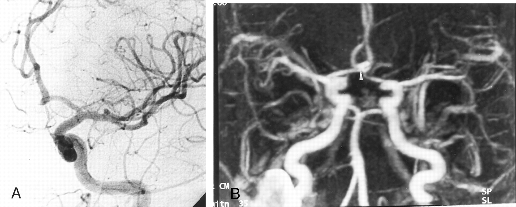
- Fig 1.
DSA and contrast-enhanced MRA show concordant findings.
A, Intracranial left carotid DSA obtained 12 months after the treatment of a 4-mm aneurysm in the AcomA. Frontal head view shows no remnant cavity at the site of the AcomA (arrow).
B, Contrast-enhanced MRA image with MIP reconstruction in the frontal view shows findings in agreement with those at DSA.
- Fig 2.
TOF-MRA and contrast-enhanced MRA show concordant findings.
A, TOF-MRA image obtained with MIP reconstruction obtained 12 months after the treatment of a 8-mm aneurysm in the AcomA. Axial image depicts a residual neck of 4-mm diameter (arrowhead).
B, Contrast-enhanced MRA image obtained with axial MIP reconstruction in the axial plane depicts a residual cavity (arrow), in agreement with the TOF-MRA findings.
- Fig 3.
Detection of aneurysmal neck remnant.
A, DSA image of the left internal carotid artery obtained 12 months after the treatment of a 5-mm aneurysm of the AcomA. Frontal head view shows a residual neck with a 3-mm diameter (arrow).
B, TOF-MRA image obtained with MIP reconstruction in the axial plane does not depict the residual neck.
C, Contrast-enhanced MRA image obtained with axial MIP reconstruction in the axial plane shows a small residual cavity (arrow), in agreement with the DSA findings.
- Fig 4.
Patient with a 6-mm aneurysm of the AcomA that was treated with GDCs. Only the left carotid angiogram showed the aneurysm at initial DSA.
A, Left carotid artery angiogram obtained 12 months after treatment. Frontal head view shows no recurrent aneurysm.
B, Contrast-enhanced MRA image obtained with MIP reconstruction shows a hyperintense area (arrowhead) at the site of the AcomA. This finding was misinterpreted as a residual aneurysmal neck.


Tables
- TABLE 1:
MR angiographic and DSA findings of intraaneurysmal flow at 12-month follow-up after GDC treatment
MR Angiographic Finding DSA Finding Complete Occlusion Small Residual Neck Large Residual Neck Not Assessable TOF image Complete occlusion 15 2 0 0 Small residual neck 0 2 0 0 Large residual neck 0 0 1 0 Not assessable 0 0 0 0 Contrast-enhanced image Complete occlusion 14 0 0 0 Small residual neck 1 4 0 0 Large residual neck 0 0 1 0 Not assessable 0 0 0 0 - TABLE 2:
Sensitivity and specificity of MR angiography and DSA in the detection of a residual aneurysmal neck at 1-year follow-up after GDC treatment
MR Angiography Sensitivity, % Specificity, % Interobserver P Value Intertechnique P Value TOF 60 100 .16 <.001 Contrast-enhanced 100 93 <.001 <.001
In this issue


Jump to section
Related Articles
Cited By...
- MRA versus DSA for the follow-up imaging of intracranial aneurysms treated using endovascular techniques: a meta-analysis
- Is Visual Evaluation of Aneurysm Coiling a Reliable Study End Point?: Systematic Review and Meta-Analysis
- MRA Versus DSA for Follow-Up of Coiled Intracranial Aneurysms: A Meta-Analysis
- Endovascular Treatment of Anterior Communicating Artery Aneurysms: A Systematic Review and Meta-Analysis
- Outcomes of Endovascular Treatments of Aneurysms: Observer Variability and Implications for Interpreting Case Series and Planning Randomized Trials
- Residual Flow After Cerebral Aneurysm Coil Occlusion: Diagnostic Accuracy of MR Angiography
- Long-Term Prospective Follow-Up of Intracranial Aneurysms Treated with Endovascular Coiling Using Contrast-Enhanced MR Angiography
- Intracranial Aneurysms Treated With Guglielmi Detachable Coils: Imaging Follow-Up With Contrast-Enhanced MR Angiography
- Matrix Detachable Coils for the Endovascular Treatment of Intracranial Aneurysms: Analysis of Early Angiographic and Clinical Outcomes