Advertisement

Main menu

  • Home
  • Content
    • Current Issue
    • Accepted Manuscripts
    • Article Preview
    • Past Issue Archive
    • Video Articles
    • AJNR Case Collection
    • Case of the Week Archive
    • Case of the Month Archive
    • Classic Case Archive
  • Special Collections
    • AJNR Awards
    • Low-Field MRI
    • Alzheimer Disease
    • ASNR Foundation Special Collection
    • Photon-Counting CT
    • View All
  • Multimedia
    • AJNR Podcasts
    • AJNR SCANtastic
    • Trainee Corner
    • MRI Safety Corner
    • Imaging Protocols
  • For Authors
    • Submit a Manuscript
    • Submit a Video Article
    • Submit an eLetter to the Editor/Response
    • Manuscript Submission Guidelines
    • Statistical Tips
    • Fast Publishing of Accepted Manuscripts
    • Graphical Abstract Preparation
    • Imaging Protocol Submission
    • Author Policies
  • About Us
    • About AJNR
    • Editorial Board
    • Editorial Board Alumni
  • More
    • Become a Reviewer/Academy of Reviewers
    • Subscribers
    • Permissions
    • Alerts
    • Feedback
    • Advertisers
    • ASNR Home

User menu

  • Alerts
  • Log in

Search

  • Advanced search
American Journal of Neuroradiology

American Journal of Neuroradiology

ASHNR American Society of Functional Neuroradiology ASHNR American Society of Pediatric Neuroradiology ASSR
  • Alerts
  • Log in

Advanced Search

  • Home
  • Content
    • Current Issue
    • Accepted Manuscripts
    • Article Preview
    • Past Issue Archive
    • Video Articles
    • AJNR Case Collection
    • Case of the Week Archive
    • Case of the Month Archive
    • Classic Case Archive
  • Special Collections
    • AJNR Awards
    • Low-Field MRI
    • Alzheimer Disease
    • ASNR Foundation Special Collection
    • Photon-Counting CT
    • View All
  • Multimedia
    • AJNR Podcasts
    • AJNR SCANtastic
    • Trainee Corner
    • MRI Safety Corner
    • Imaging Protocols
  • For Authors
    • Submit a Manuscript
    • Submit a Video Article
    • Submit an eLetter to the Editor/Response
    • Manuscript Submission Guidelines
    • Statistical Tips
    • Fast Publishing of Accepted Manuscripts
    • Graphical Abstract Preparation
    • Imaging Protocol Submission
    • Author Policies
  • About Us
    • About AJNR
    • Editorial Board
    • Editorial Board Alumni
  • More
    • Become a Reviewer/Academy of Reviewers
    • Subscribers
    • Permissions
    • Alerts
    • Feedback
    • Advertisers
    • ASNR Home
  • Follow AJNR on Twitter
  • Visit AJNR on Facebook
  • Follow AJNR on Instagram
  • Join AJNR on LinkedIn
  • RSS Feeds

AJNR Awards, New Junior Editors, and more. Read the latest AJNR updates

OtherHead and Neck Imaging

Plain Film Imaging of Baerveldt Glaucoma Drainage Implants

Elizenda M. Ceballos and Richard K. Parrish
American Journal of Neuroradiology June 2002, 23 (6) 935-937;
Elizenda M. Ceballos
aFrom the Department of Ophthalmology, University of Miami School of Medicine, Miami, FL
  • Find this author on Google Scholar
  • Find this author on PubMed
  • Search for this author on this site
Richard K. Parrish II
aFrom the Department of Ophthalmology, University of Miami School of Medicine, Miami, FL
  • Find this author on Google Scholar
  • Find this author on PubMed
  • Search for this author on this site
  • Article
  • Figures & Data
  • Info & Metrics
  • Responses
  • References
  • PDF
Loading

Abstract

Summary: A glaucoma drainage implant was detected on plain skull radiographs before MR imaging examination of the brain. The patient was denied the MR imaging for fear of dislodging the “metallic foreign body.” The Baerveldt glaucoma drainage implant was mistakenly identified as an orbital metallic object based on its radiographic characteristics. Because none of the current glaucoma drainage implants contain ferromagnetic material, patients with these devices can undergo MR imaging without special precaution.

Drainage implants are increasingly being used for the surgical treatment of glaucoma refractory to medical and standard surgical therapy. Echographic and radiographic findings may suggest the diagnosis of an orbital foreign body if the ophthalmic history is not known.

Case Report

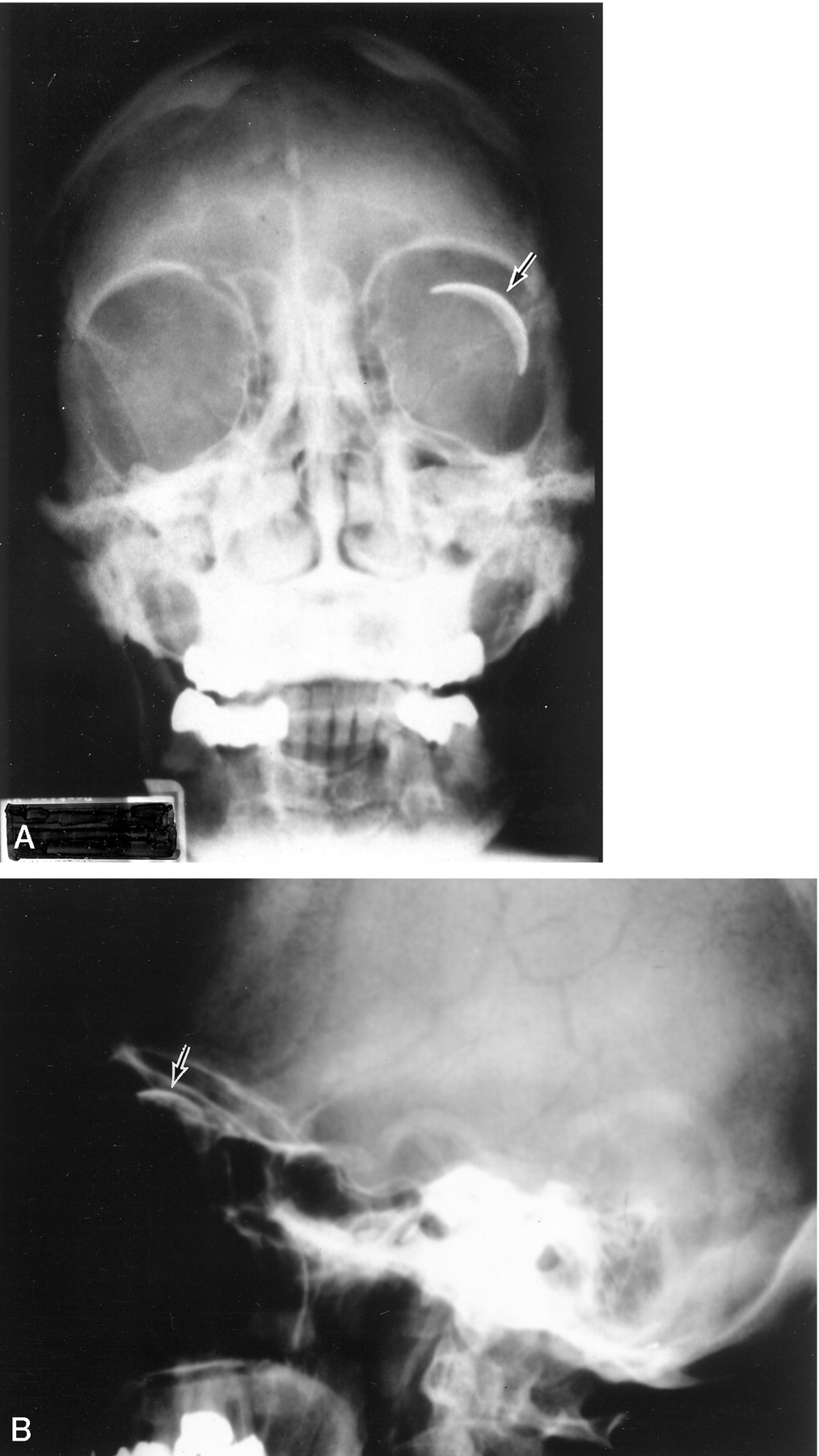
An asymptomatic 82-year-old female patient with open angle glaucoma presented to her ophthalmologist for evaluation of a “metallic object” in her left orbit (Fig 1). The object in question was identified as a 350-mm2 Baerveldt glaucoma drainage implant (Pharmacia Co., Kalamazoo, MI) that had been previously placed to lower intraocular pressure. The smooth convex radiographic profile of the device corresponded to the globe curvature in the superotemporal orbit.

Fig 1.
  • Download figure
  • Open in new tab
  • Download powerpoint
Fig 1.

Plain film radiographs obtained from an asymptomatic 82-year-old female patient with open angle glaucoma who presented to her ophthalmologist for evaluation of a “metallic object” in her left orbit.

A, Anteroposterior view shows the convex, barium-impregnated plate of the Baerveldt glaucoma drainage implant positioned superotemporally on the globe.

B, Lateral view shows the implant.

Discussion

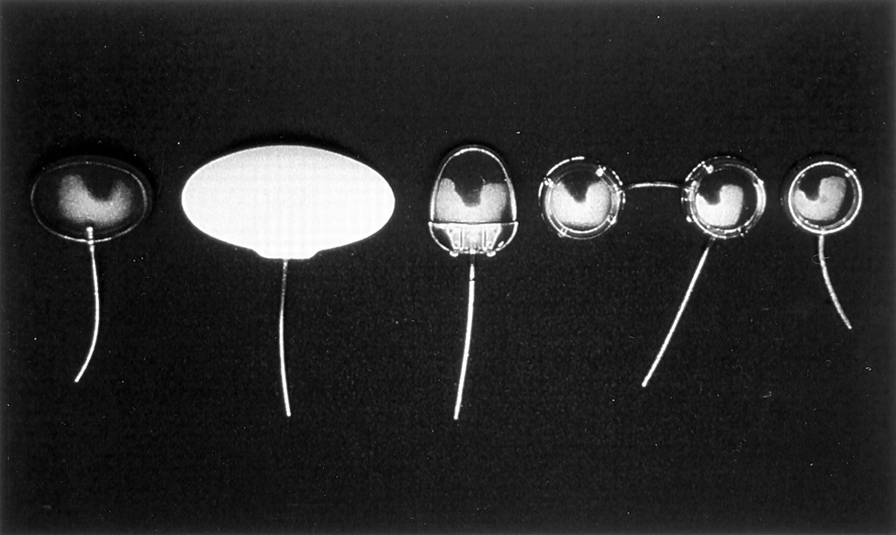
Of the commercially available glaucoma drainage implants, only the Baerveldt glaucoma drainage implant (250, 350, and 425 mm2) is radiopaque (Fig 2). Barium-impregnated silicone used in the fabrication of the Baerveldt plate increases echographic resolution and facilitates radiographic identification. The Krupin-Denver eye valve to disc implant (184 mm2) (E. Benson Hood Laboratories, Pembroke, MA), Ahmed glaucoma valve (184 mm2) (New World Medical, Rancho Cucamonga, CA), Molteno drainage device (single plate 129 mm2 and double plate 259 mm2) (Molteno Ophthalmic Ltd., Dunedin, New Zealand), and the Joseph valve (Valve Implants Limited, Hertford, England) feature a translucent silicone tube connected to a plate made of rigid plastic or flexible silicone rubber that is fixed to the sclera (Fig 3). None of these devices contain ferromagnetic material or have been impregnated with a radiopaque substance.

Fig 2.
  • Download figure
  • Open in new tab
  • Download powerpoint
Fig 2.

Baerveldt glaucoma drainage implants are shown in three sizes with surface areas of 250, 350, and 425 mm2 (left to right).

Fig 3.
  • Download figure
  • Open in new tab
  • Download powerpoint
Fig 3.

Commercially available glaucoma drainage devices are shown. From left to right, they are the Krupin drainage device, Baerveldt drainage implant (350 mm2), Ahmed implant, double plate Molteno device, and single plate Molteno device. (Note that the Joseph valve is not shown).

Intraocular pressure is lowered when aqueous humor flows from inside the eye through the tube into the space between the plate that rests on the scleral surface and surrounding fibrous capsule. The adjacent periocular tissues absorb aqueous humor from the reservoir. Although intraocular ferromagnetic foreign bodies have been shown to move during MR imaging (1–3), because neither the Baerveldt drainage implant nor any of the other commercially available devices contain ferromagnetic material, inadvertent dislocation of these devices is unlikely.

References

  1. ↵
    Lagouros PA, Langer BG, Peyman GA, Mafee MF, Spigos DG, Grisolano J. Magnetic resonance imaging and intraocular foreign bodies. Arch Ophthalmol 1987;105:551–553
    CrossRefPubMed
  2. Williams S, Char DH, Dillon WP, Lincoff N, Moseley M. Ferrous intraocular foreign bodies and magnetic resonance imaging. Am J Ophthalmol 1988;105:398–401
    CrossRefPubMed
  3. ↵
    Ta CN, Bowman RW. Hyphema caused by metallic intraocular foreign body during magnetic resonance imaging. Am J Ophthalmol 2000;129:533–534
    CrossRefPubMed
  • Received August 13, 2001.
  • Accepted after revision January 28, 2002.
  • Copyright © American Society of Neuroradiology
View Abstract
PreviousNext
Back to top

In this issue

American Journal of Neuroradiology: 23 (6)
American Journal of Neuroradiology
Vol. 23, Issue 6
1 Jun 2002
  • Table of Contents
  • Index by author
Advertisement
Print
Download PDF
Email Article

Thank you for your interest in spreading the word on American Journal of Neuroradiology.

NOTE: We only request your email address so that the person you are recommending the page to knows that you wanted them to see it, and that it is not junk mail. We do not capture any email address.

Enter multiple addresses on separate lines or separate them with commas.
Plain Film Imaging of Baerveldt Glaucoma Drainage Implants
(Your Name) has sent you a message from American Journal of Neuroradiology
(Your Name) thought you would like to see the American Journal of Neuroradiology web site.
CAPTCHA
This question is for testing whether or not you are a human visitor and to prevent automated spam submissions.
Cite this article
Elizenda M. Ceballos, Richard K. Parrish
Plain Film Imaging of Baerveldt Glaucoma Drainage Implants
American Journal of Neuroradiology Jun 2002, 23 (6) 935-937;

Citation Manager Formats

  • BibTeX
  • Bookends
  • EasyBib
  • EndNote (tagged)
  • EndNote 8 (xml)
  • Medlars
  • Mendeley
  • Papers
  • RefWorks Tagged
  • Ref Manager
  • RIS
  • Zotero
0 Responses
Respond to this article
Share
Bookmark this article
Plain Film Imaging of Baerveldt Glaucoma Drainage Implants
Elizenda M. Ceballos, Richard K. Parrish
American Journal of Neuroradiology Jun 2002, 23 (6) 935-937;
del.icio.us logo Twitter logo Facebook logo Mendeley logo
  • Tweet Widget
  • Facebook Like
  • Google Plus One

Jump to section

  • Article
    • Abstract
    • Case Report
    • Discussion
    • References
  • Figures & Data
  • Info & Metrics
  • Responses
  • References
  • PDF

Related Articles

  • No related articles found.
  • PubMed
  • Google Scholar

Cited By...

  • No citing articles found.
  • Crossref
  • Google Scholar

This article has not yet been cited by articles in journals that are participating in Crossref Cited-by Linking.

More in this TOC Section

  • ASL Sensitivity for Head and Neck Paraganglioma
  • Post SRS Peritumoral Hyperintense Signal of VSs
  • Contrast Enhanced Pituitary CISS/FIESTA
Show more Head and Neck Imaging

Similar Articles

Advertisement

Indexed Content

  • Current Issue
  • Accepted Manuscripts
  • Article Preview
  • Past Issues
  • Editorials
  • Editor's Choice
  • Fellows' Journal Club
  • Letters to the Editor
  • Video Articles

Cases

  • Case Collection
  • Archive - Case of the Week
  • Archive - Case of the Month
  • Archive - Classic Case

More from AJNR

  • Trainee Corner
  • Imaging Protocols
  • MRI Safety Corner
  • Book Reviews

Multimedia

  • AJNR Podcasts
  • AJNR Scantastics

Resources

  • Turnaround Time
  • Submit a Manuscript
  • Submit a Video Article
  • Submit an eLetter to the Editor/Response
  • Manuscript Submission Guidelines
  • Statistical Tips
  • Fast Publishing of Accepted Manuscripts
  • Graphical Abstract Preparation
  • Imaging Protocol Submission
  • Evidence-Based Medicine Level Guide
  • Publishing Checklists
  • Author Policies
  • Become a Reviewer/Academy of Reviewers
  • News and Updates

About Us

  • About AJNR
  • Editorial Board
  • Editorial Board Alumni
  • Alerts
  • Permissions
  • Not an AJNR Subscriber? Join Now
  • Advertise with Us
  • Librarian Resources
  • Feedback
  • Terms and Conditions
  • AJNR Editorial Board Alumni

American Society of Neuroradiology

  • Not an ASNR Member? Join Now

© 2025 by the American Society of Neuroradiology All rights, including for text and data mining, AI training, and similar technologies, are reserved.
Print ISSN: 0195-6108 Online ISSN: 1936-959X

Powered by HighWire