Advertisement
  • Main menu
  • User menu
  • Search
  • English ▼
    • English
    • Afrikaans
    • Albanian
    • Amharic
    • Arabic
    • Armenian
    • Azerbaijani
    • Basque
    • Belarusian
    • Bengali
    • Bosnian
    • Bulgarian
    • Catalan
    • Cebuano
    • Chichewa
    • Chinese (Simplified)
    • Chinese (Traditional)
    • Corsican
    • Croatian
    • Czech
    • Danish
    • Dutch
    • Esperanto
    • Estonian
    • Filipino
    • Finnish
    • French
    • Frisian
    • Galician
    • Georgian
    • German
    • Greek
    • Gujarati
    • Haitian Creole
    • Hausa
    • Hawaiian
    • Hebrew
    • Hindi
    • Hmong
    • Hungarian
    • Icelandic
    • Igbo
    • Indonesian
    • Irish
    • Italian
    • Japanese
    • Javanese
    • Kannada
    • Kazakh
    • Khmer
    • Korean
    • Kurdish (Kurmanji)
    • Kyrgyz
    • Lao
    • Latin
    • Latvian
    • Lithuanian
    • Luxembourgish
    • Macedonian
    • Malagasy
    • Malay
    • Malayalam
    • Maltese
    • Maori
    • Marathi
    • Mongolian
    • Myanmar (Burmese)
    • Nepali
    • Norwegian
    • Pashto
    • Persian
    • Polish
    • Portuguese
    • Punjabi
    • Romanian
    • Russian
    • Samoan
    • Scottish Gaelic
    • Serbian
    • Sesotho
    • Shona
    • Sindhi
    • Sinhala
    • Slovak
    • Slovenian
    • Somali
    • Spanish
    • Sudanese
    • Swahili
    • Swedish
    • Tajik
    • Tamil
    • Telugu
    • Thai
    • Turkish
    • Ukrainian
    • Urdu
    • Uzbek
    • Vietnamese
    • Welsh
    • Xhosa
    • Yiddish
    • Yoruba
    • Zulu

Main menu

  • Home
  • Content
    • Current Issue
    • Accepted Manuscripts
    • Article Preview
    • Past Issue Archive
    • Video Articles
    • AJNR Case Collection
    • Case of the Week Archive
    • Case of the Month Archive
    • Classic Case Archive
  • Special Collections
    • Advancing NeuroMRI with High-Relaxivity Contrast Agents
    • AJNR Awards
    • Low-Field MRI
    • Alzheimer Disease
    • ASNR Foundation Special Collection
    • Photon-Counting CT
    • View All
  • Multimedia
    • AJNR Podcasts
    • AJNR SCANtastic
    • Trainee Corner
    • MRI Safety Corner
    • Imaging Protocols
  • For Authors
    • Submit a Manuscript
    • Submit a Video Article
    • Submit an eLetter to the Editor/Response
    • Manuscript Submission Guidelines
    • Statistical Tips
    • Fast Publishing of Accepted Manuscripts
    • Graphical Abstract Preparation
    • Imaging Protocol Submission
    • Author Policies
  • About Us
    • About AJNR
    • Editorial Board
    • Editorial Board Alumni
  • More
    • Become a Reviewer/Academy of Reviewers
    • Subscribers
    • Permissions
    • Alerts
    • Feedback
    • Advertisers
    • ASNR Home

User menu

  • Alerts
  • Log in

Search

  • Advanced search
American Journal of Neuroradiology

American Journal of Neuroradiology

ASHNR American Society of Functional Neuroradiology ASHNR American Society of Pediatric Neuroradiology ASSR
  • Alerts
  • Log in

Advanced Search

English ▼
  • English
  • Afrikaans
  • Albanian
  • Amharic
  • Arabic
  • Armenian
  • Azerbaijani
  • Basque
  • Belarusian
  • Bengali
  • Bosnian
  • Bulgarian
  • Catalan
  • Cebuano
  • Chichewa
  • Chinese (Simplified)
  • Chinese (Traditional)
  • Corsican
  • Croatian
  • Czech
  • Danish
  • Dutch
  • Esperanto
  • Estonian
  • Filipino
  • Finnish
  • French
  • Frisian
  • Galician
  • Georgian
  • German
  • Greek
  • Gujarati
  • Haitian Creole
  • Hausa
  • Hawaiian
  • Hebrew
  • Hindi
  • Hmong
  • Hungarian
  • Icelandic
  • Igbo
  • Indonesian
  • Irish
  • Italian
  • Japanese
  • Javanese
  • Kannada
  • Kazakh
  • Khmer
  • Korean
  • Kurdish (Kurmanji)
  • Kyrgyz
  • Lao
  • Latin
  • Latvian
  • Lithuanian
  • Luxembourgish
  • Macedonian
  • Malagasy
  • Malay
  • Malayalam
  • Maltese
  • Maori
  • Marathi
  • Mongolian
  • Myanmar (Burmese)
  • Nepali
  • Norwegian
  • Pashto
  • Persian
  • Polish
  • Portuguese
  • Punjabi
  • Romanian
  • Russian
  • Samoan
  • Scottish Gaelic
  • Serbian
  • Sesotho
  • Shona
  • Sindhi
  • Sinhala
  • Slovak
  • Slovenian
  • Somali
  • Spanish
  • Sudanese
  • Swahili
  • Swedish
  • Tajik
  • Tamil
  • Telugu
  • Thai
  • Turkish
  • Ukrainian
  • Urdu
  • Uzbek
  • Vietnamese
  • Welsh
  • Xhosa
  • Yiddish
  • Yoruba
  • Zulu
  • Home
  • Content
    • Current Issue
    • Accepted Manuscripts
    • Article Preview
    • Past Issue Archive
    • Video Articles
    • AJNR Case Collection
    • Case of the Week Archive
    • Case of the Month Archive
    • Classic Case Archive
  • Special Collections
    • Advancing NeuroMRI with High-Relaxivity Contrast Agents
    • AJNR Awards
    • Low-Field MRI
    • Alzheimer Disease
    • ASNR Foundation Special Collection
    • Photon-Counting CT
    • View All
  • Multimedia
    • AJNR Podcasts
    • AJNR SCANtastic
    • Trainee Corner
    • MRI Safety Corner
    • Imaging Protocols
  • For Authors
    • Submit a Manuscript
    • Submit a Video Article
    • Submit an eLetter to the Editor/Response
    • Manuscript Submission Guidelines
    • Statistical Tips
    • Fast Publishing of Accepted Manuscripts
    • Graphical Abstract Preparation
    • Imaging Protocol Submission
    • Author Policies
  • About Us
    • About AJNR
    • Editorial Board
    • Editorial Board Alumni
  • More
    • Become a Reviewer/Academy of Reviewers
    • Subscribers
    • Permissions
    • Alerts
    • Feedback
    • Advertisers
    • ASNR Home
  • Follow AJNR on Twitter
  • Visit AJNR on Facebook
  • Follow AJNR on Instagram
  • Join AJNR on LinkedIn
  • RSS Feeds

AJNR Awards, New Junior Editors, and more. Read the latest AJNR updates


Improved Turnaround Times | Median time to first decision: 12 days

Research ArticleSpine Imaging and Spine Image-Guided Interventions

Is Percutaneous Vertebroplasty without Pretreatment Venography Safe? Evaluation of 205 Consecutives Procedures

Cristiana Vasconcelos, Philippe Gailloud, Norman J. Beauchamp, Donald V. Heck and Kieran J. Murphy
American Journal of Neuroradiology June 2002, 23 (6) 913-917;
Cristiana Vasconcelos
aFrom the Department of Radiology and Radiological Sciences, Division of Interventional Neuroradiology, The Johns Hopkins Hospital, Baltimore, MD
  • Find this author on Google Scholar
  • Find this author on PubMed
  • Search for this author on this site
Philippe Gailloud
aFrom the Department of Radiology and Radiological Sciences, Division of Interventional Neuroradiology, The Johns Hopkins Hospital, Baltimore, MD
  • Find this author on Google Scholar
  • Find this author on PubMed
  • Search for this author on this site
Norman J. Beauchamp
aFrom the Department of Radiology and Radiological Sciences, Division of Interventional Neuroradiology, The Johns Hopkins Hospital, Baltimore, MD
  • Find this author on Google Scholar
  • Find this author on PubMed
  • Search for this author on this site
Donald V. Heck
aFrom the Department of Radiology and Radiological Sciences, Division of Interventional Neuroradiology, The Johns Hopkins Hospital, Baltimore, MD
  • Find this author on Google Scholar
  • Find this author on PubMed
  • Search for this author on this site
Kieran J. Murphy
aFrom the Department of Radiology and Radiological Sciences, Division of Interventional Neuroradiology, The Johns Hopkins Hospital, Baltimore, MD
  • Find this author on Google Scholar
  • Find this author on PubMed
  • Search for this author on this site
  • Article
  • Figures & Data
  • Info & Metrics
  • Responses
  • References
  • PDF
Loading

Article Figures & Data

Figures

  • Fig 1.
    • Download figure
    • Open in new tab
    • Download powerpoint
    Fig 1.

    84-year-old man with osteoporosis and compression fractures at T12 and L1.

    A, Sagittal T1-weighted MR image shows a severe T12 compression fracture with minor posterior wall displacement and an inferior endplate fracture at L1 with bone marrow edema.

    B, Lateral fluoroscopic view of the T12 vertebral body confirms severe loss of vertebral height and documents a superior endplate fracture (arrowhead).

    C, Lateral fluoroscopic view of T12 after placement of an 11-gauge needle by means of a right transpedicular approach. Because of the pedicle angulation and the superior endplate fracture, a suboptimal posterior needle tip position had to be accepted. The cement distributed within the vertebral body both anteriorly and posteriorly and crossed the superior endplate fracture plane to reach the disk space.

  • Fig 2.
    • Download figure
    • Open in new tab
    • Download powerpoint
    Fig 2.

    76-year-old woman with osteoporosis and multiple compression fractures.

    A, Two-dimensional CT reconstruction in the sagittal plane shows L2 and L3 compression fractures, as well as a previously treated L5 fracture. Note the inferior and superior endplate fractures at L3.

    B, Lateral fluoroscopic view of L3 after PV. Note the extension of the cement into both the inferior and superior endplate fracture lines. The L2 fracture was treated during the same session.

  • Fig 3.
    • Download figure
    • Open in new tab
    • Download powerpoint
    Fig 3.

    81-year-old man with T7 and T8 biopsy-proved metastases from unknown primary carcinoma.

    A, Sagittal T1-weighted MR image shows a T7 metastatic lesion extending into the epidural space and into the T8 vertebral body through the T7–T8 disk space.

    B, Lateral fluoroscopic view after T8 PV. The cement injected at T8 followed the extension pathway of the lesion and reached the inferior aspect of T7 via the T7–T8 disk space.

    C, Anteroposterior fluoroscopic view shows that the cement distributed only along the inferior aspect of T7. To complete the treatment, a second needle was placed in the T7 vertebral body by means of a right transpedicular approach. Since both pedicles were involved by the metastatic process and not identifiable fluoroscopically, the medial wall of the pedicles located above and below were used as osseous landmarks (“virtual pedicle” technique).
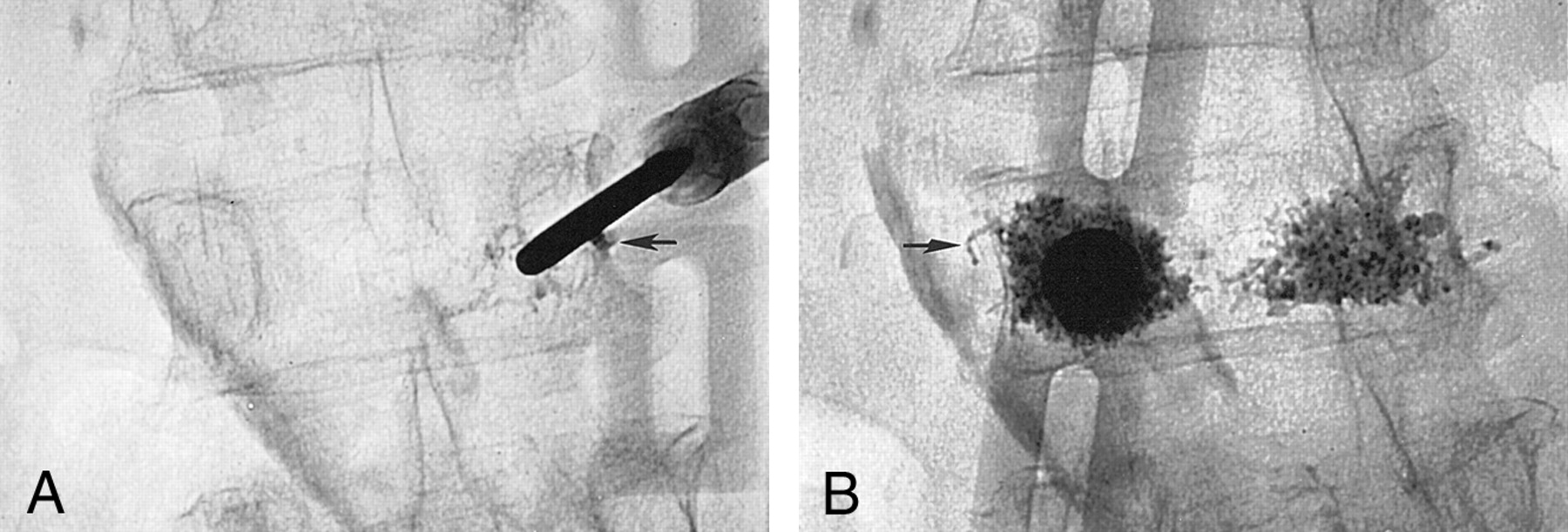
  • Fig 4.
    • Download figure
    • Open in new tab
    • Download powerpoint
    Fig 4.

    63-year-old woman with osteoporosis and a compression fracture at L3 associated with a superior endplate Schmorl’s node.

    A, Fluoroscopic view shows the 11-gauge needle that was placed by means of a right transpedicular approach. The presence of the Schmorl’s node precluded a median position of the needle tip. During PMMA injection, a leak (arrow) in the perivertebral venous system was immediately observed. The injection was resumed after a 20-second delay and completed uneventfully.

    B, Fluoroscopic view shows a second needle that was placed by means of a left transpedicular approach. Again, note a small leak (arrow) in the perivertebral venous system. The Schmorl’s node is outlined by the cement deposition.

PreviousNext
Back to top

In this issue

American Journal of Neuroradiology: 23 (6)
American Journal of Neuroradiology
Vol. 23, Issue 6
1 Jun 2002
  • Table of Contents
  • Index by author
Advertisement
Print
Download PDF
Email Article
Cite this article
0 Responses
Respond to this article
Share
Bookmark this article
Is Percutaneous Vertebroplasty without Pretreatment Venography Safe? Evaluation of 205 Consecutives Procedures
Cristiana Vasconcelos, Philippe Gailloud, Norman J. Beauchamp, Donald V. Heck, Kieran J. Murphy
American Journal of Neuroradiology Jun 2002, 23 (6) 913-917;
del.icio.us logo Twitter logo Facebook logo Mendeley logo
  • Tweet Widget

Jump to section

  • Article
    • Abstract
    • Methods
    • Results
    • Discussion
    • Conclusion
    • References
  • Figures & Data
  • Info & Metrics
  • Responses
  • References
  • PDF

Related Articles

  • No related articles found.
  • PubMed
  • Google Scholar

Cited By...

  • Cardiac Cement Embolism After Thoracic Kyphoplasty: Successful Conservative Treatment With 4-Year Follow-Up
  • Acute Paraplegia After Vertebroplasty Caused by Epidural Hemorrhage: A Case Report
  • Acute Paraplegia After Vertebroplasty Caused by Epidural Hemorrhage: A Case Report
  • Percutaneous vertebroplasty and balloon kyphoplasty for the treatment of osteoporotic vertebral compression fractures and osteolytic tumours
  • Radiation Dose to the Operator during Vertebroplasty: Prospective Comparison of the Use of 1-cc Syringes versus an Injection Device
  • Crossref
  • Google Scholar

This article has not yet been cited by articles in journals that are participating in Crossref Cited-by Linking.

More in this TOC Section

  • Dynamic CT Myelography: Patient Positioning
  • Evaluation of SIH MR Scoring Systems in Normals
  • Management Outcomes For VO Spine Biopsy
Show more SPINE IMAGING AND SPINE IMAGE-GUIDED INTERVENTIONS

Similar Articles

Advertisement

Indexed Content

  • Current Issue
  • Accepted Manuscripts
  • Article Preview
  • Past Issues
  • Editorials
  • Editor's Choice
  • Fellows' Journal Club
  • Letters to the Editor
  • Video Articles

Cases

  • Case Collection
  • Archive - Case of the Week
  • Archive - Case of the Month
  • Archive - Classic Case

More from AJNR

  • Trainee Corner
  • Imaging Protocols
  • MRI Safety Corner
  • Book Reviews

Multimedia

  • AJNR Podcasts
  • AJNR Scantastics

Resources

  • Turnaround Time
  • Submit a Manuscript
  • Submit a Video Article
  • Submit an eLetter to the Editor/Response
  • Manuscript Submission Guidelines
  • Statistical Tips
  • Fast Publishing of Accepted Manuscripts
  • Graphical Abstract Preparation
  • Imaging Protocol Submission
  • Evidence-Based Medicine Level Guide
  • Publishing Checklists
  • Author Policies
  • Become a Reviewer/Academy of Reviewers
  • News and Updates

About Us

  • About AJNR
  • Editorial Board
  • Editorial Board Alumni
  • Alerts
  • Permissions
  • Not an AJNR Subscriber? Join Now
  • Advertise with Us
  • Librarian Resources
  • Feedback
  • Terms and Conditions
  • AJNR Editorial Board Alumni

American Society of Neuroradiology

  • Not an ASNR Member? Join Now

© 2025 by the American Society of Neuroradiology All rights, including for text and data mining, AI training, and similar technologies, are reserved.
Print ISSN: 0195-6108 Online ISSN: 1936-959X

Powered by HighWire
Email this Article

Thank you for your interest in spreading the word on American Journal of Neuroradiology.

NOTE: We only request your email address so that the person you are recommending the page to knows that you wanted them to see it, and that it is not junk mail. We do not capture any email address.

Enter multiple addresses on separate lines or separate them with commas.
Is Percutaneous Vertebroplasty without Pretreatment Venography Safe? Evaluation of 205 Consecutives Procedures
(Your Name) has sent you a message from American Journal of Neuroradiology
(Your Name) thought you would like to see the American Journal of Neuroradiology web site.
CAPTCHA
This question is for testing whether or not you are a human visitor and to prevent automated spam submissions.
Cite this article
Cristiana Vasconcelos, Philippe Gailloud, Norman J. Beauchamp, Donald V. Heck, Kieran J. Murphy
Is Percutaneous Vertebroplasty without Pretreatment Venography Safe? Evaluation of 205 Consecutives Procedures
American Journal of Neuroradiology Jun 2002, 23 (6) 913-917;

Citation Manager Formats

  • BibTeX
  • Bookends
  • EasyBib
  • EndNote (tagged)
  • EndNote 8 (xml)
  • Medlars
  • Mendeley
  • Papers
  • RefWorks Tagged
  • Ref Manager
  • RIS
  • Zotero

We use cookies on this site to enhance your user experience. By clicking any link on this page you are giving your consent for us to set cookies.