
Article Figures & Data
Figures
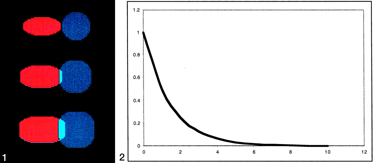
- Fig 1.
Successive iterations of morphologic dilatation operator on two simulated clusters.
Row 1 (from the top) demonstrates two non-overlapping clusters of different shapes.
Row 2 demonstrates growth of each cluster by using a morphologic dilatation operator that created an overlap region while maintaining the native shape of each cluster.
Row 3 demonstrates increasing overlap with repeated iterations of dilatation operator. During each iteration, additional areas of overlap are weighted by using an exponential weighting function and added to the total overlap volume.
Fig 2. Exponential weighting function. Additional overlapping clusters from dilatation operation are weighted on the basis of the iteration.
- Fig 3.
Group map for the word-generation paradigm (P < .001, height corrected). Images displayed in Talairach space (right side of the image is the right side of the subject). Bilateral frontal lobe activation is demonstrated (left > right), as well as distributed activations in the left basal ganglia, thalamus, midbrain, superior frontal lobe, anterior cingulate region, and bilateral cerebellum.
- Fig 4.
Group map for the FBL paradigm (P < .001, height corrected). Images displayed in Talairach space (right side of the image is the right side of the subject). Bilateral temporal lobe activation is demonstrated (left > right), and a focal area of activity in the left inferior frontal gyrus (BA 9) is present. Activation is more focal and less spatially distributed than with the word-generation paradigm. A small area of right cerebellar activity is also present.
- Fig 5.
Word generation and FBL activation maps in one subject obtained 1 week apart (P < .0001 corrected for spatial extent at P < .05). Images displayed in Talairach space (right side of the image is the right of the subject).
Row 1 (from the top) Word-generation paradigm on day 1. Images demonstrate bilateral frontal, bilateral cerebellar, bilateral thalamic, bilateral occipital left parietal, and anterior cingulate regions.
Row 2, Word-generation paradigm performed 1 week later. Although the activations are highly reproducible, numerous areas of extraneous activation are also present.
Row 3, FBL paradigm on day 1. Images demonstrates activation of bilateral temporal and left frontal regions.
Row 4, FBL paradigm performed 1 week later. Although the reproducibility indices for this paradigm are lower, the degree of extraneous activation is also markedly reduced.


Tables
Subject Word-Generation FBL 1 0.865517 0.577685 2 0.597006 0.450596 3 0.725085 0.599033 4 0.759399 0.696469 5 0.623490 0.374860 6 0.987388 0.434296 7 0.730123 0.258259 8 0.706395 0.570180 Mean (SD) 0.749 (0.127) 0.495 (0.141) ↵* P < .005
Subject Word-Generation FBL 1 0.994310 0.781844 2 0.657320 0.432581 3 0.841645 0.719868 4 0.842682 0.771116 5 0.768336 0.560528 6 0.959938 0.523434 7 0.987398 0.324236 8 0.777632 0.659780 Mean (SD) 0.854 (0.12) 0.597 (0.165) ↵* P < .008
Subject Word-Generation FBL 1 0.792541 0.433713 2 0.563576 0.424092 3 0.638156 0.472435 4 0.711660 0.617451 5 0.557372 0.269693 6 0.955450 0.380242 7 0.601508 0.213573 8 0.655076 0.491226 Mean (SD) 0.684 (0.134) 0.413 (0.127) ↵* P < .002
Subject Word-Generation FBL 1 0.282255 0.251434 2 0.112344 0.177413 3 0.377311 0.375264 4 0.237336 0.122048 5 0.235070 0.205566 6 0.256373 0.153030 7 0.313335 0.0541177 8 0.371002 0.210081 Mean (SD) 0.273 (0.085) 0.194 (0.095) ↵* P < .06
Subject Word-Generation FBL 1 0.734886 0.609496 2 0.742806 0.581206 3 0.651104 0.632916 4 0.773129 0.704500 5 0.741684 0.631687 6 0.668879 0.568074 7 0.664156 0.634116 8 0.707600 0.686904 Mean (SD) 0.711 (0.0446) 0.631 (0.047) ↵* P < .004


