
Article Figures & Data
Figures
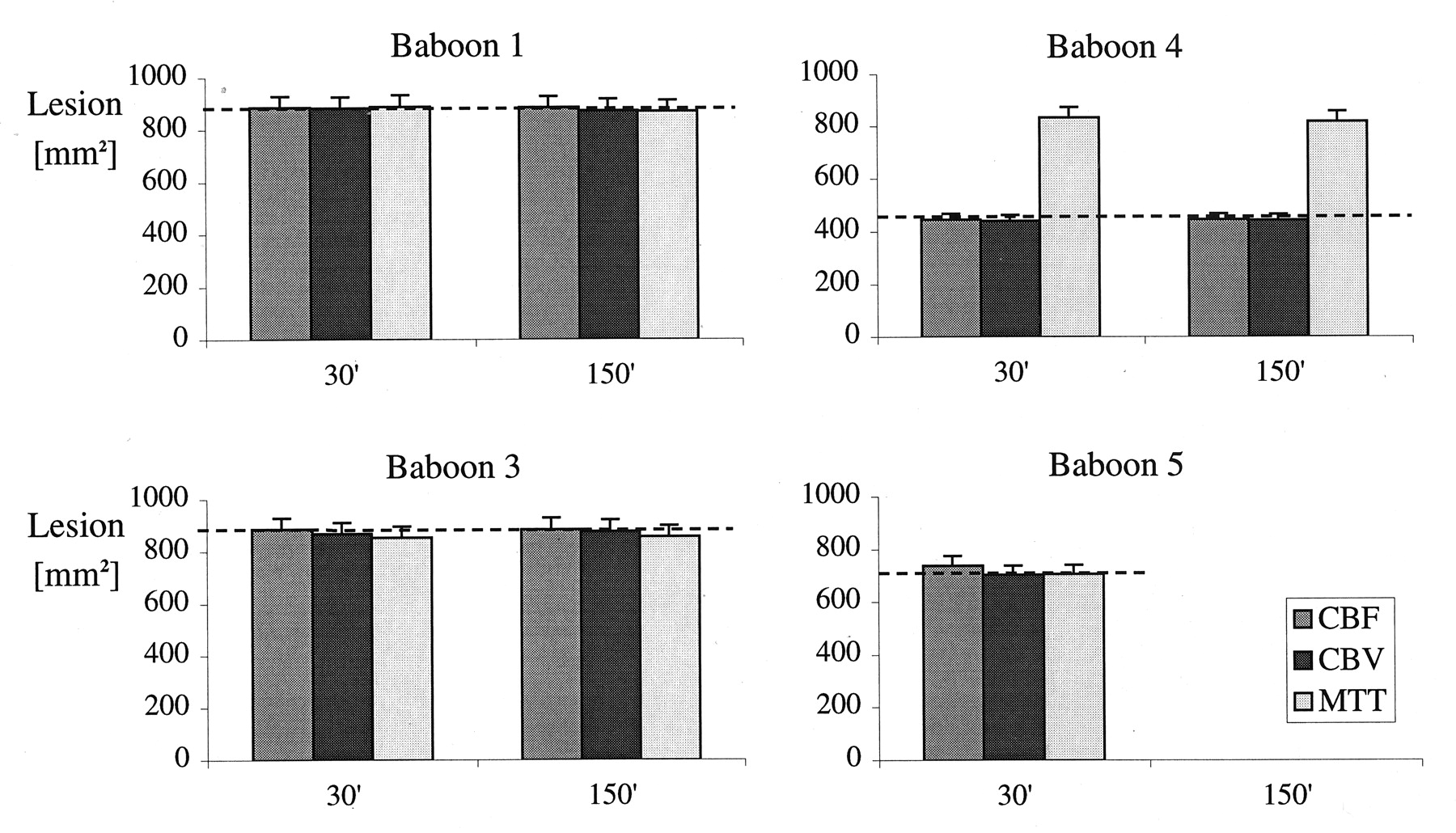
- Fig 1.
Acutely hypoperfused lesion sizes, as measured with perfusion CT, for animals 1, 3, 4, and 5 at 30- and 150-minute time points after the start of endovascular occlusion. At each time point, the leftmost column corresponds to lesion size (in square millimeters) measured on the CBF map; middle column, lesion size measured on the CBV map; and rightmost column, lesion size measured on the MTT map. The dashed line represents outcome infarct size, as determined on the ex vivo T2-weighted MR image. Data for animal 5 were not available at 150 minutes after occlusion because of technical factors.
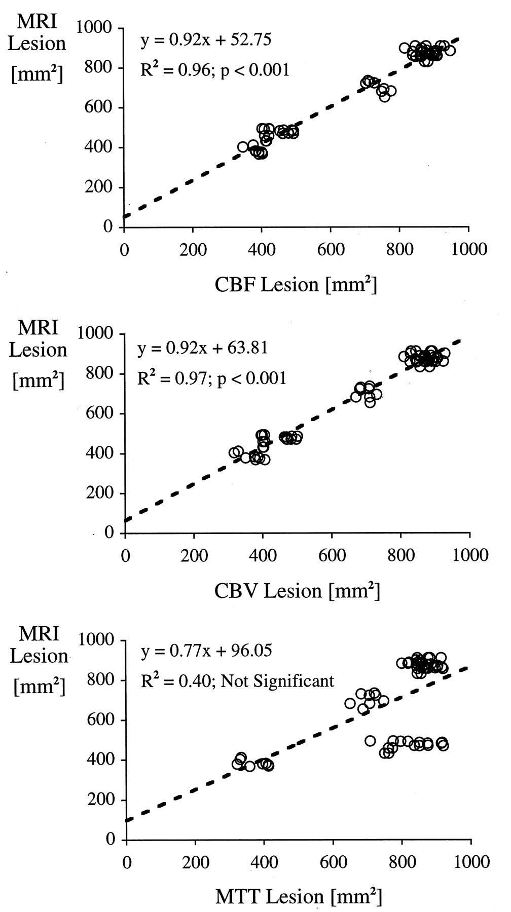
- Fig 2.
Regression statistics between the CT-determined CBF-, CBV-, and MTT-based lesion sizes and the 48-hour outcome lesion sizes, as measured at ex vivo MR imaging. Each point represents a single measurement by an observer. Scatter represents inter- and intra-observer variations. These variations were not statistically significant, as determined by using ANOVA. Data from animal 2 were not included in this particular analysis because the occluding balloon leaked, with resultant early reperfusion and subsequent reduction in the size of the infarcted region (Fig 6).
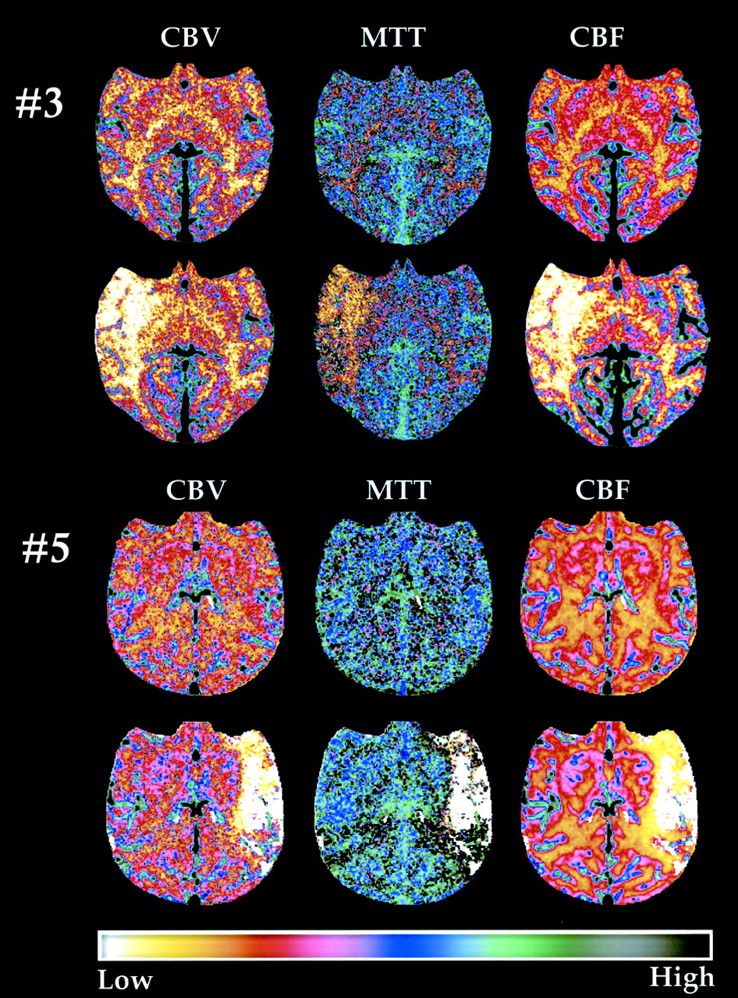
- Fig 3.
Pre- and postocclusion maps for animals 3 and 5. The first row and third row from the top present the CBV, MTT, and CBF maps from the control preocclusion study in animals 3 and 5, respectively. The second row and fourth row from the top present the maps obtained 30 minutes after the onset of MCA occlusion. In animal 3, right MCA territorial hypoperfusion is present, with involvement of the putamen and subtle involvement of the anterolateral thalamus. In animal 5, left MCA territorial hypoperfusion is clearly visible after occlusion, but the basal ganglia are spared. Note that each image is individually windowed to facilitate visualization of the lesions.
- Fig 4.
DSA images obtained immediately after MCA occlusion (left) and immediately after reperfusion (right) in animal 5. During occlusion, the left MCA vessels are absent (arrows), but they are clearly seen after reperfusion.
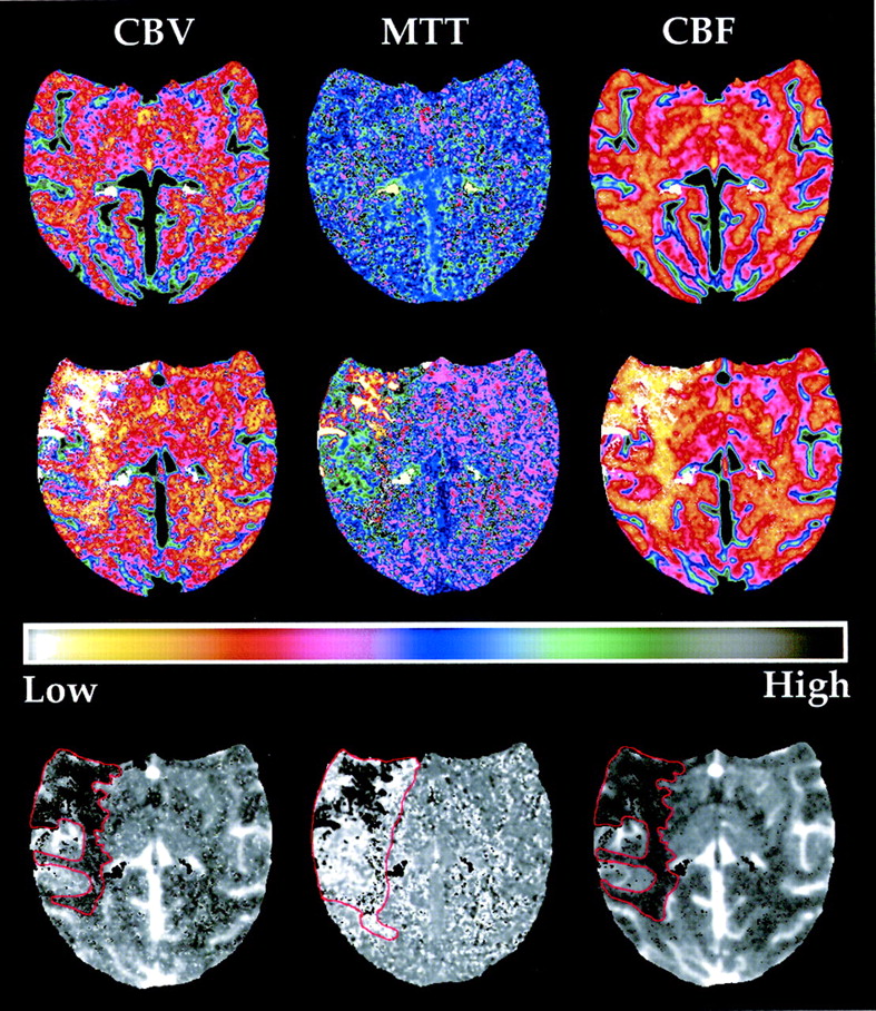
- Fig 5.
Images in animal 4. Top row, CBV, MTT, and CBF maps from the control preocclusion study. Middle row, Results from the study obtained 30 minutes after the start of occlusion. The right MCA territorial hypoperfusion is clearly visible after occlusion. Bottom row, Examples of ROIs representing the lesion on the maps. The lesion is larger on the MTT map than on the CBV and CBF maps. This is likely due to the presence of collateral circulation that results in normal perfusion on the CBV and CBF maps but prolonged transit time. Each image is individually windowed to facilitate presentation of the lesion.
- Fig 6.
In animal 2, which did not have any neurologic deficit, the balloon leaked between the 30- and 150-minute postocclusion study points. This early reperfusion reduced the outcome size of the lesion. This is an example of a desirable situation in which partial reperfusion of an ischemic territory occurs sufficiently early to substantially reduce the outcome infarct size.


Tables
Baboon Observer 1 Measurements (mm2) Observer 2 Measurements (mm2) CT Perfusion CT Perfusion 30 Minutes 150 Minutes 30 Minutes 150 Minutes CBF CBV MTT CBF CBV MTT MRI CBF CBV MTT CBF CBV MTT MRI 1 878 888 878 847 845 846 912 864 897 886 897 907 856 882 2 818 815 775 376 329 334 413 840 854 845 384 386 396 382 3 865 829 855 817 929 844 901 888 869 843 903 844 905 873 4 422 399 775 402 405 709 495 453 463 915 493 469 875 484 5 756 731 749 ND ND ND 696 704 709 726 ND ND ND 724 1 884 885 857 874 857 847 834 840 884 905 894 850 917 865 2 844 833 844 346 317 331 404 868 858 832 403 406 413 370 3 840 811 819 909 871 801 884 877 908 874 904 905 849 879 4 422 398 819 408 396 797 493 493 482 921 462 498 852 472 5 776 671 707 ND ND ND 684 715 685 683 ND ND ND 732 1 909 881 872 911 896 868 863 865 847 920 849 874 847 858 2 862 868 840 392 350 322 379 826 833 839 382 376 406 383 3 909 868 859 903 892 876 863 881 889 850 862 859 823 890 4 415 403 763 412 401 750 434 464 501 852 488 485 917 488 5 759 712 688 ND ND ND 655 710 711 722 ND ND ND 736 1 920 893 915 931 832 883 912 909 871 868 867 925 898 863 2 852 850 805 392 379 359 369 871 819 820 401 391 413 377 3 948 855 844 907 874 860 887 861 902 866 849 828 878 861 4 409 406 774 421 401 762 460 478 470 837 479 471 876 472 5 749 711 651 ND ND ND 683 729 683 707 ND ND ND 725 Coefficient of Variation (%)* Coefficient of Variation (%)* 1 2.2 0.6 2.8 4.2 3.2 2.1 4.4 3.3 2.5 2.5 2.6 3.7 3.8 1.2 2 2.3 2.7 4.0 5.8 7.9 4.7 5.3 2.5 2.1 1.3 2.8 3.2 2.0 1.7 3 5.4 3.0 2.1 5.1 3.0 3.8 1.8 1.3 2.0 1.7 3.2 3.8 4.1 1.4 4 1.5 0.9 3.1 1.9 1.0 4.8 6.2 3.7 3.5 4.9 2.9 2.7 3.1 1.7 5 1.5 3.6 5.8 ND ND ND 2.6 1.5 2.2 2.7 ND ND ND 0.8 ↵* Average variabilities were 3.7% for observer 1 and 2.1% for observer 2. No statistically significant differences in interobserver or intraobserver variabilities were present, as measured by using ANOVA (P > 0.36).
Baboon Lesion Areas* 30 Minutes 150 Minutes Outcome Size† Neurobehavior Score CBF CBV MTT CBF CBV MTT 1 884 ± 10 881 ± 6 888 ± 8 884 ± 11 873 ± 12 870 ± 9 873 ± 10 4 2 848 ± 7 841 ± 7 825 ± 9 384 ± 6 367 ± 11 372 ± 14 385 ± 6 0 3 884 ± 12 866 ± 12 851 ± 6 882 ± 12 875 ± 12 854 ± 12 880 ± 5 3 4 444 ± 11 440 ± 15 832 ± 22 445 ± 14 441 ± 16 817 ± 26 475 ± 7 0 5 737 ± 9 702 ± 7 704 ± 11 No Data No Data No Data 704 ± 10 0 Mean ± SEM‡ 737 ± 33 722 ± 32 819 ± 14 737 ± 44 730 ± 43 847 ± 11 733 ± 30


