Advertisement

Main menu

  • Home
  • Content
    • Current Issue
    • Accepted Manuscripts
    • Article Preview
    • Past Issue Archive
    • Video Articles
    • AJNR Case Collection
    • Case of the Week Archive
    • Case of the Month Archive
    • Classic Case Archive
  • Special Collections
    • AJNR Awards
    • Low-Field MRI
    • Alzheimer Disease
    • ASNR Foundation Special Collection
    • Photon-Counting CT
    • View All
  • Multimedia
    • AJNR Podcasts
    • AJNR SCANtastic
    • Trainee Corner
    • MRI Safety Corner
    • Imaging Protocols
  • For Authors
    • Submit a Manuscript
    • Submit a Video Article
    • Submit an eLetter to the Editor/Response
    • Manuscript Submission Guidelines
    • Statistical Tips
    • Fast Publishing of Accepted Manuscripts
    • Graphical Abstract Preparation
    • Imaging Protocol Submission
    • Author Policies
  • About Us
    • About AJNR
    • Editorial Board
    • Editorial Board Alumni
  • More
    • Become a Reviewer/Academy of Reviewers
    • Subscribers
    • Permissions
    • Alerts
    • Feedback
    • Advertisers
    • ASNR Home

User menu

  • Alerts
  • Log in

Search

  • Advanced search
American Journal of Neuroradiology

American Journal of Neuroradiology

ASHNR American Society of Functional Neuroradiology ASHNR American Society of Pediatric Neuroradiology ASSR
  • Alerts
  • Log in

Advanced Search

  • Home
  • Content
    • Current Issue
    • Accepted Manuscripts
    • Article Preview
    • Past Issue Archive
    • Video Articles
    • AJNR Case Collection
    • Case of the Week Archive
    • Case of the Month Archive
    • Classic Case Archive
  • Special Collections
    • AJNR Awards
    • Low-Field MRI
    • Alzheimer Disease
    • ASNR Foundation Special Collection
    • Photon-Counting CT
    • View All
  • Multimedia
    • AJNR Podcasts
    • AJNR SCANtastic
    • Trainee Corner
    • MRI Safety Corner
    • Imaging Protocols
  • For Authors
    • Submit a Manuscript
    • Submit a Video Article
    • Submit an eLetter to the Editor/Response
    • Manuscript Submission Guidelines
    • Statistical Tips
    • Fast Publishing of Accepted Manuscripts
    • Graphical Abstract Preparation
    • Imaging Protocol Submission
    • Author Policies
  • About Us
    • About AJNR
    • Editorial Board
    • Editorial Board Alumni
  • More
    • Become a Reviewer/Academy of Reviewers
    • Subscribers
    • Permissions
    • Alerts
    • Feedback
    • Advertisers
    • ASNR Home
  • Follow AJNR on Twitter
  • Visit AJNR on Facebook
  • Follow AJNR on Instagram
  • Join AJNR on LinkedIn
  • RSS Feeds

AJNR Awards, New Junior Editors, and more. Read the latest AJNR updates

OtherSpine Imaging and Spine Image-Guided Interventions

Paradoxical Cerebral Arterial Embolization of Cement during Intraoperative Vertebroplasty: Case Report

Rebecca Scroop, Joseph Eskridge and Gavin W. Britz
American Journal of Neuroradiology May 2002, 23 (5) 868-870;
Rebecca Scroop
  • Find this author on Google Scholar
  • Find this author on PubMed
  • Search for this author on this site
Joseph Eskridge
  • Find this author on Google Scholar
  • Find this author on PubMed
  • Search for this author on this site
Gavin W. Britz
  • Find this author on Google Scholar
  • Find this author on PubMed
  • Search for this author on this site
  • Article
  • Figures & Data
  • Info & Metrics
  • Responses
  • References
  • PDF
Loading

This article has been retracted. Please see:

  • Retraction - March 01, 2004

Abstract

Summary: Paradoxical cerebral embolism of cement occurred in a 78-year-old woman after cement−assisted transpedicular spinal fixation surgery. Multiple pulmonary emboli of polymethylmethacrylate precipitated pulmonary hypertension and right-to-left shunting into the systemic circulation through a patent foramen ovale. This rare complication occurred because of failure to recognize venous migration of cement during the procedure and the injection of multiple levels in one setting. Although this was an open procedure, the technical aspects were the same as for vertebroplasty and the precautions should be applied to percutaneous vertebroplasty.

Acrylic cement (polymethylmethacrylate [PMMA]) is commonly used to provide increased prosthetic fixation by conforming to macroscopic irregularities in the target bone. Most commonly, PMMA is used for peripheral fixation of orthopaedic hardware, such as in hip or knee arthroplasty. Percutaneous vertebroplasty involves injection of PMMA into a diseased vertebral body for pain relief and partial bone remodeling (1).

Pulmonary emboli of PMMA and fat are known to occur after both procedures but with uncertain frequency and are usually asymptomatic (2–4). Even more rarely, cerebral arterial emboli have been reported to occur in association with peripheral PMMA-assisted arthroplasty (5). We report a case of paradoxical cerebral embolization during intraoperative transpedicular PMMA injection.

Case Report

A 78-year-old woman was hospitalized for revision of thoracolumbar hardware because of increasing back pain. She had undergone posterior segmental hardware placement from T11–L2 after an L1 crush fracture 8 months previously. Recently obtained radiographs confirmed failure of the hardware with retraction of pedicle screws at three levels.

The old hardware was removed, and posterior segmental fusion from T4 to L4 was performed using Synthes titanium USS implants. The bone was osteoporotic, and it was elected to use PMMA to assist fixation of the pedicular screws at T4 through L4 with transpedicular cement injection. The cement consisted of simplex powder, 1.2 g of tobramycin, and 6 g of sterile barium sulfate, which was mixed with PMMA liquid acrylic to a toothpaste consistency and injected via an 11-gauge needle. The needle tip was positioned in the mid third of the vertebral body, close to midline. Approximately 1 cc of cement was injected bilaterally at each of the 13 levels. At the T3 and L5 levels, no screws were placed but transpedicular cement was used to improve bone strength. Fluoroscopic monitoring was used intermittently by the operator during the procedure, and at no time was there concern recorded of significant extravasation of cement into the venous system.

Toward the end of the operative procedure, the patient became hypotensive with elevation in pulmonary arterial pressure to 60 mmHg and her oxygenation status declined to a PaO2 of 50. After the patient’s condition was stabilized, a radiograph of the chest was obtained and revealed multiple PMMA emboli (Fig 1B). CT of the chest confirmed pulmonary emboli (Fig 2B) and the presence of PMMA within the paravertebral and epidural veins (Fig 2A and B). During the recovery phase from the general anesthetic, it was noted that the patient was hemiparetic on the right side. CT of the head revealed a left-sided middle cerebral artery territory acute infarct and a hyperattenuated focus within the left middle cerebral artery consistent with PMMA (Fig 3A). Several other emboli were seen within more peripheral cerebral vessels (Fig 3B), which were clinically silent. An echocardiogram was obtained and confirmed right heart dilation, elevated pulmonary arterial pressure, and a patent foramen ovale.

Fig 1.
  • Download figure
  • Open in new tab
  • Download powerpoint
Fig 1.

Pre- and postoperative radiographs of the chest.

A, Normal anteroposterior preoperative radiograph of the chest.

B, Anteroposterior postoperative radiograph of the chest shows numerous emboli of cement throughout the lungs.

Fig 2.
  • Download figure
  • Open in new tab
  • Download powerpoint
Fig 2.

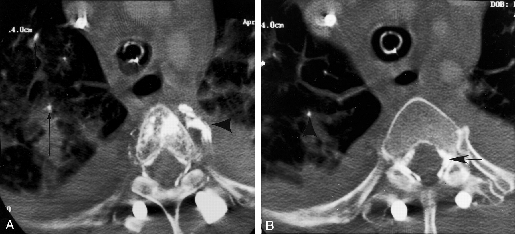
CT scans confirm pulmonary emboli.

A, CT scan obtained through the level of T4 shows cement within the paravertebral veins (arrowhead) and small emboli of cement within the lungs (arrows).

B, CT scan obtained at the T5 level shows cement in the epidural veins (arrow) and pulmonary emboli (arrowhead).

Fig 3.
  • Download figure
  • Open in new tab
  • Download powerpoint
Fig 3.

CT scans of the head.

A, Unenhanced CT scan of the head shows an embolus of cement within the left middle cerebral artery and early ischemic changes.

B, CT scan of the same patient, obtained at a more superior level, shows other emboli in more peripheral branches (arrows).

The patient was not anticoagulated because of the recent surgery, and the patient’s respiratory status slowly improved after a period of 10 days with assisted ventilation. The patient remained hemiparetic with a moderate expressive dysphasia at the 3-month follow-up examination.

Discussion

Transpedicular injection of PMMA performed with vertebroplasty is frequently associated with leaking into the external vertebral venous plexuses. From there, cement could migrate into the inferior vena cava and result in pulmonary emboli. If the needle tip is positioned within the basivertebral venous plexus, cement injection may result in more extensive leakage into the venous system and then into the lungs.

In the majority of cases, leakage is asymptomatic and the leakage occurs because of the fluid consistency of the PMMA at the time of injection. For this reason, real-time monitoring during injection and adequate opacification of the PMMA are required. If venous filling is noted, repositioning of the needle or temporary cessation of PMMA injection to allow polymerization is suggested. Although most pulmonary emboli from PMMA are asymptomatic, clinically significant disease has been reported (2).

Cerebral emboli have been reported to occur after PMMA- and non-PMMA-assisted arthroplasty and have been attributed to fat emboli resulting from raised intramedullary pressure during reaming and cementation (6, 7). When systemic emboli are seen, they have been attributed to paradoxical embolism via a patent foramen ovale (8). Transpulmonary passage of small emboli, both fat and air, has recently been shown in experimental models, suggesting that microemboli in the lungs may undergo embolization into the systemic circulation (5). In our case, cerebral embolism occurred via a patent foramen ovale. Multiple pulmonary emboli of PMMA were apparent on a postoperative CT scan of the chest. The resultant elevation in right heart pressure caused paradoxical emboli of PMMA via a patent foramen ovale into the systemic circulation.

Although this was an open procedure, cement was injected in much the same way as for percutaneous vertebroplasty, with transpedicular placement of an 11-gauge needle into the vertebral body. Therefore, the complications from this procedure could potentially occur with vertebroplasty. Symptomatic embolization in this case was thought to have resulted from two factors: venous leakage of cement that was not appreciated even with fluoroscopic monitoring and injection of multiple levels in one setting. The former is well established as good practice for vertebroplasty. The value of obtaining a vertebrogram before injection of cement is controversial and should not preclude real-time monitoring (2). The properties of iodinated contrast material differ significantly from those of PMMA. Inevitably, venous extravasation occurs, and residual vertebral opacification may interfere with visibility of PMMA. This has resulted in a shift in practice, with obtaining a vertebrogram no longer being standard (9, 10). The consistency and opacification of the cement were not thought to have contributed significantly to the complications experienced.

Assuming that venous extravasation and pulmonary emboli can and probably do occur to some degree in association with clinically uncomplicated vertebroplasty, it is prudent to limit the levels treated at a single setting. In this case, a total of 15 levels were treated with transpedicular PMMA and resulted in significantly raised pulmonary pressures. Although not based on experimental or hard clinical evidence, we recommend that in patients without underlying pulmonary disease, a maximum of three levels be treated in a single setting, even with good fluoroscopic monitoring. Unlike this case, vertebroplasty does not require general anesthesia, and it is our practice to use local anesthesia and neurolept sedation that allows early detection of any changes in the clinical condition of the patient (11).

References

  1. ↵
    Deramont H, Galibert R, Debussche-Depriester C. Percutaneous vertebroplasty with methylmethcrylate: technique, method, results [abstr]. Chicago, Radiological Society of North America,1990
  2. ↵
    Padovani B, Kasriel O, Brunner P, Peretti P. Pulmonary embolism caused by acrylic cement: a rare complication of percutaneous vertebroplasty. AJNR Am J Neuroradiol 1999;20:375–377
    Abstract/FREE Full Text
  3. Jensen ME, Avery JE, Mathis JM, Kallmes DF, Cloft HJ, Dio JE. Percutaneous polymethylmethacrylate vertebroplasty in the treatment of osteoporotic vertebral compression fractures: technical aspects. AJNR Am J Neuroradiol 1997;18:1897–1904
    Abstract
  4. ↵
    Turchin DC, Anderson GI, Schemitsch EH, Byrick RJ, Mullen BM, Richards RR. Pulmonary and systemic fat embolization following medullary canal pressurization. Trans Orthop Res Soc 1995;20:252
  5. ↵
    Byrick RJ, Mullen JB, Mazer CD, Guest CB. Transpulmonary systemic fat embolism: studies in mongrel dogs after cemented arthroplasty. Anesthesiology 1996;84:721–723
    CrossRefPubMed
  6. ↵
    Weiss SJ, Cheung AT, Stecker MM, Garino JP, Hughes JE, Murphy FL. Fatal paradoxical cerebral embolization during bilateral knee arthroplasty. Anesthesiology 1996;84:721–723
  7. ↵
    Edmonds CR, Barbut D, Hager D, Sharrock NE. Intraoperative cerebral arterial embolization during total hip arthroplasty. Anesthesiology 2000;93:315–318
    CrossRefPubMed
  8. ↵
    Christie J, Burnett R, Potts HR, Pell AC. Echocardiography of transarterial embolism during cemented and uncemented hemiarthroplasty of the hip. J Bone Joint Surg Br 1994;74:409–412
  9. ↵
    Weill A, Chiras J, Simon JM, Rose M, Sola-Martinez MT, Enkaoua E. Spinal metastases: indications for and results of percutaneous injection of acrylic surgical cement. Radiology 1996;99:241–247
  10. ↵
    Gangi A, Kastler BA, Dietemann JL. Percutaneous vertebroplasty guided by a combination CT and fluoroscopy. AJNR Am J Neuroradiol 1994;15:83–86
    Abstract/FREE Full Text
  11. ↵
    Chiras J, Depriester C, Weill A, Sola-Martinez MT, Deramond H. Vertebroplasties percutanees. J Neuroradiol 1997;24:45–59
    PubMed
  • Received June 28, 2001.
  • Accepted after revision October 16, 2001.
  • Copyright © American Society of Neuroradiology
View Abstract
PreviousNext
Back to top

In this issue

American Journal of Neuroradiology: 23 (5)
American Journal of Neuroradiology
Vol. 23, Issue 5
1 May 2002
  • Table of Contents
  • Index by author
Advertisement
Print
Download PDF
Email Article

Thank you for your interest in spreading the word on American Journal of Neuroradiology.

NOTE: We only request your email address so that the person you are recommending the page to knows that you wanted them to see it, and that it is not junk mail. We do not capture any email address.

Enter multiple addresses on separate lines or separate them with commas.
Paradoxical Cerebral Arterial Embolization of Cement during Intraoperative Vertebroplasty: Case Report
(Your Name) has sent you a message from American Journal of Neuroradiology
(Your Name) thought you would like to see the American Journal of Neuroradiology web site.
CAPTCHA
This question is for testing whether or not you are a human visitor and to prevent automated spam submissions.
Cite this article
Rebecca Scroop, Joseph Eskridge, Gavin W. Britz
Paradoxical Cerebral Arterial Embolization of Cement during Intraoperative Vertebroplasty: Case Report
American Journal of Neuroradiology May 2002, 23 (5) 868-870;

Citation Manager Formats

  • BibTeX
  • Bookends
  • EasyBib
  • EndNote (tagged)
  • EndNote 8 (xml)
  • Medlars
  • Mendeley
  • Papers
  • RefWorks Tagged
  • Ref Manager
  • RIS
  • Zotero
0 Responses
Respond to this article
Share
Bookmark this article
Paradoxical Cerebral Arterial Embolization of Cement during Intraoperative Vertebroplasty: Case Report
Rebecca Scroop, Joseph Eskridge, Gavin W. Britz
American Journal of Neuroradiology May 2002, 23 (5) 868-870;
del.icio.us logo Twitter logo Facebook logo Mendeley logo
  • Tweet Widget
  • Facebook Like
  • Google Plus One

Jump to section

  • Article
    • Abstract
    • Case Report
    • Discussion
    • References
  • Figures & Data
  • Info & Metrics
  • Responses
  • References
  • PDF

Related Articles

  • PubMed
  • Google Scholar

Cited By...

  • Paradoxical embolus to the brain from embolization of a carotid body tumor
  • Treatment of painful osteoporotic vertebral compression fractures: A BRIEF REVIEW OF THE EVIDENCE FOR PERCUTANEOUS VERTEBROPLASTY
  • Minimally Invasive Techniques for the Treatment of Osteoporotic Vertebral Fractures
  • Percutaneous vertebroplasty and balloon kyphoplasty for the treatment of osteoporotic vertebral compression fractures and osteolytic tumours
  • Two patients with the same type of iatrogenic disease
  • Crossref
  • Google Scholar

This article has not yet been cited by articles in journals that are participating in Crossref Cited-by Linking.

More in this TOC Section

  • Management Outcomes For VO Spine Biopsy
  • Cone Beam CT Myelography
  • Post-Procedural Brachial Neuritis Features
Show more Spine Imaging and Spine Image-Guided Interventions

Similar Articles

Advertisement

Indexed Content

  • Current Issue
  • Accepted Manuscripts
  • Article Preview
  • Past Issues
  • Editorials
  • Editor's Choice
  • Fellows' Journal Club
  • Letters to the Editor
  • Video Articles

Cases

  • Case Collection
  • Archive - Case of the Week
  • Archive - Case of the Month
  • Archive - Classic Case

More from AJNR

  • Trainee Corner
  • Imaging Protocols
  • MRI Safety Corner
  • Book Reviews

Multimedia

  • AJNR Podcasts
  • AJNR Scantastics

Resources

  • Turnaround Time
  • Submit a Manuscript
  • Submit a Video Article
  • Submit an eLetter to the Editor/Response
  • Manuscript Submission Guidelines
  • Statistical Tips
  • Fast Publishing of Accepted Manuscripts
  • Graphical Abstract Preparation
  • Imaging Protocol Submission
  • Evidence-Based Medicine Level Guide
  • Publishing Checklists
  • Author Policies
  • Become a Reviewer/Academy of Reviewers
  • News and Updates

About Us

  • About AJNR
  • Editorial Board
  • Editorial Board Alumni
  • Alerts
  • Permissions
  • Not an AJNR Subscriber? Join Now
  • Advertise with Us
  • Librarian Resources
  • Feedback
  • Terms and Conditions
  • AJNR Editorial Board Alumni

American Society of Neuroradiology

  • Not an ASNR Member? Join Now

© 2025 by the American Society of Neuroradiology All rights, including for text and data mining, AI training, and similar technologies, are reserved.
Print ISSN: 0195-6108 Online ISSN: 1936-959X

Powered by HighWire