
Article Figures & Data
Figures
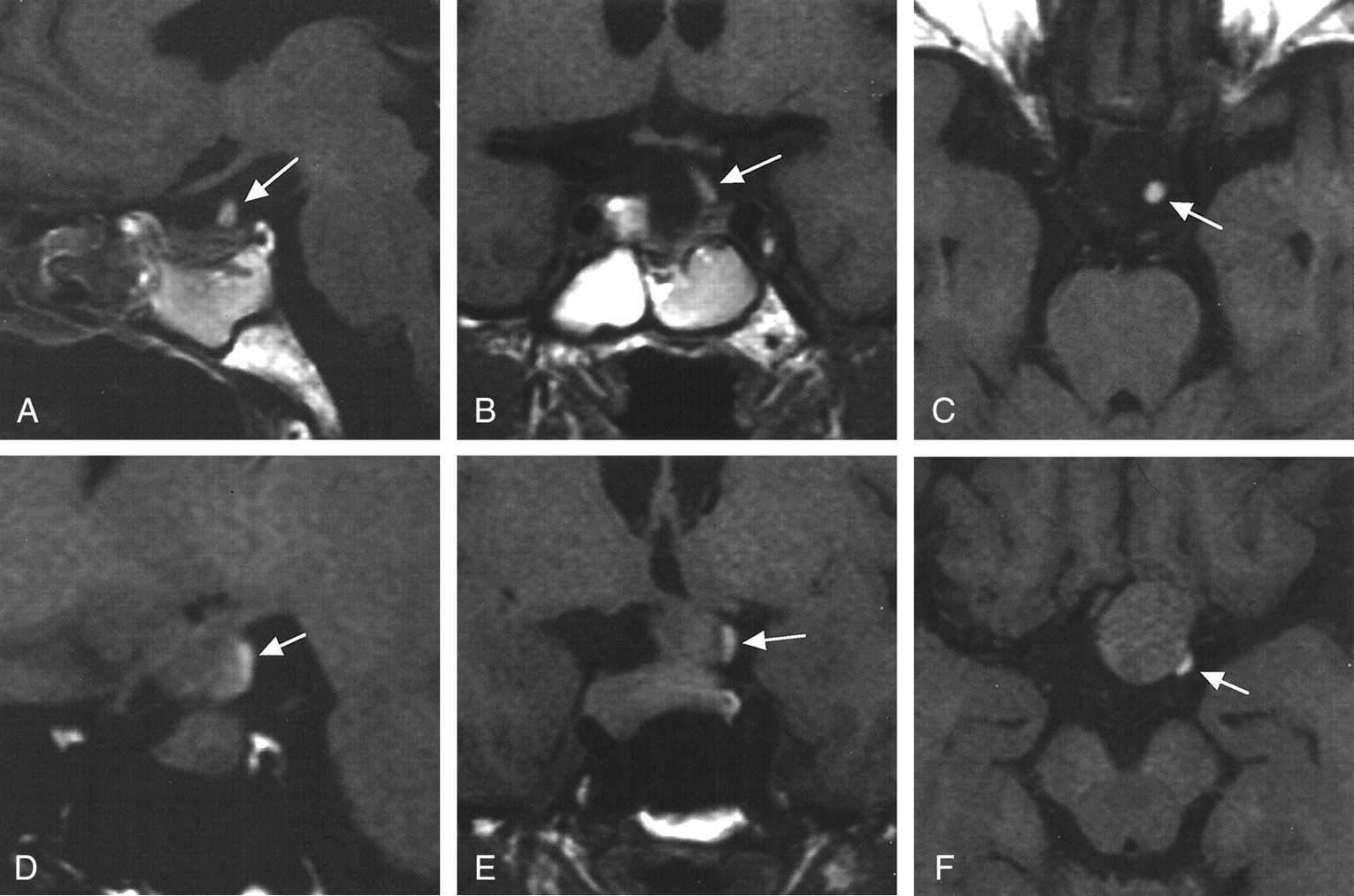
- Fig 1.
Case 5, group A. Images in a 63-year-old man with a nonfunctioning adenoma show that the HSI (arrow) was present both before and 2 weeks after surgery.
A–C, At 2 weeks after surgery, ovoid HSI is visible at the level of the diaphragm sellae on sagittal (A), coronal (B), and axial (C) sections.
D–F, Before surgery, linear HSI is visible at the posterolateral surface of adenoma, above the indentation formed by a diaphragma sellae, on the sagittal (D), coronal (E), and axial (F) sections.
- Fig 2.
Case 16, group A. In this patient, a 35 year–old woman with a nonfunctioning adenoma, MR studies were repeated at 3, 12, and 24 months after surgery and showed essentially the same findings, including HSI (arrow).
A and B, At 2 years after surgery, HSI is visible at the tip of pituitary stalk at the level of the diaphragma sellae on the coronal (A) and sagittal (B) sections.
C–F, Before surgery, linear HSI is visible at the posterior surface of adenoma, above the indentation formed by the diaphragma sellae, on the sagittal (C), coronal (D), contrast-enhanced sagittal (E), and axial (F) sections. HSI is identifiable at the stalk stump. The enhanced image shows HSI with more remarkable enhancing effect due to normal pituitary tissue; this finding suggests its connection to the pituitary stalk.
- Fig 3.
Case 17, group A. Images in a 74 year-old-woman with a growth hormone–secreting adenoma show HSI (arrow).
A, Coronal section 6 months after surgery. Postoperatively, the adenoma was debulked, and the HSI was below the chiasm and deviated to the left. It was continuous with the proximal portion of the pituitary stalk.
B, Coronal section before surgery. Preoperatively, an ovoid HSI is visible on the supradiaphragmatic level on the left side. The optic chiasm is compressed superiorly.
- Fig 4.
Sagittal sections obtained before surgery show HSI (arrows).
A, Case 9, group A 39-year-old woman with nonfunctioning adenoma. HSI was noted in both supra- and intrasellar areas. The indentation at the level of the diaphragma sellae was noted but not prominent.
B, Case 14, group A. A 54-year-old woman with nonfunctioning adenoma. HSI was noted close to the median eminence. Indentation of diaphragma sellae was not evident in this case.


Tables
Case No./Patient Age (y)/Patient Sex Signs and Symptoms* Adenoma Type† Height × Width × Thickness (mm) HSI Site§ HSI Shape‖ Postoperative MR Study Interval (mo) Postoperative HSI Postoperative DI Group A 1/63/M VFD NF 31 × 28 × 33 SD Ovoid 5 Yes No 2/63/F Headache NF 20 × 18 × 19 SD Round 36 Yes No 3/61/M VFD NF 39 × 26 × 30 SD Ovoid 6 Yes No 4/55/M VFD NF 32 × 26 × 25 SD Linear 10 Yes No 5/63/M VFD NF 26 × 22 × 20 SD Linear 0.5 Yes No 6/35/M Impotence PRL 35 × 25 × 22 SD Linear 44 Yes No 7/74/F VFD NF 35 × 22 × 26 SD Round 5 Yes No 8/59/F VFD NF 38 × 31 × 23 SD Linear 3 Yes No 9/39/F VFD NF 27 × 21 × 18 IS Linear 21 Yes No 10/39/F VFD PRL 22 × 18 × 18 SD Linear 44 Yes No 11/17/F VFD PRL 27 × 18 × 18 SD Linear 48 Yes No 12/72/F VFD NF 29 × 26 × 24 SD Linear 32 Yes No 13/61/F VFD NF 34 × 26 × 24 SD Linear 31 Yes No 14/54/F VFD NF 24 × 20 × 18 ME Linear 1 Yes No 15/28/F Amenorrhea PRL 22 × 18 × 15 SD Linear 26 Yes No 16/35/F VFD NF 29 × 26 × 20 SD Linear 24 Yes No 17/74/F Acromegaly GH 24 × 23 × 21 SD Ovoid 6 Yes No 18/33/F VFD PRL 52 × 38 × 36 None NA 27 Yes No Group B 19/60/M VFD NF 19 × 18 × 20 None NA 4 No No 20/70/M VFD NF‡ 22 × 20 × 18 None NA 7 No Yes 21/48/F VFD NF 29 × 20 × 20 None NA 27 No Yes 22/30/M VFD NF 34 × 30 × 26 None NA 22 No Yes * VFD indicates visual field defect.
† GH indicates growth hormone–secreting adenoma; NF, nonfunctioning adenoma; and PRL, prolactinoma.
‡ Pituitary apoplexy.
§ IS indicates intrasellar; ME, median eminence; SD, supradiaphragmatic.
‖ NA indicates not applicable.
Postoperative HSI Preoperative HSI Permanent DI Present Absent Present Absent Present 17 1 0 18 Absent 0 4 3 1
In this issue


Jump to section
Related Articles
Cited By...
- No citing articles found.