
Article Figures & Data
Figures
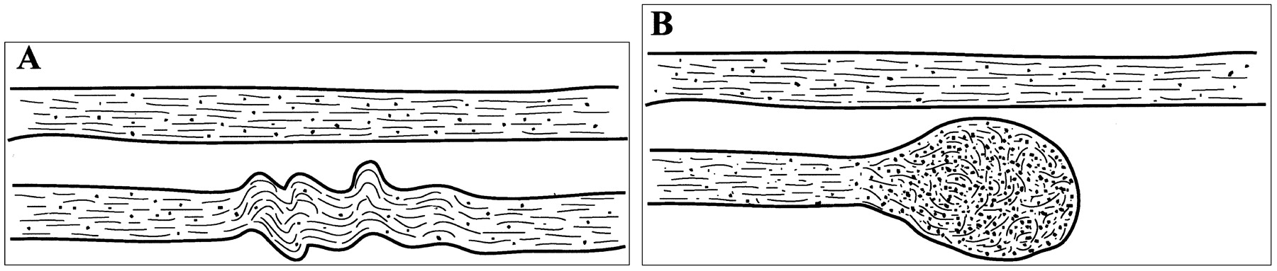
- Fig 1.
Illustration of the changes that axons undergo owing to cytoskeletal perturbation from mild traumatic brain injury.
A, The top neuron is healthy. In the bottom neuron, neurofilamentous and, generally, cytoskeletal misalignment is visible a short time after injury. This impairs axonal transport.
B, Organelles accumulate in the injured region, causing the axon to swell locally and subsequently disconnect from the rest. In this Figure, the dimensions of the axons relative to the interaxonal space do not necessarily correspond to reality.
- Fig 2.
Example of the selection of voxels from the five structures under study, in one section of a control subject. The background image is an LI map. The red dots correspond to the selected voxels. The dots were made larger than the in-plane dimensions of a single voxel for visualization purposes.
- Fig 3.
A, LI map in a control subject. The border between the anterior (AIC) and posterior (PIC) limbs of the internal capsule is not obvious from this image.
B, Absolute value color map of the same section as in A. Arrows indicate the exact level of the border between AIC and PIC. Color representation of directions in 3D space helps to clearly separate the two structures.
C, Color circle demonstrates the correspondence of colors and directions in 3D space. This circle should be thought of as a 3D dome that is viewed from below.
- Fig 4.
A, T2-weighted image in a patient with mild traumatic brain injury. Small hemorrhagic lesions are visible in the left occipital lobe and right frontal lobe.
B, Diffusion-weighted map of the same section in the same patient, for one of the 23 diffusion-weighted gradient directions. The abnormal regions from A are also visible in B.
C, Corresponding trace map. The same lesions from A and B can be detected.
D, LI map of the same section in the same patient. Decreased anisotropy is visible in the left internal capsule and left anterior corpus callosum. No abnormality was demonstrated in these regions with any other type of imaging. On this LI map, however, the hemorrhagic lesions from A, B, and C are hard to identify. R indicates right; L, left.
- Fig 5.
Graphs show distributions of FA results from 10 selected ROIs (5 structures x 2 hemispheres) for the control subjects (continuous lines) combined with the corresponding FA values for the five patients with mild traumatic brain injuries (vertical lines), in the left (A) and right (B) hemispheres. All horizontal axes represent FA values. P values are reported only for cases in which FA results for the patients were significantly different (P < .05) from the mean FA results of the control subjects. For the patients, anisotropy was found to be significantly reduced in several structures compared with normal values. Reduction of FA was more often in the internal capsule and corpus callosum and less often in the external capsule of the patients. LACC and RACC indicate left and right anterior corpus callosum, respectively; LPCC and RPCC, left and right posterior corpus callosum; LEC and REC, left and right external capsule; LAIC and RAIC, left and right anterior internal capsule; LPIC and RPIC, left and right posterior internal capsule.
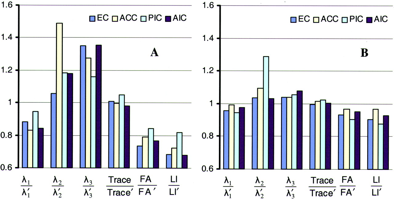
- Fig 6.
Bar graphs of ratios of diffusion characteristics between the two sides of four structures in a patient with mild traumatic brain injury, using the immediate (A) and short-term follow-up (B) data. The quantities λ1, λ2, λ3, trace, FA, and LI correspond to that side of the structure that has the lowest anisotropy on the immediate image. The quantities λ1’, λ2’, λ3’, trace’, FA’, and LI’ correspond to the contralateral side of these structures. The four structures are external capsule (EC), anterior corpus callosum (ACC), posterior internal capsule (PIC), and anterior internal capsule (AIC). Each structure corresponds to a specific color. For the immediate data set, one side of the four structures has lower λ1, higher λ2 and λ3, and lower FA and LI than its contralateral homologous side. Also, trace is similar for both sides. For the follow-up data set, λ1, λ2, λ3, FA, and LI have become more similar between the two sides of each structure, therefore giving ratios that are closer to 1. The ratios of the trace have remained at values almost equal to 1. When looking at the absolute values of the diffusion characteristics, we notice that the ratios change because the side of each structure that had low anisotropy on the immediate dataset has higher anisotropy on the follow-up dataset.


Tables
Diffusion characteristics of five structures from both hemispheres in a patient with mild traumatic brain injury and a healthy control subject
Structure and Parameters Patient Control Subject Left Hemisphere Right Hemisphere P Value Left Hemisphere Right Hemisphere P Value Anterior corpus callosum λ1 1.50 ± 0.15 1.81 ± 0.25 <0.005 1.84 ± 0.27 1.88 ± 0.21 <0.4 λ2 0.67 ± 0.12 0.45 ± 0.11 <0.005 0.43 ± 0.12 0.42 ± 0.09 <0.4 λ3 0.37 ± 0.11 0.29 ± 0.08 <0.005 0.20 ± 0.07 0.22 ± 0.07 <0.3 Trace 2.54 ± 0.21 2.55 ± 0.21 ≈1 2.46 ± 0.21 2.52 ± 0.23 <0.3 FA 0.6 ± 0.09 0.76 ± 0.09 <0.005 0.80 ± 0.08 0.81 ± 0.06 <0.4 LI 0.47 ± 0.10 0.65 ± 0.1 <0.005 0.72 ± 0.11 0.72 ± 0.07 <1 Posterior corpus callosum λ1 1.74 ± 0.27 1.64 ± 0.22 <0.2 1.71 ± 0.12 1.62 ± 0.14 <0.05 λ2 0.47 ± 0.15 0.49 ± 0.12 <0.4 0.47 ± 0.10 0.45 ± 0.11 <0.4 λ3 0.27 ± 0.10 0.24 ± 0.07 <0.2 0.23 ± 0.08 0.16 ± 0.09 <0.025 Trace 2.49 ± 0.25 2.36 ± 0.19 <0.1 2.41 ± 0.15 2.23 ± 0.19 <0.005 FA 0.75 ± 0.10 0.74 ± 0.08 <0.4 0.77 ± 0.07 0.79 ± 0.07 <0.3 LI 0.65 ± 0.12 0.65 ± 0.09 <1 0.69 ± 0.08 0.70 ± 0.08 <0.4 External capsule λ1 1.28 ± 0.13 1.13 ± 0.13 <0.005 1.26 ± 0.12 1.25 ± 0.11 <0.5 λ2 0.69 ± 0.10 0.73 ± 0.10 <0.1 0.65 ± 0.11 0.66 ± 0.08 <0.4 λ3 0.37 ± 0.14 0.50 ± 0.10 <0.005 0.34 ± 0.10 0.37 ± 0.05 <0.2 Trace 2.34 ± 0.14 2.36 ± 0.16 <0.4 2.26 ± 0.19 2.28 ± 0.12 <0.4 FA 0.53 ± 0.10 0.39 ± 0.10 <0.005 0.56 ± 0.08 0.53 ± 0.06 <0.2 LI 0.38 ± 0.09 0.26 ± 0.08 <0.005 0.41 ± 0.08 0.37 ± 0.06 <0.1 Posterior internal capsule λ1 1.23 ± 0.17 1.30 ± 0.16 <0.1 1.42 ± 0.11 1.41 ± 0.12 <0.5 λ2 0.64 ± 0.07 0.54 ± 0.11 <0.005 0.49 ± 0.08 0.46 ± 0.07 <0.2 λ3 0.36 ± 0.08 0.31 ± 0.04 <0.01 0.25 ± 0.04 0.24 ± 0.07 <0.4 Trace 2.24 ± 0.17 2.14 ± 0.13 <0.025 2.16 ± 0.09 2.12 ± 0.15 <0.2 FA 0.53 ± 0.09 0.63 ± 0.08 <0.005 0.70 ± 0.06 0.71 ± 0.07 <0.4 LI 0.41 ± 0.09 0.50 ± 0.08 <0.005 0.59 ± 0.06 0.60 ± 0.07 <0.4 Anterior internal capsule λ1 1.24 ± 0.13 1.47 ± 0.06 <0.005 1.38 ± 0.15 1.55 ± 0.23 <0.025 λ2 0.59 ± 0.09 0.50 ± 0.09 <0.01 0.56 ± 0.10 0.52 ± 0.10 <0.2 λ3 0.42 ± 0.08 0.31 ± 0.06 <0.005 0.33 ± 0.09 0.35 ± 0.10 <0.3 Trace 2.25 ± 0.13 2.29 ± 0.12 <0.2 2.26 ± 0.24 2.41 ± 0.29 <0.1 FA 0.52 ± 0.09 0.68 ± 0.06 <0.005 0.63 ± 0.05 0.67 ± 0.09 <0.1 LI 0.38 ± 0.09 0.56 ± 0.06 <0.005 0.51 ± 0.06 0.55 ± 0.10 <0.1 Note.—Data are the mean ± SD. The values of λ1, λ2, λ3, and trace were multiplied by 103 before they were included in the table and are in mm2/s.
In this issue


Jump to section
Related Articles
Cited By...
- Volumetric and Diffusion Tensor Imaging biomarkers indicating long lasting post-concussion abnormalities in a youth pig model of mild Traumatic Brain Injury
- Neuroprotective Effects of Naltrexone in a Mouse Model of Post-Traumatic Epilepsy
- Differences in white matter detected by ex vivo 9.4T MRI are associated with axonal changes in the R6/1 model of Huntingtons Disease
- Repetitive mild traumatic brain injury impairs resting state fMRI connectivity and alters protein profile signaling networks
- White matter abnormalities characterise the acute stage of sports-related mild Traumatic Brain Injury
- Automated detection of axonal damage along white matter tracts in acute severe traumatic brain injury
- Personalized Connectome-Based Modeling in Patients with Semi-Acute Phase TBI: Relationship to Acute Neuroimaging and 6 Month Follow-Up
- Correlation between Cranial Nerve Microstructural Characteristics and Vestibular Schwannoma Tumor Volume
- Acute and Chronic Effects of Multiple Concussions on Midline Brain Structures
- Effects of unilateral cortical resection of the visual cortex on bilateral human white matter
- In utero diffusion tensor imaging of the fetal brain: a reproducibility study
- Defining an Analytic Framework to Evaluate Quantitative MRI Markers of Traumatic Axonal Injury: Preliminary Results in a Mouse Closed Head Injury Model
- Biomarkers of Traumatic Brain Injury: Temporal Changes in Body Fluids
- Concussion is confusing us all
- Traumatic Brain Injury Imaging Research Roadmap
- Imaging Evidence and Recommendations for Traumatic Brain Injury: Advanced Neuro- and Neurovascular Imaging Techniques
- Guidelines for the Ethical Use of Neuroimages in Medical Testimony: Report of a Multidisciplinary Consensus Conference
- Docosahexaenoic Acid Reduces ER Stress and Abnormal Protein Accumulation and Improves Neuronal Function Following Traumatic Brain Injury
- A Decade of DTI in Traumatic Brain Injury: 10 Years and 100 Articles Later
- A Forensic Neuropsychiatric Approach to Traumatic Brain Injury, Aggression, and Suicide
- Cognitive Impairment in Mild Traumatic Brain Injury: A Longitudinal Diffusional Kurtosis and Perfusion Imaging Study
- Diffusion Abnormalities in Pediatric Mild Traumatic Brain Injury
- Diffusion tensor imaging studies of mild traumatic brain injury: a meta-analysis
- Diffusion Tensor Imaging in Mild Traumatic Brain Injury Litigation
- Longitudinal study of carbon monoxide intoxication by diffusion tensor imaging with neurospsychiatric correlation
- A prospective diffusion tensor imaging study in mild traumatic brain injury
- Significant Temporal Evolution of Diffusion Anisotropy for Evaluating Early Response to Radiosurgery in Patients with Vestibular Schwannoma: Findings from Functional Diffusion Maps
- Diffusion Tensor Imaging of the Subcortical Auditory Tract in Subjects with Congenital Cochlear Nerve Deficiency
- Evaluation of Delayed Neuronal and Axonal Damage Secondary to Moderate and Severe Traumatic Brain Injury Using Quantitative MR Imaging Techniques
- MRI Identification of White Matter Reorganization Enhanced by Erythropoietin Treatment in a Rat Model of Focal Ischemia
- Diffusion tractography of axonal degeneration following shear injury
- VERBAL MEMORY DEFICIT FOLLOWING TRAUMATIC BRAIN INJURY: ASSESSMENT USING ADVANCED MRI METHODS
- Diffusion tensor imaging of acute mild traumatic brain injury in adolescents
- The Toronto traumatic brain injury study: Injury severity and quantified MRI
- Neuroradiological characterization of normal adult ageing
- Diffusion Tensor Imaging Reliably Detects Experimental Traumatic Axonal Injury and Indicates Approximate Time of Injury
- Proton MR Spectroscopy and MRI-Volumetry in Mild Traumatic Brain Injury
- Evidence for white matter disruption in traumatic brain injury without macroscopic lesions
- Enhanced visualization and quantification of magnetic resonance diffusion tensor imaging using the p:q tensor decomposition