
Article Figures & Data
Figures
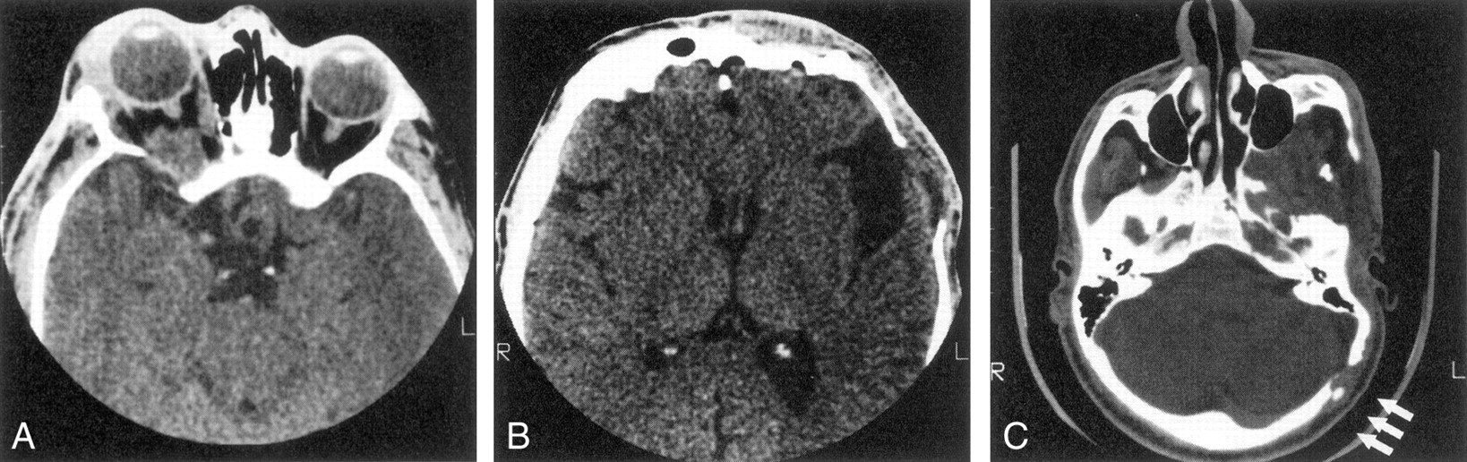
- Fig 1.
Series of axial CT scans.
A, Infiltration and decalcification of the sphenoid wing underlying neurofibromas.
B, Infiltration and decalcification of the temporal bone underlying neurofibromas.
C, Infiltration and decalcification of the occipital bone underlying neurofibromas.
- Fig 2.
Axial contrast-enhanced CT scan shows bilateral facial soft-tissue tumor infiltration, bilateral enlarged middle cranial fossae (arrowheads), bilateral large globes (buphthalmos), and infiltration of both optic nerve sheaths.
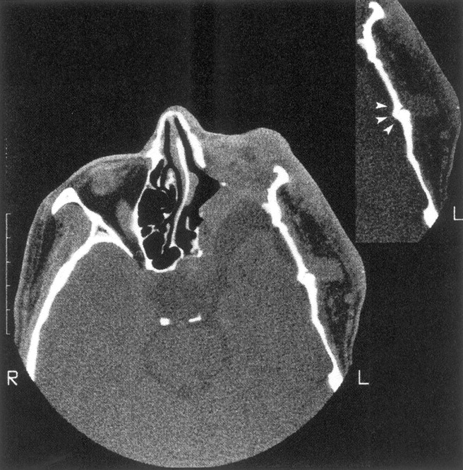
- Fig 3.
Enlargement of the left middle cranial fossa with temporal arachnoid cyst, absence of the left sphenoid wing, and flattening of the temporal bone. There is tumor invasion of the orbit with reduced orbital volume, and the left eye was enucleated. The insert shows an abnormal temporal squamosa suture (arrowheads) underlying tumor in the left superficial temporal fossa.
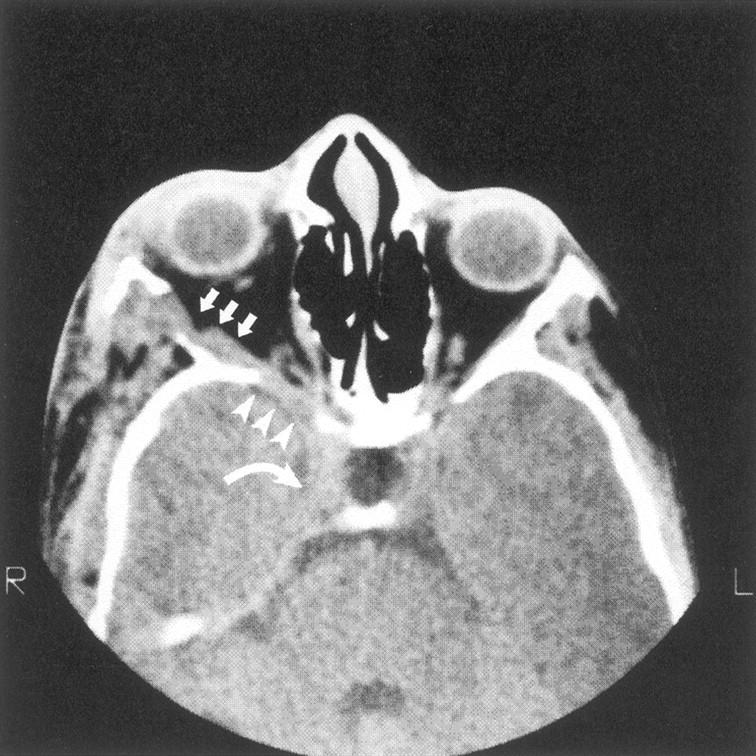
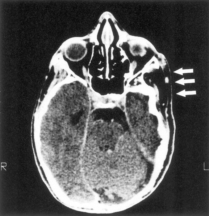
- Fig 4.
Axial CT scan shows decalcification of sphenoid bone (arrowheads) adjacent to neurofibroma infiltration of the lateral rectus muscle (arrows) in the absence of middle cranial fossa enlargement. Tumor also enlarges the superior orbital fissure and extends into the cavernous sinus (curved arrow).
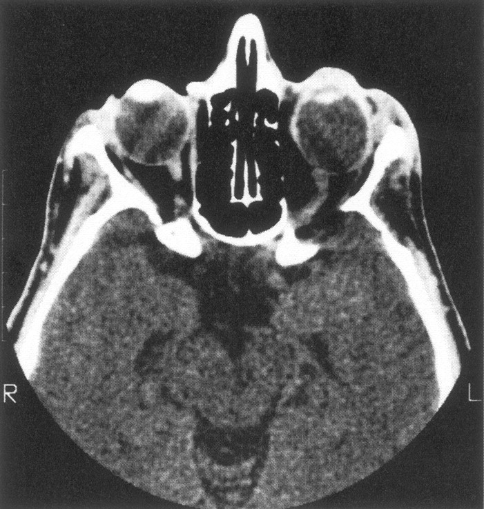
- Fig 5.
CT scan of our one patient with minimal expansion of the middle cranial fossa and abnormally large decalcification of the ipsilateral sphenoid wing, with no detectable adjacent tumor. The left globe is also enlarged.
- Fig 6.
Axial CT scan of a patient with smaller middle cranial fossa and flattened temporal bone ipsilateral to extracranial tumor (arrows).
- Fig 7.
Series of axial CT scans (soft-tissue and bone windows) obtained during three scanning sessions over several years show progression of sphenoid bone abnormalities over time. Note that the soft-tissue mass increases over time (original, A and D; 5 years later, B and E; and 10 years later, C and F). and the ipsilateral middle cranial fossa becomes progressively more enlarged.


In this issue


Jump to section
Related Articles
Cited By...
- Unilateral Creeping Destruction of Deformed Mandibular Ramus and Angle Associated with Extensive Facial Plexiform Neurofibroma in Neurofibromatosis Type 1: A Case Report with Analysis of the Literature for Diagnosing Osteolytic Events of the Mandible in Tumor-suppressor Gene Syndrome
- Clinical Images: Imaging Manifestations of Orbital Neurofibromatosis Type 1
- Analysis of Orbital Plain Radiographs for Orbital Deformities in Neurofibromatosis Type 1 Patients, with Special Reference to Alterations of the Orbital Rim as Indicators of Adjacent Plexiform Neurofibroma
- Dysplasia of the Orbit and Adjacent Bone Associated with Plexiform Neurofibroma and Ocular Disease in 42 NF-1 Patients
- Pulsating enophthalmos in an adult patient with type 1 neurofibromatosis
- Bone deformity showing a deep coronoid notch of the mandible in a patient with neurofibromatosis type 1