Advertisement

Main menu

  • Home
  • Content
    • Current Issue
    • Accepted Manuscripts
    • Article Preview
    • Past Issue Archive
    • Video Articles
    • AJNR Case Collection
    • Case of the Week Archive
    • Case of the Month Archive
    • Classic Case Archive
  • Special Collections
    • AJNR Awards
    • Low-Field MRI
    • Alzheimer Disease
    • ASNR Foundation Special Collection
    • Photon-Counting CT
    • View All
  • Multimedia
    • AJNR Podcasts
    • AJNR SCANtastic
    • Trainee Corner
    • MRI Safety Corner
    • Imaging Protocols
  • For Authors
    • Submit a Manuscript
    • Submit a Video Article
    • Submit an eLetter to the Editor/Response
    • Manuscript Submission Guidelines
    • Statistical Tips
    • Fast Publishing of Accepted Manuscripts
    • Graphical Abstract Preparation
    • Imaging Protocol Submission
    • Author Policies
  • About Us
    • About AJNR
    • Editorial Board
    • Editorial Board Alumni
  • More
    • Become a Reviewer/Academy of Reviewers
    • Subscribers
    • Permissions
    • Alerts
    • Feedback
    • Advertisers
    • ASNR Home

User menu

  • Alerts
  • Log in

Search

  • Advanced search
American Journal of Neuroradiology

American Journal of Neuroradiology

ASHNR American Society of Functional Neuroradiology ASHNR American Society of Pediatric Neuroradiology ASSR
  • Alerts
  • Log in

Advanced Search

  • Home
  • Content
    • Current Issue
    • Accepted Manuscripts
    • Article Preview
    • Past Issue Archive
    • Video Articles
    • AJNR Case Collection
    • Case of the Week Archive
    • Case of the Month Archive
    • Classic Case Archive
  • Special Collections
    • AJNR Awards
    • Low-Field MRI
    • Alzheimer Disease
    • ASNR Foundation Special Collection
    • Photon-Counting CT
    • View All
  • Multimedia
    • AJNR Podcasts
    • AJNR SCANtastic
    • Trainee Corner
    • MRI Safety Corner
    • Imaging Protocols
  • For Authors
    • Submit a Manuscript
    • Submit a Video Article
    • Submit an eLetter to the Editor/Response
    • Manuscript Submission Guidelines
    • Statistical Tips
    • Fast Publishing of Accepted Manuscripts
    • Graphical Abstract Preparation
    • Imaging Protocol Submission
    • Author Policies
  • About Us
    • About AJNR
    • Editorial Board
    • Editorial Board Alumni
  • More
    • Become a Reviewer/Academy of Reviewers
    • Subscribers
    • Permissions
    • Alerts
    • Feedback
    • Advertisers
    • ASNR Home
  • Follow AJNR on Twitter
  • Visit AJNR on Facebook
  • Follow AJNR on Instagram
  • Join AJNR on LinkedIn
  • RSS Feeds

AJNR Awards, New Junior Editors, and more. Read the latest AJNR updates

OtherBRAIN

Astroblastoma: Radiologic-Pathologic Correlation and Distinction from Ependymoma

John D. Port, Daniel J. Brat, Peter C. Burger and Martin G. Pomper
American Journal of Neuroradiology February 2002, 23 (2) 243-247;
John D. Port
  • Find this author on Google Scholar
  • Find this author on PubMed
  • Search for this author on this site
Daniel J. Brat
  • Find this author on Google Scholar
  • Find this author on PubMed
  • Search for this author on this site
Peter C. Burger
  • Find this author on Google Scholar
  • Find this author on PubMed
  • Search for this author on this site
Martin G. Pomper
  • Find this author on Google Scholar
  • Find this author on PubMed
  • Search for this author on this site
  • Article
  • Figures & Data
  • Info & Metrics
  • Responses
  • References
  • PDF
Loading

Article Figures & Data

Figures

  • Tables
  • Fig 1.
    • Download figure
    • Open in new tab
    • Download powerpoint
    Fig 1.

    Histopathologic specimens of a well-differentiated and malignant astroblastoma.

    A, Well-differentiated astroblastomas are composed of astroblastic pseudorosettes, which are elongated tumor cells with broad or tapering processes extending to a central vessel (arrow).

    B, Malignant astroblastomas contain hypercellular mitotically active regions that often display vascular proliferation or necrosis (arrowheads).

  • Fig 2.
    • Download figure
    • Open in new tab
    • Download powerpoint
    Fig 2.

    The classic MR imaging appearance of an astroblastoma in a 3-year-old female patient (patient 5).

    A and B, Axial T2-weighted images [2500/90/1 (TR/TE/excitations)] depict a large, well-circumscribed, heterogeneous supratentorial mass with peripheral cystic changes (arrows). Note that the solid component of the mass has a heterogeneous bubbly appearance in its center.

    C and D, Axial postcontrast T1-weighted images (500/20/1) show rim enhancement of the cystic component (arrows) and heterogeneous enhancement of the solid component. In this case, the tumor was malignant.
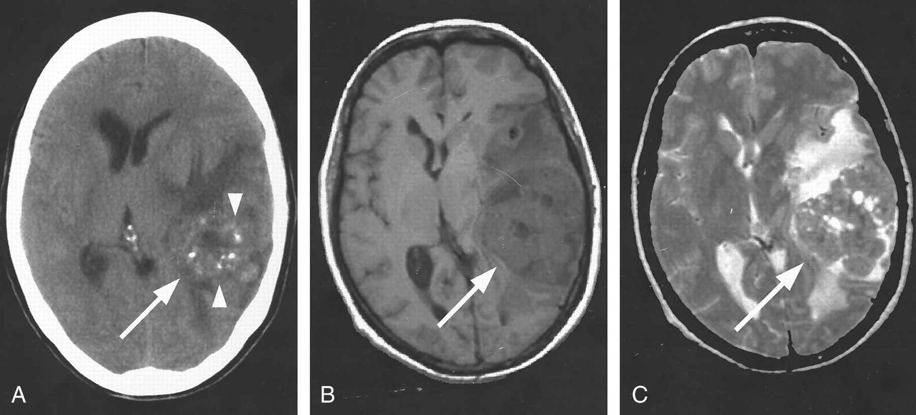
  • Fig 3.
    • Download figure
    • Open in new tab
    • Download powerpoint
    Fig 3.

    The classic CT and MR imaging appearance of an astroblastoma in a 42-year-old female patient (patient 2).

    A, Axial non-contrast-enhanced CT scan shows punctate calcifications (arrowheads) within the solid portion of the large left temporal lobe mass (arrow).

    B, Axial non-contrast-enhanced T1-weighted image (500/8/1) shows the solid portion of the tumor (arrow) to be relatively hypointense to gray matter.

    C, Axial T2-weighted image (2640/98/1) shows the solid portion of the tumor (arrow) as isointense to gray matter. This tumor was well differentiated.
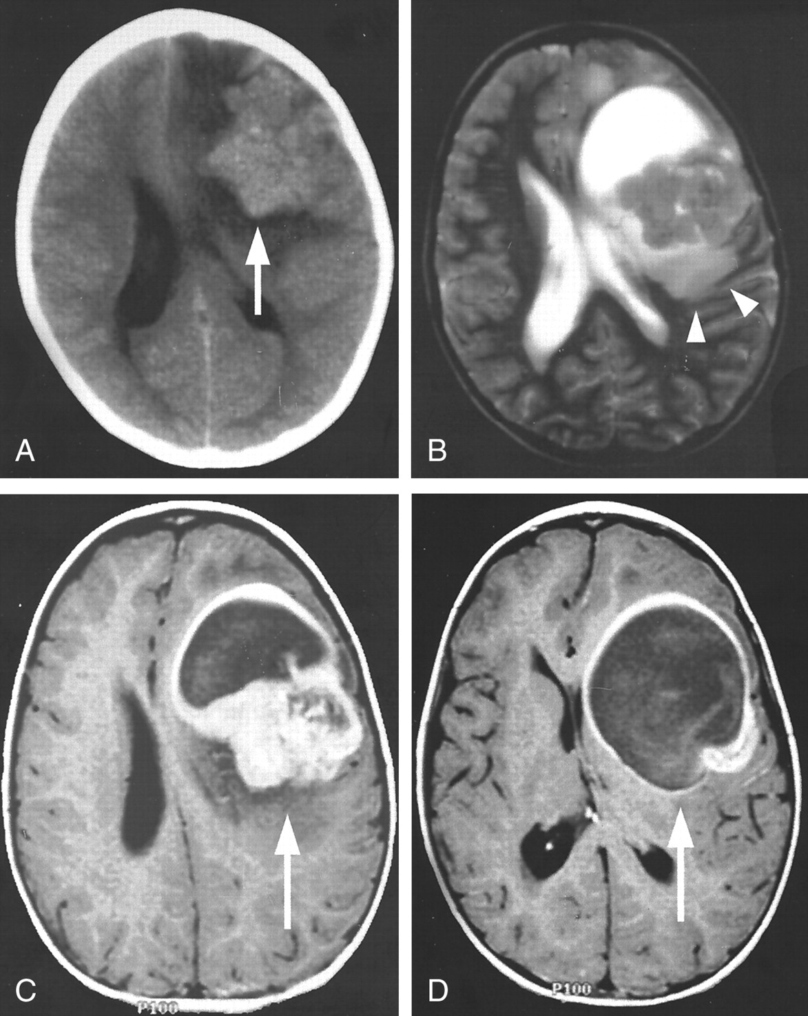
  • Fig 4.
    • Download figure
    • Open in new tab
    • Download powerpoint
    Fig 4.

    The classic CT and MR imaging appearance of an astroblastoma in a 5-year-old female patient (patient 4).

    A, Axial non-contrast-enhanced CT scan shows the solid component of the mass has a slightly higher attenuation than that of the adjacent gray matter (arrow).

    B, Axial T2-weighted image (7935/115/1) depicts the relative lack of peritumoral T2 hyperintensity (representing vasogenic edema or tumor infiltration or both) given the large tumor size (arrowheads).

    C and D, Axial postcontrast T1-weighted images (450/18/1) depict intense heterogeneous enhancement of the solid portion (C, arrow) and rim enhancement of the cystic portion (D, arrow). The diagnosis of a malignant astroblastoma was confirmed on the basis of histologic findings.

  • Fig 5.
    • Download figure
    • Open in new tab
    • Download powerpoint
    Fig 5.

    MR images of an aggressive astroblastoma in a 15-year-old female patient (patient 6).

    A, Sagittal T1-weighted image (810/14/1) shows an aggressive mass that has invaded the anterior body of the corpus callosum (arrowhead).

    B, Axial T2-weighted image (5000/93/1) shows the lesion to have slightly higher T2 signal intensity than that typically seen with an astroblastoma, and the mass appears centered within the anterior horn of the left lateral ventricle (arrow), suggesting the diagnosis of ependymoma.

    C, Coronal postcontrast T1-weighted image (950/14/1) depicts heterogeneous enhancement of the solid portion (arrowhead) and rim enhancement around the cystic portion (arrow). The tumor was a malignant astroblastoma.

  • Fig 6.
    • Download figure
    • Open in new tab
    • Download powerpoint
    Fig 6.

    MR images of a relatively small astroblastoma in a 30-year-old male patient (patient 1).

    A, Axial T2-weighted image (3466/84/1) shows the solid component of this left frontal lesion has the characteristic bubbly appearance of an astroblastoma (arrow), but a moderate amount of peritumoral T2 hyperintensity associated with the lesion (arrowheads) is present. Furthermore, the cystic component is absent.

    B, Axial postcontrast T1-weighted image (550/8) shows the typically intense heterogeneous enhancement of the solid component. The overall imaging characteristics were nonspecific; histologic analysis confirmed the diagnosis of a well-differentiated astroblastoma.

Tables

  • Figures
  • Patient Demographics

    PatientAge (y)/SexHistologic FindingsSurgeryRadiation TherapyFollow-up
    130/maleWell-differentiatedGross total resectionNoNo recurrence at 11 months
    242/femaleWell-differentiatedGross total resectionNoLost
    324/femaleWell-differentiatedGross total resectionNoLost
    45/femaleMalignantGross total resection5400 cGyRecurrence at 11 months
    53/femaleMalignantGross total resection3800 cGyNo recurrence at 64 months
    615/femaleMalignantSubtotal resection7200 cGyRecurrence at 30 months
PreviousNext
Back to top

In this issue

American Journal of Neuroradiology: 23 (2)
American Journal of Neuroradiology
Vol. 23, Issue 2
1 Feb 2002
  • Table of Contents
  • Index by author
Advertisement
Print
Download PDF
Email Article

Thank you for your interest in spreading the word on American Journal of Neuroradiology.

NOTE: We only request your email address so that the person you are recommending the page to knows that you wanted them to see it, and that it is not junk mail. We do not capture any email address.

Enter multiple addresses on separate lines or separate them with commas.
Astroblastoma: Radiologic-Pathologic Correlation and Distinction from Ependymoma
(Your Name) has sent you a message from American Journal of Neuroradiology
(Your Name) thought you would like to see the American Journal of Neuroradiology web site.
CAPTCHA
This question is for testing whether or not you are a human visitor and to prevent automated spam submissions.
Cite this article
John D. Port, Daniel J. Brat, Peter C. Burger, Martin G. Pomper
Astroblastoma: Radiologic-Pathologic Correlation and Distinction from Ependymoma
American Journal of Neuroradiology Feb 2002, 23 (2) 243-247;

Citation Manager Formats

  • BibTeX
  • Bookends
  • EasyBib
  • EndNote (tagged)
  • EndNote 8 (xml)
  • Medlars
  • Mendeley
  • Papers
  • RefWorks Tagged
  • Ref Manager
  • RIS
  • Zotero
0 Responses
Respond to this article
Share
Bookmark this article
Astroblastoma: Radiologic-Pathologic Correlation and Distinction from Ependymoma
John D. Port, Daniel J. Brat, Peter C. Burger, Martin G. Pomper
American Journal of Neuroradiology Feb 2002, 23 (2) 243-247;
del.icio.us logo Twitter logo Facebook logo Mendeley logo
  • Tweet Widget
  • Facebook Like
  • Google Plus One

Jump to section

  • Article
    • Abstract
    • Case Reports
    • Discussion
    • Conclusion
    • Footnotes
    • References
  • Figures & Data
  • Info & Metrics
  • Responses
  • References
  • PDF

Related Articles

  • No related articles found.
  • PubMed
  • Google Scholar

Cited By...

  • Cortically Based Brain Tumors in Children: A Decision-Tree Approach in the Radiology Reading Room
  • Radiology-Pathology and Surgical Correlation in Astroblastoma
  • Molecular Profiling Reclassifies Adult Astroblastoma into Known and Clinically Distinct Tumor Entities with Frequent Mitogen-Activated Protein Kinase Pathway Alterations
  • Crossref
  • Google Scholar

This article has not yet been cited by articles in journals that are participating in Crossref Cited-by Linking.

More in this TOC Section

  • Multimodal CT Provides Improved Performance for Lacunar Infarct Detection
  • Optimal MRI Sequence for Identifying Occlusion Location in Acute Stroke: Which Value of Time-Resolved Contrast-Enhanced MRA?
  • Evaluating the Effects of White Matter Multiple Sclerosis Lesions on the Volume Estimation of 6 Brain Tissue Segmentation Methods
Show more BRAIN

Similar Articles

Advertisement

Indexed Content

  • Current Issue
  • Accepted Manuscripts
  • Article Preview
  • Past Issues
  • Editorials
  • Editor's Choice
  • Fellows' Journal Club
  • Letters to the Editor
  • Video Articles

Cases

  • Case Collection
  • Archive - Case of the Week
  • Archive - Case of the Month
  • Archive - Classic Case

More from AJNR

  • Trainee Corner
  • Imaging Protocols
  • MRI Safety Corner
  • Book Reviews

Multimedia

  • AJNR Podcasts
  • AJNR Scantastics

Resources

  • Turnaround Time
  • Submit a Manuscript
  • Submit a Video Article
  • Submit an eLetter to the Editor/Response
  • Manuscript Submission Guidelines
  • Statistical Tips
  • Fast Publishing of Accepted Manuscripts
  • Graphical Abstract Preparation
  • Imaging Protocol Submission
  • Evidence-Based Medicine Level Guide
  • Publishing Checklists
  • Author Policies
  • Become a Reviewer/Academy of Reviewers
  • News and Updates

About Us

  • About AJNR
  • Editorial Board
  • Editorial Board Alumni
  • Alerts
  • Permissions
  • Not an AJNR Subscriber? Join Now
  • Advertise with Us
  • Librarian Resources
  • Feedback
  • Terms and Conditions
  • AJNR Editorial Board Alumni

American Society of Neuroradiology

  • Not an ASNR Member? Join Now

© 2025 by the American Society of Neuroradiology All rights, including for text and data mining, AI training, and similar technologies, are reserved.
Print ISSN: 0195-6108 Online ISSN: 1936-959X

Powered by HighWire