
Article Figures & Data
Figures
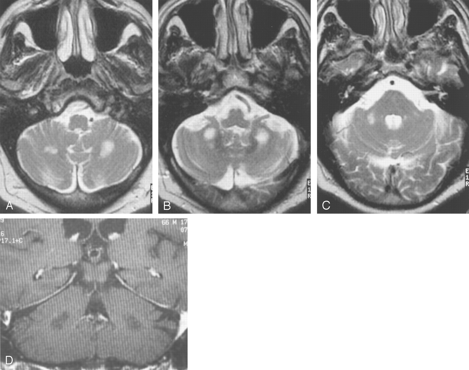
- Fig 1.
Images from the case of a 66-year-old man with fragile X premutation.
A–C, Axial view T2-weighted images show high signal intensity in white matter inferior and lateral to the deep cerebellar nuclei. Only slightly increased signal intensity can be seen in the MCPs.
D, Coronal view T1-weighted contrast-enhanced image shows white matter regions of low signal intensity that do not show enhancement.
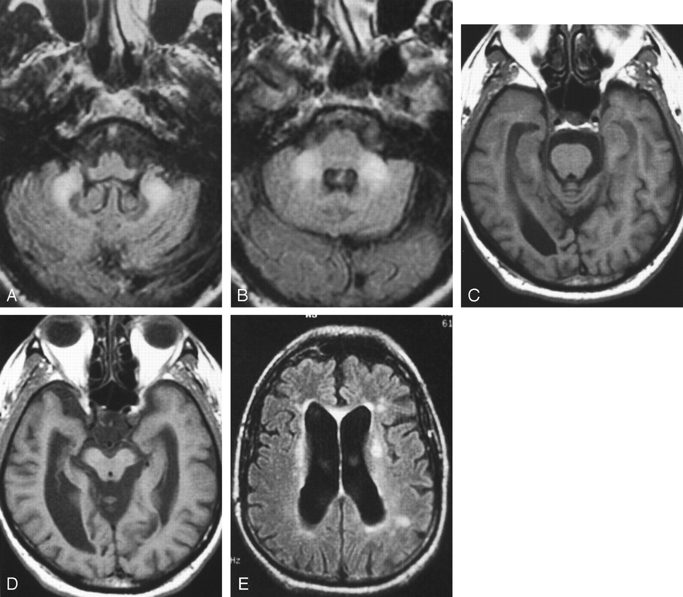
- Fig 2.
Images from the case of a 70-year-old man with fragile X premutation.
A and B, Axial view T2-weighted images show symmetrically increased T2 signal intensity in the cerebellum with mildly increased signal intensity in the MCPs.
C, Coronal view T2-weighted image. Moderate cerebellar volume loss and severe parietal cortical volume loss can be seen with thinning of the corpus callosum. Punctate areas of cerebral white matter increased signal intensity.
D, Sagittal view T1-weighted image shows mild pontine volume loss, decreased anteroposterior dimension of the pons, prominence in prepontine cistern, and severe thinning of the corpus callosum.
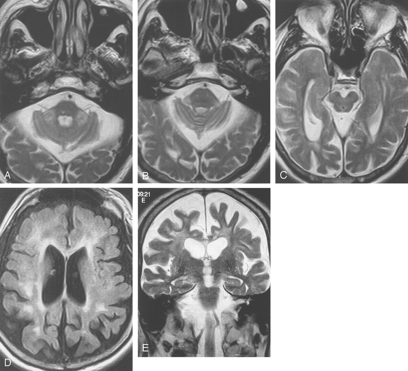
- Fig 3.
Images from the case of a 61-year-old man with fragile X premutation.
A and B, Increased signal intensity is shown in inferior cerebellar white matter on inversion recovery images. The deep cerebellar nuclei are normal in size and signal intensity. The pons is normal in signal intensity.
C and D, Axial view T1-weighted images show volume loss involving the pontomesencephalic junction with enlargement of the temporal horns and ambient cisterns. The cerebral peduncles are moderately decreased in area (D). Moderate enlargement of the lateral ventricles and cerebral sulci can be seen.
E, Axial view inversion recovery image shows increased signal intensity in the thinned corpus callosum and in periventricular white matter.
- Fig 4.
Images from the case of a 69-year-old man with fragile X premutation.
A and B, Axial view T2-weighted images show increased signal intensity in the MCPs, which are slightly thinned in the oblique transverse dimension. Prominence in size of the subarachnoid spaces can be seen. The pons shows a nonspecific right paramedian punctate area of increased signal intensity.
C, Axial view T2-weighted image. Mesencephalon and middle cerebral peduncles are mildly decreased in size, with prominence of the ambient cisterns.
D, Axial view inversion recovery image shows volume loss with increased signal intensity in frontal and parietal white matter and in the genu of the thinned corpus callosum.
E, Coronal view T2-weighted image shows volume loss and increased T2 signal intensity in the frontal white matter and in the thinned corpus callosum.


Tables
Mean or % SD No. of Patients Age at MR imaging 68 years 5.5 17 Age at tremor onset 58.3 years 3.9 15 Age at ataxia onset 61.6 years 5.6 15 Significant impairment Writing 89% 17 Walking 50% 17 Tremor 89% 17 Character of tremor Kinetic > postural 100% 15 Discrete resting tremor 58% 15 Gait ataxia 89% 17 Dyssynergia 72% 17 Tone, mild increase 47% 14 Neuropsychologic data VIQ 98 16 6 PIQ 83 17 6 FSIQ 91 18 6 Executive function deficits 100% 6 VIQ < 85 33% 6 PIQ < 85 66% 6 Impotence 100% 6 CGG repeat 86 10 17 FMRP level 77.5%* 9% 6 mRNA level 2.53-fold† 0.48 6 * Percentage of lymphocytes that are positive for fragile X mental retardation 1 protein immuno-cytochemical staining for fragile X mental retardation 1 protein (19,46).
† 2.53-fold increase over normal (16,46).
Note.—VIQ indicates verbal intelligence quotient; PIQ, performance intelligence level; FSIQ, full scale intelligence quotient; FMRP, fragile X mental retardation 1 protein; mRNA, messenger RNA.
- TABLE 2:
Age and MR measurements from symptomatic fragile X premutation carriers and control participants
Age (yr) Pons AP (mm) Pons Trans (mm) Pons RC (mm) MCP R (mm) MCP L (mm) IV Height (mm) Ant Horn (mm) Fr. Cereb-Vent Index Evan’s Index III Width (mm) Control participants Average 66.1 26 31 29 18 18 11 36 0.32 0.28 6.1 Maximum 76.0 28 33 31 19 22 14 40 0.35 0.31 9.0 Minimum 56.0 23 27 26 15 14 10 31 0.28 0.24 4.0 SD 6.1 1.7 1.7 1.4 1.1 1.9 1.2 2.7 0.02 0.02 1.4 Patients Average 67.6 22 25 26 13 13 12 44 0.40 0.35 11.2 Maximum 77.0 24 31 29 17 17 16 60 0.50 0.46 15.0 Minimum 59.0 19 19 23 9 9 8 23 0.30 0.25 7.0 SD 4.7 1.2 2.9 1.8 2.0 2.0 2.5 8.0 0.05 0.05 2.7 P value 0.46 <.005 <.005 <.005 <.005 <.005 0.21 <.005 <.005 <.005 <.005 Note.—AP indicates anteroposterior; Trans, transverse; RC, rostral-caudal; MCP, middle cerebellar peduncle; R, right; L, left; IV Height, vertical dimension of the fourth ventricle measured from the floor of the fourth ventricle to its apex on a midline sagittal image; Ant Horn, maximal width of both anterior horns; Fr. Cereb-Vent Index, ratio of maximal width of both anterior horns to the diameter of inner table of skull along the line of measurement of anterior horn transverse dimension; Evan’s index, ratio of maximal width of both anterior horns to the diameter of inner table of skull at its point of greatest transverse diameter on any image; III Width, width of third ventricle; Note that MCP R and MCP L are the narrowest oblique transverse dimension of the right and left middle cerebellar peduncles measured from an axial image. P values were derived from Student’s t test of control versus patient populations.
In this issue


Jump to section
Related Articles
Cited By...
- Glial dysregulation in human brain in Fragile X-related disorders
- Oculodentodigital Dysplasia: A Hypomyelinating Leukodystrophy with a Characteristic MRI Pattern of Brain Stem Involvement
- ASFMR1 splice variant: A predictor of fragile X-associated tremor/ataxia syndrome
- MR Imaging Features of the Cerebellum in Adult-Onset Neuronal Intranuclear Inclusion Disease: 8 Cases
- Peripheral neuropathy in complex inherited diseases: an approach to diagnosis
- FXTAS: New insights and the need for revised diagnostic criteria
- Motor and mental dysfunction in mother-daughter transmitted FXTAS
- Molecular Pathogenesis of Fragile X-Associated Tremor/Ataxia Syndrome
- Fragile X-Associated Tremor/Ataxia Syndrome: Clinical Phenotype, Diagnosis, and Treatment
- Penetrance of marked cognitive impairment in older male carriers of the FMR1 gene premutation
- Invited Article: An MRI-based approach to the diagnosis of white matter disorders
- Fragile X associated tremor/ataxia syndrome (FXTAS) with dementia in a female harbouring FMR1 premutation
- Detection of early FXTAS motor symptoms using the CATSYS computerised neuromotor test battery
- FMR1 CGG repeat length predicts motor dysfunction in premutation carriers
- Volumetric brain changes in females with fragile X-associated tremor/ataxia syndrome (FXTAS)
- Molecular and imaging correlates of the fragile X-associated tremor/ataxia syndrome
- Size bias of fragile X premutation alleles in late-onset movement disorders
- The roles of Sp1, Sp3, USF1/USF2 and NRF-1 in the regulation and three-dimensional structure of the Fragile X mental retardation gene promoter
- Spastic paraparesis, cerebellar ataxia, and intention tremor: a severe variant of FXTAS?
- FMR1 gene premutation is a frequent genetic cause of late-onset sporadic cerebellar ataxia
- Intranuclear inclusions in neural cells with premutation alleles in fragile X associated tremor/ataxia syndrome