
Article Figures & Data
Figures
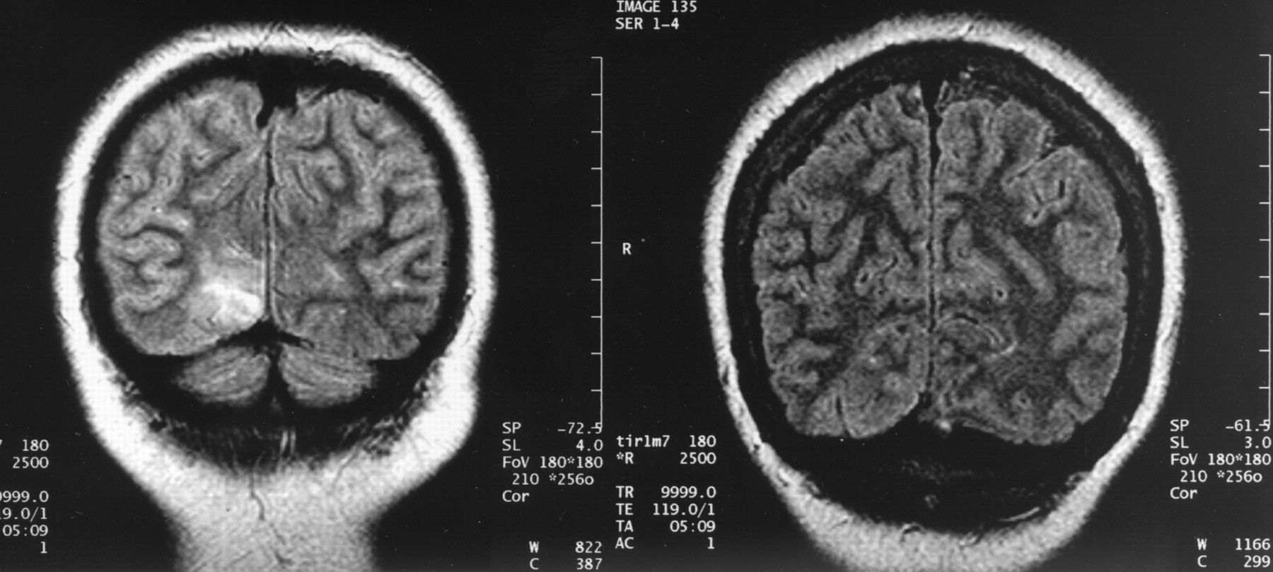
- Fig 1.
Coronal view FLAIR images from case 1 (left) and case 2 (right) (9999/119/1 [TR/TE/NEX]) show hyperintensity in the ganglioglioma within the right occipital lobe in case 1 and isointensity to normal gray matter in the benign cortical dysplasia in case 2.
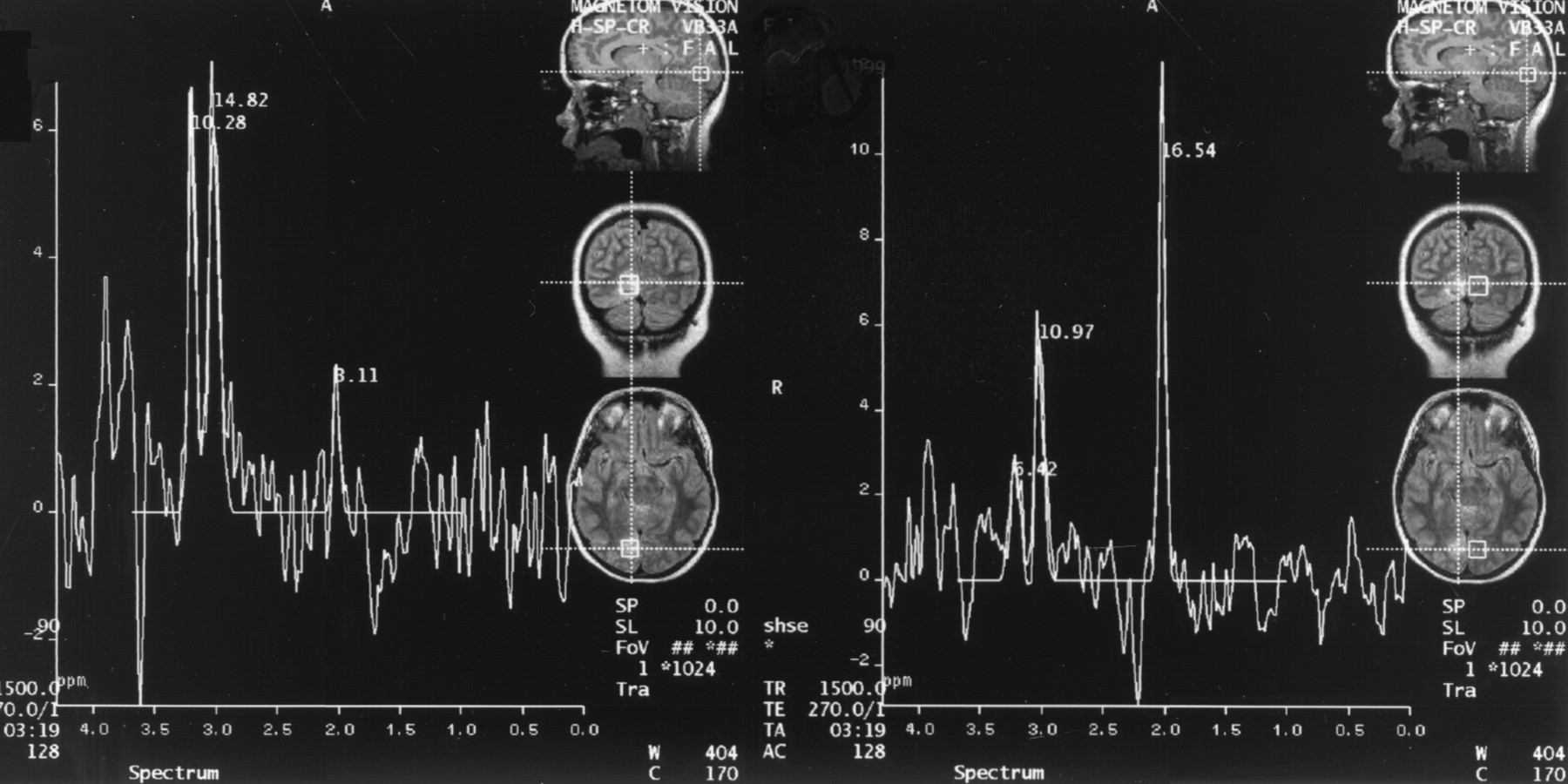
- Fig 2.
MR spectra from case 1 (1500/270; number of signals acquired, 128), obtained by using a 2 × 2 × 2 cm voxel centered within abnormal right occipital tissue (left) and within normal left occipital tissue (right), show reduced NAA/Cr and NAA/Cho ratios in the abnormal right occipital tissue compared with the normal left occipital tissue.
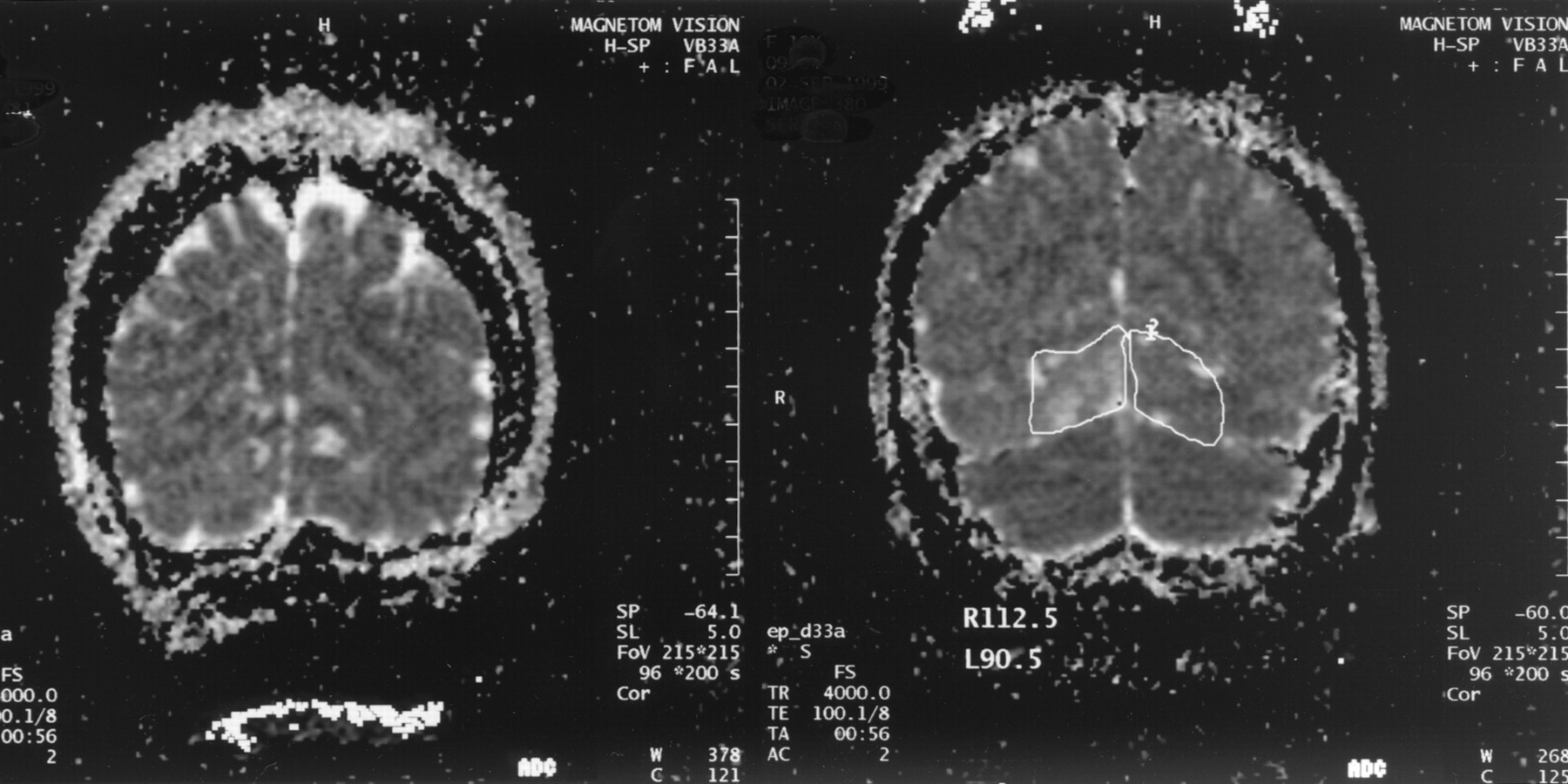
- Fig 3.
ADC maps (4000/100/2) from case 2 (left) and case 1 (right). Mean ADC measurements within regions of interest encompassing the dysplastic right occipital cortex and normal contralateral homologous region in case 1 are displayed. Note the hyperintensity (indicative of increased ADC values) in the right occipital cortex in case 1 and the isointensity in case 2.
- Fig 4.
A binucleate neoplastic neuron (arrow) is seen in the ganglioglioma (hematoxylin and eosin; original magnification, ×400) in case 1.
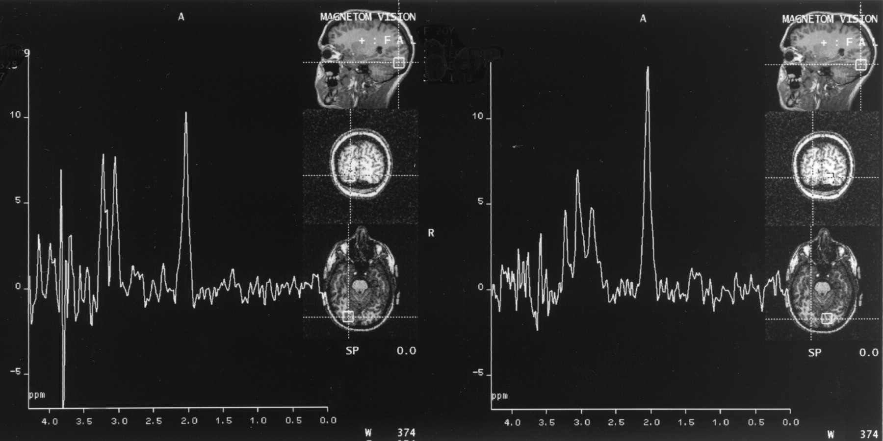
- Fig 5.
Case 2 spectra (1500/270/128), obtained by using a 2 × 2 × 2 cm voxel size (left, right occipital lobe voxel; right, left occipital lobe voxel), show reduced NAA/Cr and NAA/Cho ratios in the voxel placed within the abnormal right occipital tissue compared with those obtained in the contralateral normal occipital lobe. However, the degree of the relative reduction is less than that seen in case 1.
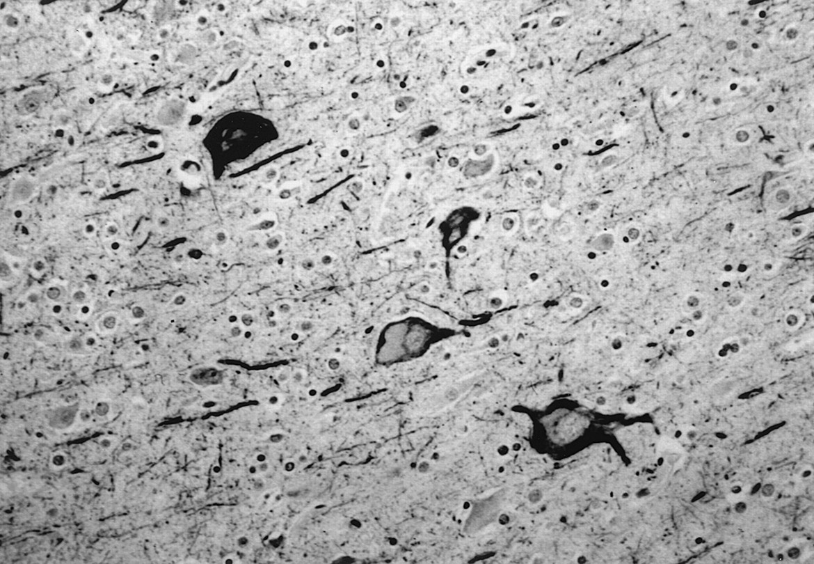
- Fig 6.
Neurofilament immunohistochemistry shows the enlarged, atypical neurons characteristic of severe cortical dysplasia with neuronal cytoskeletal abnormalities (neurofilament stain with hematoxylin counterstain; original magnification, ×400) in case 2.


Tables
Metabolite ratios and ratios of mean ADC values in abnormal occipital cortex versus those in contralateral homologous normal occipital cortex
Case 1 Case 2 NAA/Cr, normal side 1.51 1.52 NAA/Cr, abnormal side 0.21 1.43 Ratio of NAA/Cr, normal side to ratio of NAA/Cr, abnormal side 7.19 1.06 NAA/Cho, normal side 2.58 4.01 NAA/Cho, abnormal side 0.30 1.34 Ratio of NAA/Cho, normal side to ratio of NAA/Cho, abnormal side 8.60 2.99 Cho/Cr, normal side 0.58 0.38 Cho/Cr, abnormal side 0.70 1.06 Ratio of Cho/Cr, normal side to ratio of Cho/Cr, abnormal side 1.21 2.79 Ratio of mean ADC value, abnormal side to mean ADC value, normal side 1.22 1.04
In this issue


Jump to section
Related Articles
Cited By...
- No citing articles found.