Advertisement

Main menu

  • Home
  • Content
    • Current Issue
    • Accepted Manuscripts
    • Article Preview
    • Past Issue Archive
    • Video Articles
    • AJNR Case Collection
    • Case of the Week Archive
    • Case of the Month Archive
    • Classic Case Archive
  • Special Collections
    • AJNR Awards
    • Low-Field MRI
    • Alzheimer Disease
    • ASNR Foundation Special Collection
    • Photon-Counting CT
    • View All
  • Multimedia
    • AJNR Podcasts
    • AJNR SCANtastic
    • Trainee Corner
    • MRI Safety Corner
    • Imaging Protocols
  • For Authors
    • Submit a Manuscript
    • Submit a Video Article
    • Submit an eLetter to the Editor/Response
    • Manuscript Submission Guidelines
    • Statistical Tips
    • Fast Publishing of Accepted Manuscripts
    • Graphical Abstract Preparation
    • Imaging Protocol Submission
    • Author Policies
  • About Us
    • About AJNR
    • Editorial Board
    • Editorial Board Alumni
  • More
    • Become a Reviewer/Academy of Reviewers
    • Subscribers
    • Permissions
    • Alerts
    • Feedback
    • Advertisers
    • ASNR Home

User menu

  • Alerts
  • Log in

Search

  • Advanced search
American Journal of Neuroradiology

American Journal of Neuroradiology

ASHNR American Society of Functional Neuroradiology ASHNR American Society of Pediatric Neuroradiology ASSR
  • Alerts
  • Log in

Advanced Search

  • Home
  • Content
    • Current Issue
    • Accepted Manuscripts
    • Article Preview
    • Past Issue Archive
    • Video Articles
    • AJNR Case Collection
    • Case of the Week Archive
    • Case of the Month Archive
    • Classic Case Archive
  • Special Collections
    • AJNR Awards
    • Low-Field MRI
    • Alzheimer Disease
    • ASNR Foundation Special Collection
    • Photon-Counting CT
    • View All
  • Multimedia
    • AJNR Podcasts
    • AJNR SCANtastic
    • Trainee Corner
    • MRI Safety Corner
    • Imaging Protocols
  • For Authors
    • Submit a Manuscript
    • Submit a Video Article
    • Submit an eLetter to the Editor/Response
    • Manuscript Submission Guidelines
    • Statistical Tips
    • Fast Publishing of Accepted Manuscripts
    • Graphical Abstract Preparation
    • Imaging Protocol Submission
    • Author Policies
  • About Us
    • About AJNR
    • Editorial Board
    • Editorial Board Alumni
  • More
    • Become a Reviewer/Academy of Reviewers
    • Subscribers
    • Permissions
    • Alerts
    • Feedback
    • Advertisers
    • ASNR Home
  • Follow AJNR on Twitter
  • Visit AJNR on Facebook
  • Follow AJNR on Instagram
  • Join AJNR on LinkedIn
  • RSS Feeds

AJNR Awards, New Junior Editors, and more. Read the latest AJNR updates

Research ArticlePediatric Neuroimaging

Terminal Zones of Myelination: MR Evaluation of Children Aged 20–40 Months

Cecilia Parazzini, Cristina Baldoli, Giuseppe Scotti and Fabio Triulzi
American Journal of Neuroradiology November 2002, 23 (10) 1669-1673;
Cecilia Parazzini
  • Find this author on Google Scholar
  • Find this author on PubMed
  • Search for this author on this site
Cristina Baldoli
  • Find this author on Google Scholar
  • Find this author on PubMed
  • Search for this author on this site
Giuseppe Scotti
  • Find this author on Google Scholar
  • Find this author on PubMed
  • Search for this author on this site
Fabio Triulzi
  • Find this author on Google Scholar
  • Find this author on PubMed
  • Search for this author on this site
  • Article
  • Figures & Data
  • Info & Metrics
  • Responses
  • References
  • PDF
Loading

Article Figures & Data

Figures

  • Tables
  • Fig 1.
    • Download figure
    • Open in new tab
    • Download powerpoint
    Fig 1.

    Axial T2-weighted MR images.

    A, Image in a 21-month-old girl with grade 0 myelination in the frontal lobe. The subcortical white matter is clearly hyperintense, so not myelinated, in the prerolandic area and along the first and second convolutions bilaterally. Subcortical hyperintensity was also evident in the frontopolar regions.

    B, Example of grade 2 myelination in the frontal area. A T2 hyperintensity is present along the first and second convolutions, while the prerolandic area is myelinated.

    C, Example of grade 3 myelination. T2 hyperintensity is no longer evident, and myelination appears complete.
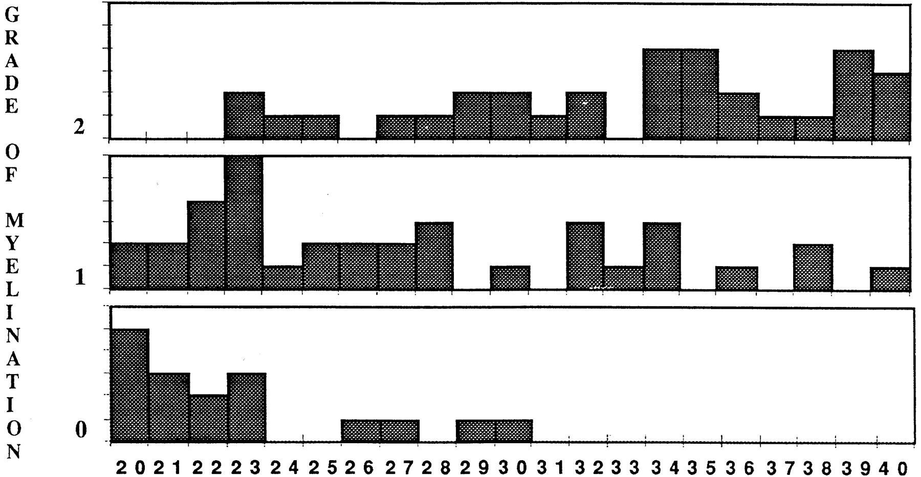
  • Fig 2.
    • Download figure
    • Open in new tab
    • Download powerpoint
    Fig 2.

    Histogram of frontal lobe myelination. Numbers on x axis are ages in months.
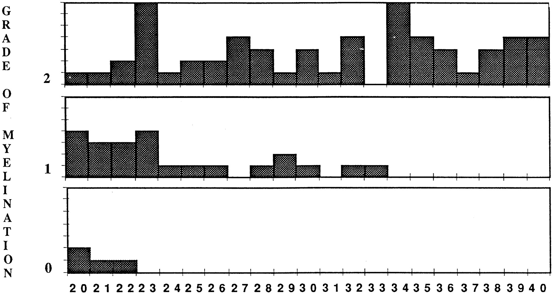
  • Fig 3.
    • Download figure
    • Open in new tab
    • Download powerpoint
    Fig 3.

    Histogram of temporal lobe myelination. Numbers on x axis are ages in months.

  • Fig 4.
    • Download figure
    • Open in new tab
    • Download powerpoint
    Fig 4.

    Histogram of parietal lobe myelination. Numbers on x axis are ages in months.

  • Fig 5.
    • Download figure
    • Open in new tab
    • Download powerpoint
    Fig 5.

    Histogram of peritrigonal area myelination. Numbers on x axis are ages in months.
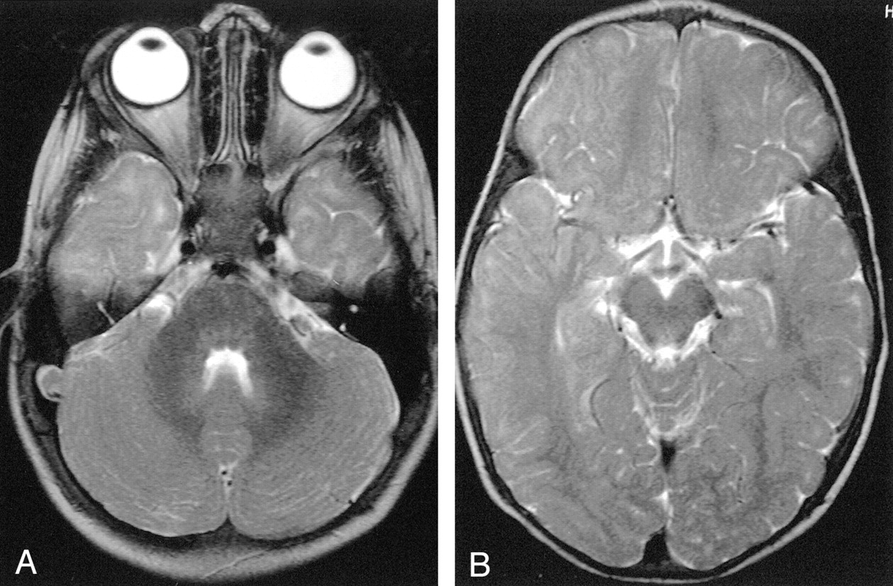
  • Fig 6.
    • Download figure
    • Open in new tab
    • Download powerpoint
    Fig 6.

    A and B, Axial T2-weighted MR images show grade 0 myelination in the temporal lobe. Subcortical T2 hyperintensity is recognizable in both temporopolar (A) and temporolateral (B) areas.
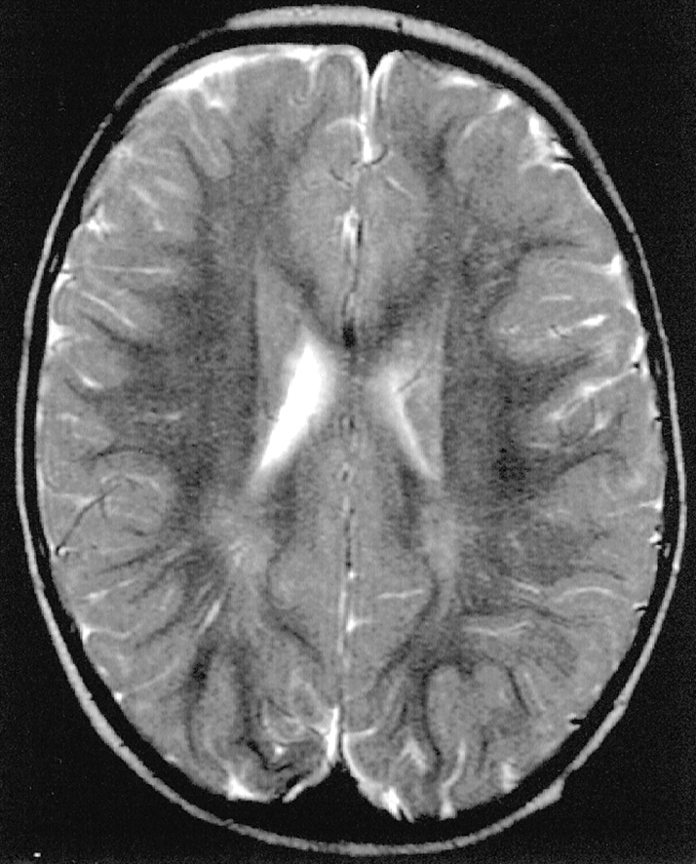
  • Fig 7.
    • Download figure
    • Open in new tab
    • Download powerpoint
    Fig 7.

    Axial T2-weighted MR image shows peritrigonal linear areas of hyperintensity that can be referred to perivascular spaces.

Tables

  • Figures
  • Subdivision of marker sites and summary of grading system

    GradeMarker Sites
    Frontal LobeTemporal LobeParietal LobePeritrigonal Area
    Pre-RPolarI–IILateralPolarPost-RGyriPosteriorSuperior
    0−−−−−−−−−
    1+−−+−+−+−
    2++−++++++
    3+++
    • Note.—R indicates rolandic; I–II, first and second convolutions; +, presence of myelination (absence of T2 hyperintensity); −, absence of myelination (presence of T2 hyperintensity).

PreviousNext
Back to top

In this issue

American Journal of Neuroradiology: 23 (10)
American Journal of Neuroradiology
Vol. 23, Issue 10
1 Nov 2002
  • Table of Contents
  • Index by author
Advertisement
Print
Download PDF
Email Article

Thank you for your interest in spreading the word on American Journal of Neuroradiology.

NOTE: We only request your email address so that the person you are recommending the page to knows that you wanted them to see it, and that it is not junk mail. We do not capture any email address.

Enter multiple addresses on separate lines or separate them with commas.
Terminal Zones of Myelination: MR Evaluation of Children Aged 20–40 Months
(Your Name) has sent you a message from American Journal of Neuroradiology
(Your Name) thought you would like to see the American Journal of Neuroradiology web site.
CAPTCHA
This question is for testing whether or not you are a human visitor and to prevent automated spam submissions.
Cite this article
Cecilia Parazzini, Cristina Baldoli, Giuseppe Scotti, Fabio Triulzi
Terminal Zones of Myelination: MR Evaluation of Children Aged 20–40 Months
American Journal of Neuroradiology Nov 2002, 23 (10) 1669-1673;

Citation Manager Formats

  • BibTeX
  • Bookends
  • EasyBib
  • EndNote (tagged)
  • EndNote 8 (xml)
  • Medlars
  • Mendeley
  • Papers
  • RefWorks Tagged
  • Ref Manager
  • RIS
  • Zotero
0 Responses
Respond to this article
Share
Bookmark this article
Terminal Zones of Myelination: MR Evaluation of Children Aged 20–40 Months
Cecilia Parazzini, Cristina Baldoli, Giuseppe Scotti, Fabio Triulzi
American Journal of Neuroradiology Nov 2002, 23 (10) 1669-1673;
del.icio.us logo Twitter logo Facebook logo Mendeley logo
  • Tweet Widget
  • Facebook Like
  • Google Plus One

Jump to section

  • Article
    • Abstract
    • Methods
    • Results
    • Discussion
    • Acknowledgments
    • References
  • Figures & Data
  • Info & Metrics
  • Responses
  • References
  • PDF

Related Articles

  • No related articles found.
  • PubMed
  • Google Scholar

Cited By...

  • Effects of diffusion MRI spatial resolution on human brain short-range association fiber reconstruction and structural connectivity estimation
  • A Unified Imaging-Histology Framework for Superficial White Matter Architecture Studies in the Human Brain
  • Clinical Reasoning: A case of abnormal eye movements in an infant: More than meets the eye
  • Crossref
  • Google Scholar

This article has not yet been cited by articles in journals that are participating in Crossref Cited-by Linking.

More in this TOC Section

  • Clinical SVR of Fetal Brain MRI
  • FRACTURE MR in Congenital Vertebral Anomalies
  • Comparing MRI Perfusion in Pediatric Brain Tumors
Show more Pediatric Neuroimaging

Similar Articles

Advertisement

Indexed Content

  • Current Issue
  • Accepted Manuscripts
  • Article Preview
  • Past Issues
  • Editorials
  • Editor's Choice
  • Fellows' Journal Club
  • Letters to the Editor
  • Video Articles

Cases

  • Case Collection
  • Archive - Case of the Week
  • Archive - Case of the Month
  • Archive - Classic Case

More from AJNR

  • Trainee Corner
  • Imaging Protocols
  • MRI Safety Corner
  • Book Reviews

Multimedia

  • AJNR Podcasts
  • AJNR Scantastics

Resources

  • Turnaround Time
  • Submit a Manuscript
  • Submit a Video Article
  • Submit an eLetter to the Editor/Response
  • Manuscript Submission Guidelines
  • Statistical Tips
  • Fast Publishing of Accepted Manuscripts
  • Graphical Abstract Preparation
  • Imaging Protocol Submission
  • Evidence-Based Medicine Level Guide
  • Publishing Checklists
  • Author Policies
  • Become a Reviewer/Academy of Reviewers
  • News and Updates

About Us

  • About AJNR
  • Editorial Board
  • Editorial Board Alumni
  • Alerts
  • Permissions
  • Not an AJNR Subscriber? Join Now
  • Advertise with Us
  • Librarian Resources
  • Feedback
  • Terms and Conditions
  • AJNR Editorial Board Alumni

American Society of Neuroradiology

  • Not an ASNR Member? Join Now

© 2025 by the American Society of Neuroradiology All rights, including for text and data mining, AI training, and similar technologies, are reserved.
Print ISSN: 0195-6108 Online ISSN: 1936-959X

Powered by HighWire