Advertisement
  • Main menu
  • User menu
  • Search
  • English ▼
    • English
    • Afrikaans
    • Albanian
    • Amharic
    • Arabic
    • Armenian
    • Azerbaijani
    • Basque
    • Belarusian
    • Bengali
    • Bosnian
    • Bulgarian
    • Catalan
    • Cebuano
    • Chichewa
    • Chinese (Simplified)
    • Chinese (Traditional)
    • Corsican
    • Croatian
    • Czech
    • Danish
    • Dutch
    • Esperanto
    • Estonian
    • Filipino
    • Finnish
    • French
    • Frisian
    • Galician
    • Georgian
    • German
    • Greek
    • Gujarati
    • Haitian Creole
    • Hausa
    • Hawaiian
    • Hebrew
    • Hindi
    • Hmong
    • Hungarian
    • Icelandic
    • Igbo
    • Indonesian
    • Irish
    • Italian
    • Japanese
    • Javanese
    • Kannada
    • Kazakh
    • Khmer
    • Korean
    • Kurdish (Kurmanji)
    • Kyrgyz
    • Lao
    • Latin
    • Latvian
    • Lithuanian
    • Luxembourgish
    • Macedonian
    • Malagasy
    • Malay
    • Malayalam
    • Maltese
    • Maori
    • Marathi
    • Mongolian
    • Myanmar (Burmese)
    • Nepali
    • Norwegian
    • Pashto
    • Persian
    • Polish
    • Portuguese
    • Punjabi
    • Romanian
    • Russian
    • Samoan
    • Scottish Gaelic
    • Serbian
    • Sesotho
    • Shona
    • Sindhi
    • Sinhala
    • Slovak
    • Slovenian
    • Somali
    • Spanish
    • Sudanese
    • Swahili
    • Swedish
    • Tajik
    • Tamil
    • Telugu
    • Thai
    • Turkish
    • Ukrainian
    • Urdu
    • Uzbek
    • Vietnamese
    • Welsh
    • Xhosa
    • Yiddish
    • Yoruba
    • Zulu

Main menu

  • Home
  • Content
    • Current Issue
    • Accepted Manuscripts
    • Article Preview
    • Past Issue Archive
    • Video Articles
    • AJNR Case Collection
    • Case of the Week Archive
    • Case of the Month Archive
    • Classic Case Archive
  • Special Collections
    • Advancing NeuroMRI with High-Relaxivity Contrast Agents
    • AJNR Awards
    • Low-Field MRI
    • Alzheimer Disease
    • ASNR Foundation Special Collection
    • Photon-Counting CT
    • View All
  • Multimedia
    • AJNR Podcasts
    • AJNR SCANtastic
    • Trainee Corner
    • MRI Safety Corner
    • Imaging Protocols
  • For Authors
    • Submit a Manuscript
    • Submit a Video Article
    • Submit an eLetter to the Editor/Response
    • Manuscript Submission Guidelines
    • Statistical Tips
    • Fast Publishing of Accepted Manuscripts
    • Graphical Abstract Preparation
    • Imaging Protocol Submission
    • Author Policies
  • About Us
    • About AJNR
    • Editorial Board
    • Editorial Board Alumni
  • More
    • Become a Reviewer/Academy of Reviewers
    • Subscribers
    • Permissions
    • Alerts
    • Feedback
    • Advertisers
    • ASNR Home

User menu

  • Alerts
  • Log in

Search

  • Advanced search
American Journal of Neuroradiology

American Journal of Neuroradiology

ASHNR American Society of Functional Neuroradiology ASHNR American Society of Pediatric Neuroradiology ASSR
  • Alerts
  • Log in

Advanced Search

English ▼
  • English
  • Afrikaans
  • Albanian
  • Amharic
  • Arabic
  • Armenian
  • Azerbaijani
  • Basque
  • Belarusian
  • Bengali
  • Bosnian
  • Bulgarian
  • Catalan
  • Cebuano
  • Chichewa
  • Chinese (Simplified)
  • Chinese (Traditional)
  • Corsican
  • Croatian
  • Czech
  • Danish
  • Dutch
  • Esperanto
  • Estonian
  • Filipino
  • Finnish
  • French
  • Frisian
  • Galician
  • Georgian
  • German
  • Greek
  • Gujarati
  • Haitian Creole
  • Hausa
  • Hawaiian
  • Hebrew
  • Hindi
  • Hmong
  • Hungarian
  • Icelandic
  • Igbo
  • Indonesian
  • Irish
  • Italian
  • Japanese
  • Javanese
  • Kannada
  • Kazakh
  • Khmer
  • Korean
  • Kurdish (Kurmanji)
  • Kyrgyz
  • Lao
  • Latin
  • Latvian
  • Lithuanian
  • Luxembourgish
  • Macedonian
  • Malagasy
  • Malay
  • Malayalam
  • Maltese
  • Maori
  • Marathi
  • Mongolian
  • Myanmar (Burmese)
  • Nepali
  • Norwegian
  • Pashto
  • Persian
  • Polish
  • Portuguese
  • Punjabi
  • Romanian
  • Russian
  • Samoan
  • Scottish Gaelic
  • Serbian
  • Sesotho
  • Shona
  • Sindhi
  • Sinhala
  • Slovak
  • Slovenian
  • Somali
  • Spanish
  • Sudanese
  • Swahili
  • Swedish
  • Tajik
  • Tamil
  • Telugu
  • Thai
  • Turkish
  • Ukrainian
  • Urdu
  • Uzbek
  • Vietnamese
  • Welsh
  • Xhosa
  • Yiddish
  • Yoruba
  • Zulu
  • Home
  • Content
    • Current Issue
    • Accepted Manuscripts
    • Article Preview
    • Past Issue Archive
    • Video Articles
    • AJNR Case Collection
    • Case of the Week Archive
    • Case of the Month Archive
    • Classic Case Archive
  • Special Collections
    • Advancing NeuroMRI with High-Relaxivity Contrast Agents
    • AJNR Awards
    • Low-Field MRI
    • Alzheimer Disease
    • ASNR Foundation Special Collection
    • Photon-Counting CT
    • View All
  • Multimedia
    • AJNR Podcasts
    • AJNR SCANtastic
    • Trainee Corner
    • MRI Safety Corner
    • Imaging Protocols
  • For Authors
    • Submit a Manuscript
    • Submit a Video Article
    • Submit an eLetter to the Editor/Response
    • Manuscript Submission Guidelines
    • Statistical Tips
    • Fast Publishing of Accepted Manuscripts
    • Graphical Abstract Preparation
    • Imaging Protocol Submission
    • Author Policies
  • About Us
    • About AJNR
    • Editorial Board
    • Editorial Board Alumni
  • More
    • Become a Reviewer/Academy of Reviewers
    • Subscribers
    • Permissions
    • Alerts
    • Feedback
    • Advertisers
    • ASNR Home
  • Follow AJNR on Twitter
  • Visit AJNR on Facebook
  • Follow AJNR on Instagram
  • Join AJNR on LinkedIn
  • RSS Feeds

AJNR Awards, New Junior Editors, and more. Read the latest AJNR updates


Improved Turnaround Times | Median time to first decision: 12 days

Research ArticleHead and Neck Imaging

Lymph Nodes of Patients with Regional Metastases from Head and Neck Squamous Cell Carcinoma as a Predictor of Pathologic Outcome: Size Changes at CT before and after Radiation Therapy

Hiroya Ojiri, Anthony A. Mancuso, William M. Mendenhall and Scott P. Stringer
American Journal of Neuroradiology November 2002, 23 (10) 1627-1631;
Hiroya Ojiri
  • Find this author on Google Scholar
  • Find this author on PubMed
  • Search for this author on this site
Anthony A. Mancuso
  • Find this author on Google Scholar
  • Find this author on PubMed
  • Search for this author on this site
William M. Mendenhall
  • Find this author on Google Scholar
  • Find this author on PubMed
  • Search for this author on this site
Scott P. Stringer
  • Find this author on Google Scholar
  • Find this author on PubMed
  • Search for this author on this site
  • Article
  • Figures & Data
  • Info & Metrics
  • Responses
  • References
  • PDF
Loading

Article Figures & Data

Figures

  • Tables
  • Fig 1.
    • Download figure
    • Open in new tab
    • Download powerpoint
    Fig 1.

    Images in a 46-year-old man with a carcinoma at the base of his tongue (T4N3[3, 3]). (The percentage decrease ratio was calculated as in follows: % decrease ratio = (1 - b/a) × 100, where a and b are the dimensions shown in images A and B, respectively.

    A, Pre-RT contrast-enhanced CT image reveals enlarged level 2 lymph nodes (N) on both sides. The one on the right measures 54 × 33 mm, and that on the left, 57 × 45 mm. The nodes contain focal areas of low attenuation. An infiltrative mass involves the lower part of the base of the tongue.

    B, Post-RT contrast-enhanced CT image shows a marked decrease in the size of the lymph nodes. The decrease ratio of the largest dimension of the nodes is 59% on the right and 51% on the left. The patient underwent planned post-RT neck dissection on both sides. The surgical specimens were negative on both sides.
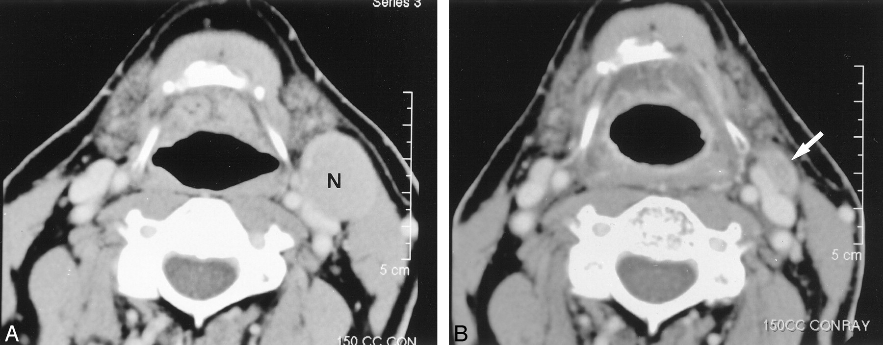
  • Fig 2.
    • Download figure
    • Open in new tab
    • Download powerpoint
    Fig 2.

    Images in a 64-year-old man with metastatic neck disease from an unknown primary squamous cell carcinoma (TXN2A).

    A, Pre-RT contrast-enhanced CT image reveals an enlarged (27 × 22 mm) level 2 lymph node (N) on the left.

    B, Post-RT contrast-enhanced CT image shows a small residual mass (arrow) (decrease ratio of the largest dimension of the node, 52%). The patient underwent left neck dissection after this study. The specimen from the left hemineck was negative.

  • Fig 3.
    • Download figure
    • Open in new tab
    • Download powerpoint
    Fig 3.

    Images in a 52-year-old man with tongue base carcinoma (T2N2B).

    A, Pre-RT contrast-enhanced CT image shows the enlarged (34 × 24 mm) level 2 lymph node (N) on the right. The node contains a focal area of low attenuation.

    B, Post-RT contrast-enhanced CT image reveals a minimal decrease in the size of the lymph node (decrease ratio of the largest dimension of the node, 6%). The surgical specimen from planned post-RT neck dissection at level 2 of the right hemineck was positive.
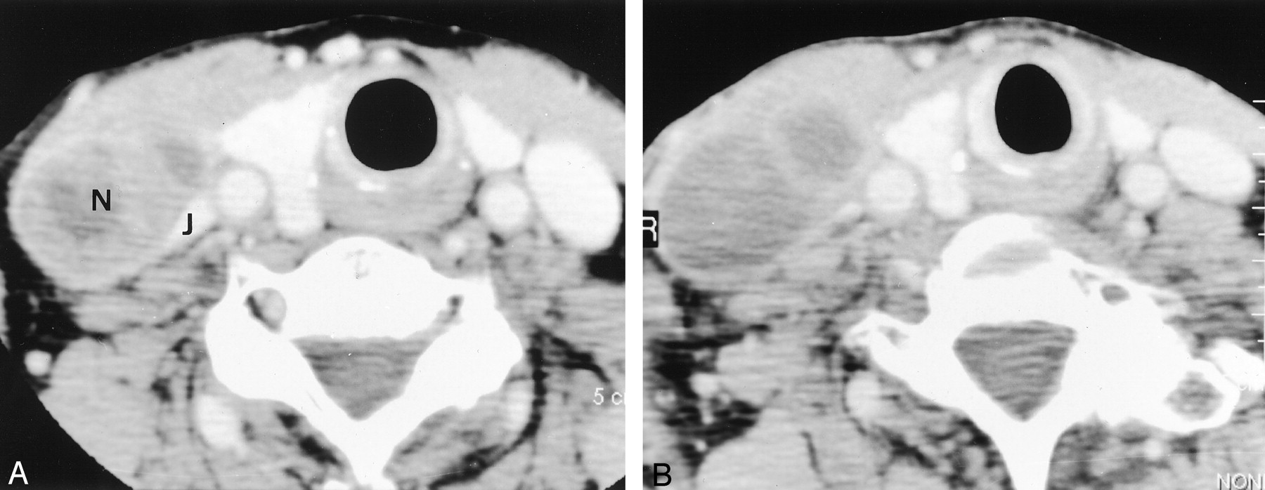
  • Fig 4.
    • Download figure
    • Open in new tab
    • Download powerpoint
    Fig 4.

    Images in a 78-year-old woman with pyriform sinus carcinoma (T2N2A).

    A, Pre-RT contrast enhanced CT image shows an enlarged (40 × 25 mm) level 4 lymph node (N) on the right. The nodal mass distorts the adjacent right internal jugular vein (J).

    B, Post-RT contrast-enhanced CT image shows a slight increase in the size of the lymph node (decrease ratio of the largest dimension, −20%). The internal attenuation is generally lower on this image than on the image in A. The specimen from the right hemineck was positive.

Tables

  • Figures
    • View popup
    TABLE 1:

    Number of heminecks in each primary site, and T- and N-stages

    CharacteristicNo. of Heminecks (N = 37)
    Primary site
     Oral cavity2
     Oropharynx22
     Hypopharynx3
     Larynx4
     Unknown6
    T stage
     T06
     T18
     T213
     T33
     T47
    N stage
     N00
     N11
     N2A8
     N2B19
     N39
    • View popup
    TABLE 2:

    Largest axial dimension of the largest node in the 37 heminecks on pre- and post-RT CT scans

    Largest Dimension, mmNo. of Heminecks
    On Pre-RT CT ScansOn Post-RT CT Scans
    <19014
    20–291418
    30–39102
    40–4973
    >5060
    • View popup
    TABLE 3:

    Pathologic outcome and percentage decrease ratio in the largest axial dimension of the largest node in each hemineck

    Decrease Ratio, %No. of Negative Specimens (n = 24)No. of Positive Specimens (n = 13)Total (N = 37)
    <304610
    31–40639
    41–508412
    >50606
PreviousNext
Back to top

In this issue

American Journal of Neuroradiology: 23 (10)
American Journal of Neuroradiology
Vol. 23, Issue 10
1 Nov 2002
  • Table of Contents
  • Index by author
Advertisement
Print
Download PDF
Email Article
Cite this article
0 Responses
Respond to this article
Share
Bookmark this article
Lymph Nodes of Patients with Regional Metastases from Head and Neck Squamous Cell Carcinoma as a Predictor of Pathologic Outcome: Size Changes at CT before and after Radiation Therapy
Hiroya Ojiri, Anthony A. Mancuso, William M. Mendenhall, Scott P. Stringer
American Journal of Neuroradiology Nov 2002, 23 (10) 1627-1631;
del.icio.us logo Twitter logo Facebook logo Mendeley logo
  • Tweet Widget

Jump to section

  • Article
    • Abstract
    • Methods
    • Results
    • Discussion
    • References
  • Figures & Data
  • Info & Metrics
  • Responses
  • References
  • PDF

Related Articles

  • No related articles found.
  • PubMed
  • Google Scholar

Cited By...

  • Improving Imaging Diagnosis of Persistent Nodal Metastases after Definitive Therapy for Oropharyngeal Carcinoma: Specific Signs for CT and Best Performance of Combined Criteria
  • PET in the Assessment of Therapy Response in Patients with Carcinoma of the Head and Neck and of the Esophagus
  • Crossref
  • Google Scholar

This article has not yet been cited by articles in journals that are participating in Crossref Cited-by Linking.

More in this TOC Section

  • Diagnostic Performance of US in Neck Node NIRADS 2
  • Hydrops Herniation into the Semicircular Canals
  • ASL Sensitivity for Head and Neck Paraganglioma
Show more HEAD AND NECK IMAGING

Similar Articles

Advertisement

Indexed Content

  • Current Issue
  • Accepted Manuscripts
  • Article Preview
  • Past Issues
  • Editorials
  • Editor's Choice
  • Fellows' Journal Club
  • Letters to the Editor
  • Video Articles

Cases

  • Case Collection
  • Archive - Case of the Week
  • Archive - Case of the Month
  • Archive - Classic Case

More from AJNR

  • Trainee Corner
  • Imaging Protocols
  • MRI Safety Corner
  • Book Reviews

Multimedia

  • AJNR Podcasts
  • AJNR Scantastics

Resources

  • Turnaround Time
  • Submit a Manuscript
  • Submit a Video Article
  • Submit an eLetter to the Editor/Response
  • Manuscript Submission Guidelines
  • Statistical Tips
  • Fast Publishing of Accepted Manuscripts
  • Graphical Abstract Preparation
  • Imaging Protocol Submission
  • Evidence-Based Medicine Level Guide
  • Publishing Checklists
  • Author Policies
  • Become a Reviewer/Academy of Reviewers
  • News and Updates

About Us

  • About AJNR
  • Editorial Board
  • Editorial Board Alumni
  • Alerts
  • Permissions
  • Not an AJNR Subscriber? Join Now
  • Advertise with Us
  • Librarian Resources
  • Feedback
  • Terms and Conditions
  • AJNR Editorial Board Alumni

American Society of Neuroradiology

  • Not an ASNR Member? Join Now

© 2025 by the American Society of Neuroradiology All rights, including for text and data mining, AI training, and similar technologies, are reserved.
Print ISSN: 0195-6108 Online ISSN: 1936-959X

Powered by HighWire
Email this Article

Thank you for your interest in spreading the word on American Journal of Neuroradiology.

NOTE: We only request your email address so that the person you are recommending the page to knows that you wanted them to see it, and that it is not junk mail. We do not capture any email address.

Enter multiple addresses on separate lines or separate them with commas.
Lymph Nodes of Patients with Regional Metastases from Head and Neck Squamous Cell Carcinoma as a Predictor of Pathologic Outcome: Size Changes at CT before and after Radiation Therapy
(Your Name) has sent you a message from American Journal of Neuroradiology
(Your Name) thought you would like to see the American Journal of Neuroradiology web site.
CAPTCHA
This question is for testing whether or not you are a human visitor and to prevent automated spam submissions.
Cite this article
Hiroya Ojiri, Anthony A. Mancuso, William M. Mendenhall, Scott P. Stringer
Lymph Nodes of Patients with Regional Metastases from Head and Neck Squamous Cell Carcinoma as a Predictor of Pathologic Outcome: Size Changes at CT before and after Radiation Therapy
American Journal of Neuroradiology Nov 2002, 23 (10) 1627-1631;

Citation Manager Formats

  • BibTeX
  • Bookends
  • EasyBib
  • EndNote (tagged)
  • EndNote 8 (xml)
  • Medlars
  • Mendeley
  • Papers
  • RefWorks Tagged
  • Ref Manager
  • RIS
  • Zotero

We use cookies on this site to enhance your user experience. By clicking any link on this page you are giving your consent for us to set cookies.