
Article Figures & Data
Figures
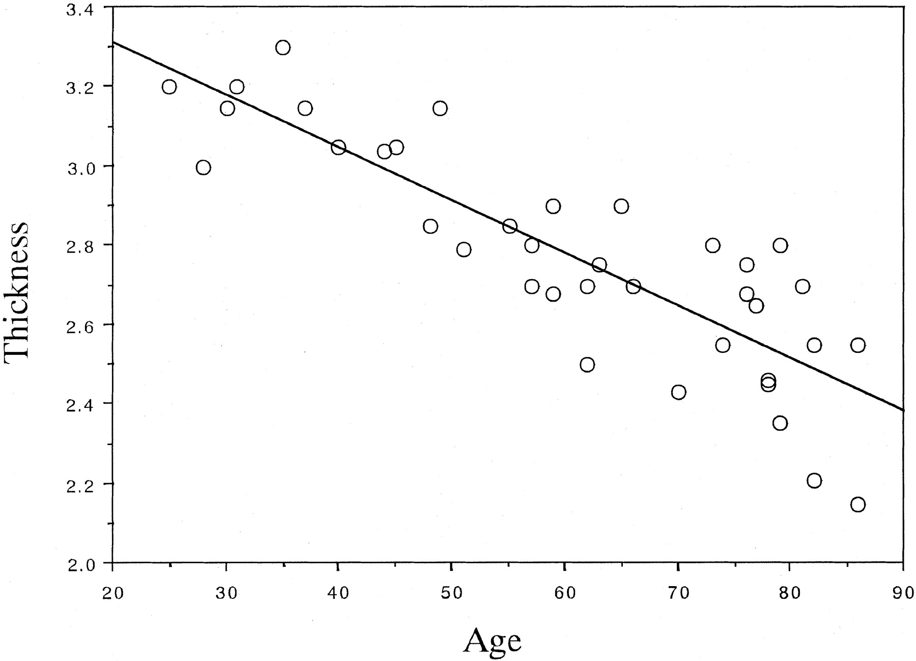
- Fig 1.
Plot shows the correlation between age and thickness of the substantia innominata in control subjects. The thickness of the substantia innominata significantly decreased with normal aging (y = 3.576 − 0.013x, r = −.86, P < .0001).
- Fig 2.
Plots show a significant correlation between MMSE scores and thickness of the substantia innominata in patients with AD but not in those with non-AD dementia. NS = not significant (P > .05).
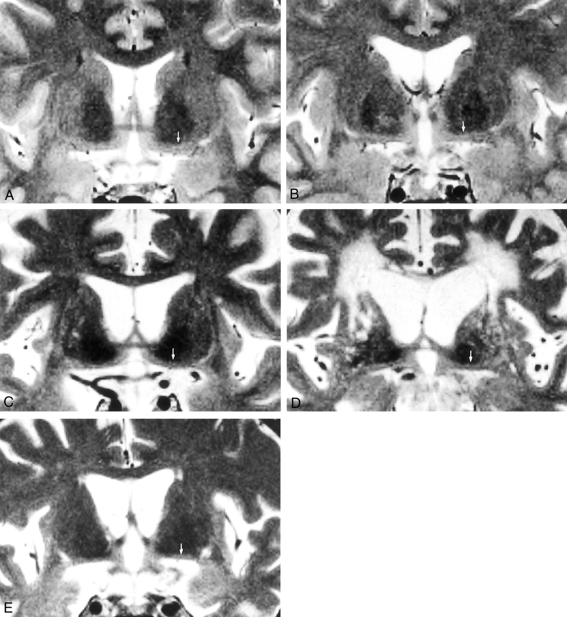
- Fig 3.
Coronal T2-weighted MR images (3000/100/3; section thickness, 3 mm; intersection gap, 0.9 mm; matrix, 256 × 231; field of view, 20 cm) shows the substantia innominata in five subjects (arrows, A–E).
A, Image in a 37-year-old healthy female control subject.
B, Image in a 70-year-old healthy male control subject shows thinning of the substantia innominata.
C, Image in a 67-year-old man with AD shows prominent atrophy of the substantia innominata.
D, Image in a 74-year-old man with vascular dementia shows prominent atrophy of the substantia innominata.
E, Image in a 64-year-old woman with frontotemporal dementia shows prominent atrophy of the substantia innominata.


Tables
Thickness of the substantia innominata in elderly control subjects and patients with dementia
Patient Group Substantia Innominata Thickness (mm) Elderly control subjects (n = 21) 2.57 ± 0.19 AD (n = 39) 1.78 ± 0.28* Vascular dementia (n = 23) 1.94 ± 0.22* Frontotemporal dementia (n = 5) 1.79 ± 0.38* Parkinson disease with dementia (n = 8) 1.93 ± 0.19* * P < .0001, compared with thickness in elderly control subjects.
In this issue


Jump to section
Related Articles
Cited By...
- Xanomeline restores endogenous nicotinic acetylcholine receptor signaling in mouse prefrontal cortex
- Are Linear Measurements of the Nucleus Basalis of Meynert Suitable as a Diagnostic Biomarker in Mild Cognitive Impairment and Alzheimer Disease?
- Grey matter atrophy of basal forebrain and hippocampus in mild cognitive impairment
- The Magic Measurement
- Correlates of response to acetylcholinesterase inhibitor therapy in Alzheimers disease