
Article Figures & Data
Figures
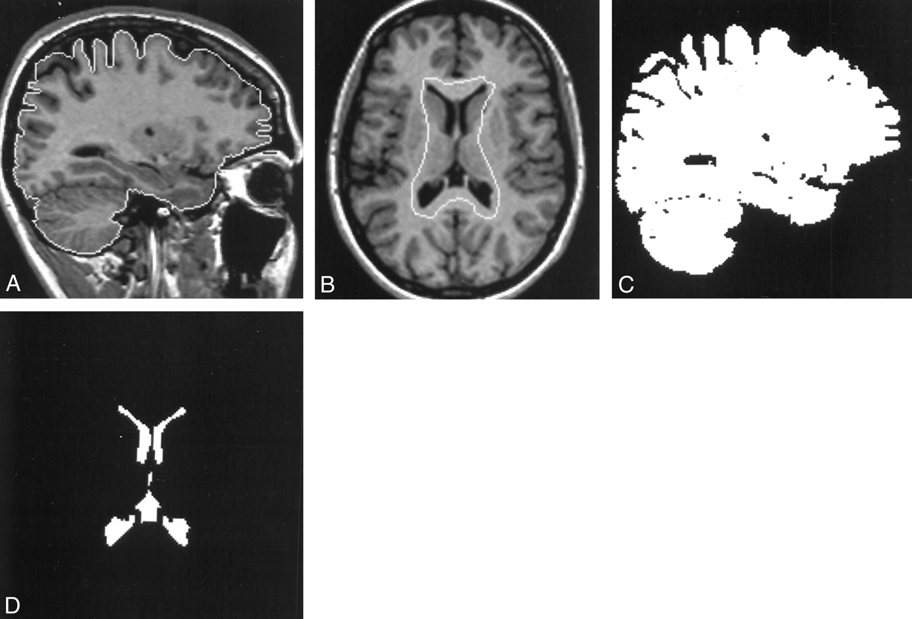
- Fig 1.
Images illustrate the contour and thresholding technique.
A, Parasagittal 3D T1-weighted MR image of the brain with the brain contour.
B, Transverse 3D T1-weighted MR image of the brain showing the initial ventricular contour.
C, Binary image of the corresponding sections of the brain.
D, Final binary image of the ventricles.
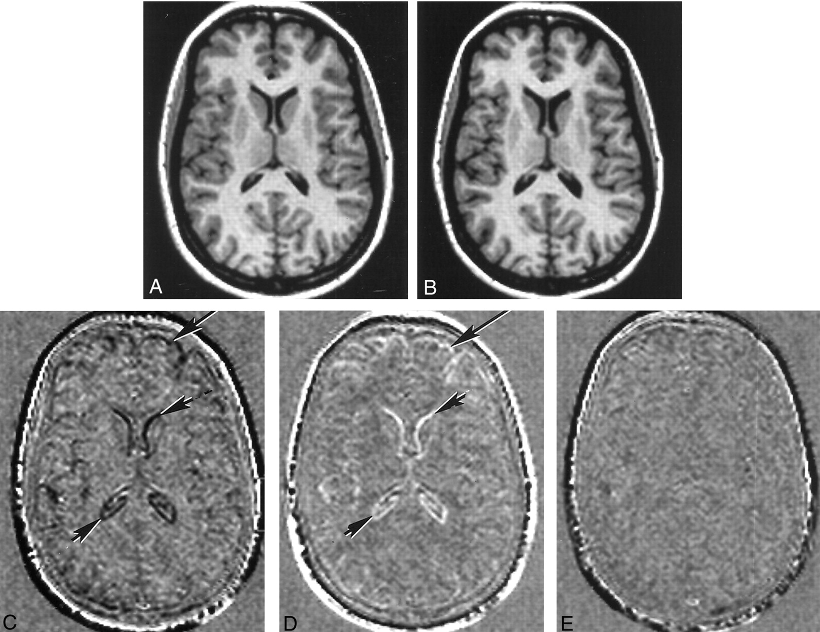
- Fig 2.
Registered and subtraction images.
A, Registered T1-weighted MR image obtained before conception.
B, Registered T1-weighted MR image obtained at term. Slight ventricular enlargement can be seen, compared with the image in A.
C, Subtraction image formed from the term image minus the preconception image. Ventricular enlargement is apparent as a dark line (small arrows). Decrease in the size of the brain is apparent at the external surface as dark lines (large arrow).
D, Subtraction image formed from the image obtained at 24 weeks minus the term image. An increase in the size of the ventricles between term and 24 weeks produces a white line (small arrows), and an increase in the size of the brain produces a white line at the external surface (large arrow).
E, Subtraction image formed from the image obtained at 24 weeks minus the preconception image. This largely featureless image is consistent with the brain and ventricles returning to their original preconception size at 24 weeks after delivery.
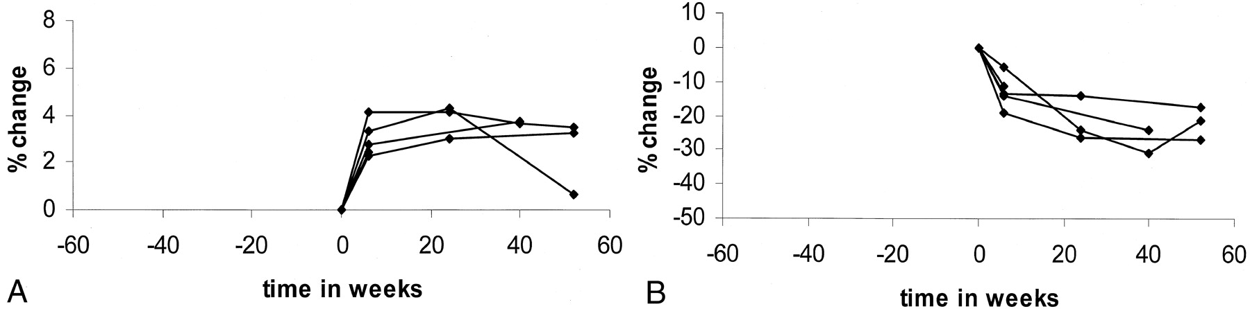
- Fig 3.
Healthy group.
A, Percentage changes in brain size before, during, and after pregnancy. The brain decreases in size until delivery and then increases in size again after delivery.
B, Percentage changes in ventricular size before, during, and after pregnancy. Ventricles increase in size until delivery and decrease in size after delivery.
- Fig 4.
Preeclamptic group.
A, Percentage changes in brain size before and after delivery. Brain increases in size after delivery. Exception is the last image of one patient who was treated with diuretics.
B, Percentage changes in ventricular size before and after delivery. Ventricles decrease in size after delivery.


Tables
Subjects Imaging Times Before Conception 15–30 Weeks’ Gestation Before Delivery 6 Weeks after Delivery 24 Weeks after Delivery 40 Weeks after Delivery 52 Weeks after Delivery Healthy group (n = 9) 2 4 9 9 7 3 3 Preeclamptic group (n = 5) 0 0 5 5 3 2 3 - TABLE 2:
Median grades of change in brain and ventricles in size in both groups assessed on subtraction images
Subjects 6-week Postdelivery Size-Predelivery Size 24-week Postdelivery Size-Predelivery Size 40-week Postdelivery Size-Predelivery Size 52-week Postdelivery Size-Predelivery Size Healthy group 1.8 1.6 2.0 2.0 (+1 to +3) (+1 to +2) (+1 to +3) (+2 to +2) −3.2 −2.8 −2.8 −2.8 (−2 to −4) (−2 to −4) (−2 to −3) (−3 to −3) Preeclamptic group 1.4 1.6 1.8 2.3 (+1 to +3) (+1 to +2) (+1 to +3) (+2 to +3) −3.0 −3.3 −3.0 −3.4 (−2 to −4) (−3 to −4) (−3 to −4) (−3 to −4) Note.—+ indicates increase in size; −, reduction in size.
Subjects Before Pregnancy 15 Weeks’ Gestation 20 Weeks’ Gestation 25 Weeks’ Gestation 30 Weeks’ Gestation 35 Weeks’ Gestation Before Delivery (Term) 6 Weeks after Delivery 24 Weeks after Delivery 40 Weeks after Delivery 52 Weeks after Delivery Healthy group 1 1437.4 1494.5 1494.7 2 1089.2 1109.7 1122.0 3 1201.7 1245.4 1267.4 1252.5 1260.2 4 1371.8 1383.6 1415.1 1420.2 1415.8 5 1207.9 1237.7 1246.2 1253.6 1244.0 6 1197.4 1195.5 1184.0 1187.3 1172.2 1163.0 1150.7 1180.1 1205.7 7 1277.5 1265.9 1255.7 1238.0 1221.9 1268.8 1290.5 8 1288.9 1247.1 1233.5 1241.5 1208.7 1248.3 1269.9 9 965.5 953.9 946.2 975.2 Preeclamptic group 1 942.7 981.7 981.9 977.3 975.5 2* 1036.2 1070.3 1080.6 1043.1 3 999.9 1027.5 1037.1 4 1274.3 1303.9 1312.0 1315.2 5 1183.4 1238.7 * Patient treated with diuretics.
Subjects Before Pregnancy 15 Weeks’ Gestation 20 Weeks’ Gestation 25 Weeks’ Gestation 30 Weeks’ Gestation 35 Weeks’ Gestation Before Delivery (Term) 6 Weeks after Delivery 24 Weeks after Delivery 40 Weeks after Delivery 52 Weeks after Delivery Healthy group 1 29.9 27.4 27.9 28.1 2 14.9 12.2 11.1 3 11.2 7.8 6.9 7.6 6.7 4 17.5 17 15.4 15.1 15.3 5 17.4 15.4 15.1 14.1 15.4 6 6.7 6.7 7.4 7.1 7.1 8.6 9.5 6.8 6.3 7 14.8 15.6 16.1 16.8 18.1 14.0 11.6 8 17.6 19.8 20.1 20.0 21.3 18.6 17.3 9 17.1 17.9 17.8 15.6 Preeclamptic group 1 11.5 10.9 8.7 7.9 9.0 2* 20.7 16.7 15.2 15.1 3 12.1 10.4 9.2 4 35.2 30.4 30.3 29.0 5 13.0 11.5 * Patient treated with diuretics.


