Advertisement
  • Main menu
  • User menu
  • Search
  • English ▼
    • English
    • Afrikaans
    • Albanian
    • Amharic
    • Arabic
    • Armenian
    • Azerbaijani
    • Basque
    • Belarusian
    • Bengali
    • Bosnian
    • Bulgarian
    • Catalan
    • Cebuano
    • Chichewa
    • Chinese (Simplified)
    • Chinese (Traditional)
    • Corsican
    • Croatian
    • Czech
    • Danish
    • Dutch
    • Esperanto
    • Estonian
    • Filipino
    • Finnish
    • French
    • Frisian
    • Galician
    • Georgian
    • German
    • Greek
    • Gujarati
    • Haitian Creole
    • Hausa
    • Hawaiian
    • Hebrew
    • Hindi
    • Hmong
    • Hungarian
    • Icelandic
    • Igbo
    • Indonesian
    • Irish
    • Italian
    • Japanese
    • Javanese
    • Kannada
    • Kazakh
    • Khmer
    • Korean
    • Kurdish (Kurmanji)
    • Kyrgyz
    • Lao
    • Latin
    • Latvian
    • Lithuanian
    • Luxembourgish
    • Macedonian
    • Malagasy
    • Malay
    • Malayalam
    • Maltese
    • Maori
    • Marathi
    • Mongolian
    • Myanmar (Burmese)
    • Nepali
    • Norwegian
    • Pashto
    • Persian
    • Polish
    • Portuguese
    • Punjabi
    • Romanian
    • Russian
    • Samoan
    • Scottish Gaelic
    • Serbian
    • Sesotho
    • Shona
    • Sindhi
    • Sinhala
    • Slovak
    • Slovenian
    • Somali
    • Spanish
    • Sudanese
    • Swahili
    • Swedish
    • Tajik
    • Tamil
    • Telugu
    • Thai
    • Turkish
    • Ukrainian
    • Urdu
    • Uzbek
    • Vietnamese
    • Welsh
    • Xhosa
    • Yiddish
    • Yoruba
    • Zulu

Main menu

  • Home
  • Content
    • Current Issue
    • Accepted Manuscripts
    • Article Preview
    • Past Issue Archive
    • Video Articles
    • AJNR Case Collection
    • Case of the Week Archive
    • Case of the Month Archive
    • Classic Case Archive
  • Special Collections
    • Advancing NeuroMRI with High-Relaxivity Contrast Agents
    • AJNR Awards
    • Low-Field MRI
    • Alzheimer Disease
    • ASNR Foundation Special Collection
    • Photon-Counting CT
    • View All
  • Multimedia
    • AJNR Podcasts
    • AJNR SCANtastic
    • Trainee Corner
    • MRI Safety Corner
    • Imaging Protocols
  • For Authors
    • Submit a Manuscript
    • Submit a Video Article
    • Submit an eLetter to the Editor/Response
    • Manuscript Submission Guidelines
    • Statistical Tips
    • Fast Publishing of Accepted Manuscripts
    • Graphical Abstract Preparation
    • Imaging Protocol Submission
    • Author Policies
  • About Us
    • About AJNR
    • Editorial Board
    • Editorial Board Alumni
  • More
    • Become a Reviewer/Academy of Reviewers
    • Subscribers
    • Permissions
    • Alerts
    • Feedback
    • Advertisers
    • ASNR Home

User menu

  • Alerts
  • Log in

Search

  • Advanced search
American Journal of Neuroradiology

American Journal of Neuroradiology

ASHNR American Society of Functional Neuroradiology ASHNR American Society of Pediatric Neuroradiology ASSR
  • Alerts
  • Log in

Advanced Search

English ▼
  • English
  • Afrikaans
  • Albanian
  • Amharic
  • Arabic
  • Armenian
  • Azerbaijani
  • Basque
  • Belarusian
  • Bengali
  • Bosnian
  • Bulgarian
  • Catalan
  • Cebuano
  • Chichewa
  • Chinese (Simplified)
  • Chinese (Traditional)
  • Corsican
  • Croatian
  • Czech
  • Danish
  • Dutch
  • Esperanto
  • Estonian
  • Filipino
  • Finnish
  • French
  • Frisian
  • Galician
  • Georgian
  • German
  • Greek
  • Gujarati
  • Haitian Creole
  • Hausa
  • Hawaiian
  • Hebrew
  • Hindi
  • Hmong
  • Hungarian
  • Icelandic
  • Igbo
  • Indonesian
  • Irish
  • Italian
  • Japanese
  • Javanese
  • Kannada
  • Kazakh
  • Khmer
  • Korean
  • Kurdish (Kurmanji)
  • Kyrgyz
  • Lao
  • Latin
  • Latvian
  • Lithuanian
  • Luxembourgish
  • Macedonian
  • Malagasy
  • Malay
  • Malayalam
  • Maltese
  • Maori
  • Marathi
  • Mongolian
  • Myanmar (Burmese)
  • Nepali
  • Norwegian
  • Pashto
  • Persian
  • Polish
  • Portuguese
  • Punjabi
  • Romanian
  • Russian
  • Samoan
  • Scottish Gaelic
  • Serbian
  • Sesotho
  • Shona
  • Sindhi
  • Sinhala
  • Slovak
  • Slovenian
  • Somali
  • Spanish
  • Sudanese
  • Swahili
  • Swedish
  • Tajik
  • Tamil
  • Telugu
  • Thai
  • Turkish
  • Ukrainian
  • Urdu
  • Uzbek
  • Vietnamese
  • Welsh
  • Xhosa
  • Yiddish
  • Yoruba
  • Zulu
  • Home
  • Content
    • Current Issue
    • Accepted Manuscripts
    • Article Preview
    • Past Issue Archive
    • Video Articles
    • AJNR Case Collection
    • Case of the Week Archive
    • Case of the Month Archive
    • Classic Case Archive
  • Special Collections
    • Advancing NeuroMRI with High-Relaxivity Contrast Agents
    • AJNR Awards
    • Low-Field MRI
    • Alzheimer Disease
    • ASNR Foundation Special Collection
    • Photon-Counting CT
    • View All
  • Multimedia
    • AJNR Podcasts
    • AJNR SCANtastic
    • Trainee Corner
    • MRI Safety Corner
    • Imaging Protocols
  • For Authors
    • Submit a Manuscript
    • Submit a Video Article
    • Submit an eLetter to the Editor/Response
    • Manuscript Submission Guidelines
    • Statistical Tips
    • Fast Publishing of Accepted Manuscripts
    • Graphical Abstract Preparation
    • Imaging Protocol Submission
    • Author Policies
  • About Us
    • About AJNR
    • Editorial Board
    • Editorial Board Alumni
  • More
    • Become a Reviewer/Academy of Reviewers
    • Subscribers
    • Permissions
    • Alerts
    • Feedback
    • Advertisers
    • ASNR Home
  • Follow AJNR on Twitter
  • Visit AJNR on Facebook
  • Follow AJNR on Instagram
  • Join AJNR on LinkedIn
  • RSS Feeds

AJNR Awards, New Junior Editors, and more. Read the latest AJNR updates


Improved Turnaround Times | Median time to first decision: 12 days

Research ArticlePediatric Neuroimaging

Analysis of the Cerebral Cortex in Holoprosencephaly with Attention to the Sylvian Fissures

A. James Barkovich, Erin M. Simon, Nancy J. Clegg, Steven L. Kinsman and Jin S. Hahn
American Journal of Neuroradiology January 2002, 23 (1) 143-150;
A. James Barkovich
  • Find this author on Google Scholar
  • Find this author on PubMed
  • Search for this author on this site
Erin M. Simon
  • Find this author on Google Scholar
  • Find this author on PubMed
  • Search for this author on this site
Nancy J. Clegg
  • Find this author on Google Scholar
  • Find this author on PubMed
  • Search for this author on this site
Steven L. Kinsman
  • Find this author on Google Scholar
  • Find this author on PubMed
  • Search for this author on this site
Jin S. Hahn
  • Find this author on Google Scholar
  • Find this author on PubMed
  • Search for this author on this site
  • Article
  • Figures & Data
  • Info & Metrics
  • Responses
  • References
  • PDF
Loading

Article Figures & Data

Figures

  • Fig 1.
    • Download figure
    • Open in new tab
    • Download powerpoint
    Fig 1.

    Axial spin-echo (3000/120 [TR/TE]) MR image shows the sylvian angle in a healthy neonate. Black lines are drawn tangential to the sylvian fissures at the level of the basal ganglia. The anterior angle is the sylvian angle.
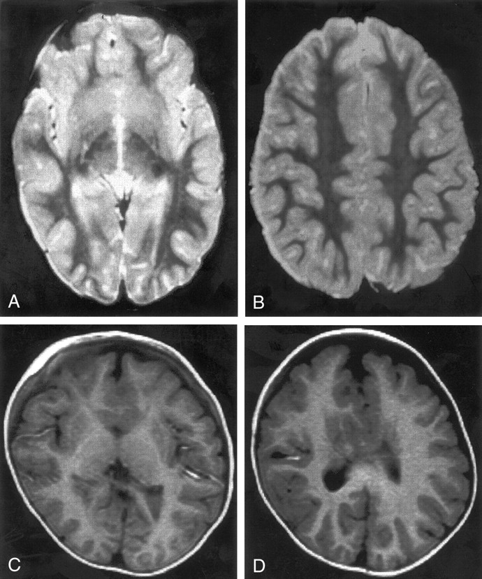
  • Fig 2.
    • Download figure
    • Open in new tab
    • Download powerpoint
    Fig 2.

    MR images show two cases of mild HPE. Both cases were classified as lobar HPE and have sylvian angles of 25°. Both have small, dysplastic frontal horns and both have a large amount of brain tissue anterior to the sylvian fissures.

    A and B, Spin-echo (2500/120) MR images show normal-appearing sulci and gyri in the posterior two-thirds.

    C and D, Inversion-recovery (2000/800/inversion time, 12 ms) MR images show normal-appearing sulci and gyri in the posterior half. The medial frontal sulci appear dysplastic.

  • Fig 3.
    • Download figure
    • Open in new tab
    • Download powerpoint
    Fig 3.

    Spin-echo (3000/120) MR image shows HPE of moderate severity, classified as less dysplastic semilobar HPE. The sylvian angle is 95°, and little brain tissue lies anterior to the sylvian fissures.
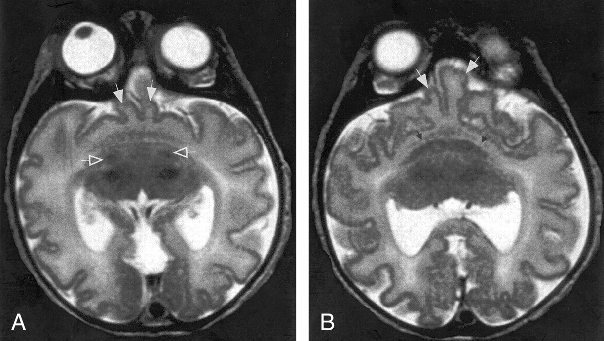
  • Fig 4.
    • Download figure
    • Open in new tab
    • Download powerpoint
    Fig 4.

    A and B, Spin-echo (3000/120) MR images show HPE of moderate severity, classified as less dysplastic semilobar HPE. The sylvian angle is 110°. Only two gyri (solid white arrows) are positioned between the sylvian fissures anteriorly. The basal ganglia are less well developed than those shown in Figure 3, being only a crescent of gray matter anterior to the thalami in B. A thin curvilinear area of gray matter hypointensity (small black arrows, B) is seen between the basal ganglia and presumed insula; this is thought to represent claustrum. Two foci of hypointensity (open white arrows, A) are seen posterior to the basal ganglia crescent but anterior to the thalami in A. These are thought to represent myelinated white matter in the internal capsules.

  • Fig 5.
    • Download figure
    • Open in new tab
    • Download powerpoint
    Fig 5.

    Fast spin-echo (3600/95) MR image shows HPE of moderate severity, classified as more dysplastic semilobar HPE. The anteriormost portion of the cerebrum appears to be a single continuous sylvian fissure. This fissure appears to be composed of the posterior halves of two sylvian fissures, with bilateral posterior opercula (arrows) forming the lateral borders, as if the anterior halves of the fissures had never formed and the posterior halves merged together.
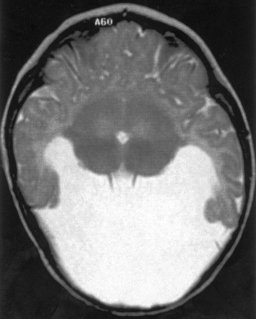
  • Fig 6.
    • Download figure
    • Open in new tab
    • Download powerpoint
    Fig 6.

    Spin-echo (2500/70) MR image shows severe HPE. No sylvian fissures are identified and, thus, no sylvian angle measured. Gyri have normal, uniform width, and sulci have normal, uniform depth. No specific gyri or sulci are identified.
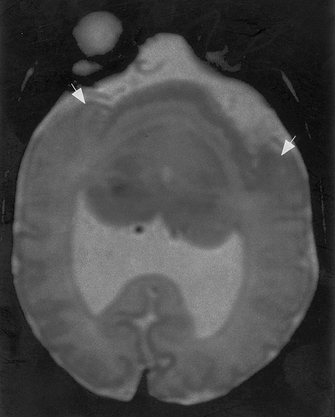
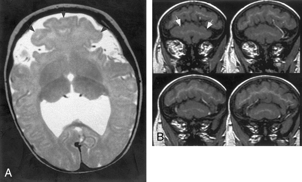
  • Fig 7.
    • Download figure
    • Open in new tab
    • Download powerpoint
    Fig 7.

    MR images show HPE of moderate severity, classified as less dysplastic semilobar. The sylvian angle is 55°.

    A, Axial fast spin-echo (4000/84) MR image shows that a mushroom-shaped region of brain (arrowheads) grows anteriorly from the region between the anterior aspects of the sylvian fissures.

    B, Coronal spoiled gradient-recalled acquisition in a steady state (36/13) MR images show the large mushroom-shaped region (arrows, B) lying below the remainder of the cerebrum in the most anterior aspect of the calvaria.

  • Fig 8.
    • Download figure
    • Open in new tab
    • Download powerpoint
    Fig 8.

    MR images show severe HPE, classified as alobar, with sylvian angle of 135°. First impression of the cerebral cortex is one of pachygyria, but the cortical thickness measures only 3 mm. The cortex appears thick because of the profound microcephaly. This case illustrates some of the difficulties of the DeMyer classification, as the interhemispheric fissure and ventricular systems are much better developed than the basal ganglia and white matter. The basal ganglia, might be considered alobar HPE, but the interhemispheric development suggests mild semilobar HPE.

    A, Axial fast spin-echo (4000/90) MR image shows apparently thickened cortex with few gyri and shallow sulci. Thalami are incompletely separated, and no basal ganglia are seen, nor were they seen at other levels.

    B, Axial fast spin-echo (4000/90) MR image obtained at a higher level shows an interhemispheric fissure spanning the posterior two-thirds of the cerebrum; the falx cerebri (arrows) is present posteriorly.

  • Fig 9.
    • Download figure
    • Open in new tab
    • Download powerpoint
    Fig 9.

    A and B, Spin-echo (700/16) MR images show mild HPE, classified as lobar, with a sylvian angle of 25°. Axial (A) and sagittal (B) views show curvilinear subcortical heterotopia (solid arrows) in the anterior cerebrum. The heterotopia connect with the overlying cortex in several places (open arrow). The overlying cortex is thin with shallow sulci.

PreviousNext
Back to top

In this issue

American Journal of Neuroradiology: 23 (1)
American Journal of Neuroradiology
Vol. 23, Issue 1
1 Jan 2002
  • Table of Contents
  • Index by author
Advertisement
Print
Download PDF
Email Article
Cite this article
0 Responses
Respond to this article
Share
Bookmark this article
Analysis of the Cerebral Cortex in Holoprosencephaly with Attention to the Sylvian Fissures
A. James Barkovich, Erin M. Simon, Nancy J. Clegg, Steven L. Kinsman, Jin S. Hahn
American Journal of Neuroradiology Jan 2002, 23 (1) 143-150;
del.icio.us logo Twitter logo Facebook logo Mendeley logo
  • Tweet Widget

Jump to section

  • Article
    • Abstract
    • Methods
    • Results
    • Discussion
    • Conclusion
    • Footnotes
    • References
  • Figures & Data
  • Info & Metrics
  • Responses
  • References
  • PDF

Related Articles

  • No related articles found.
  • PubMed
  • Google Scholar

Cited By...

  • In Utero MR Imaging of Fetal Holoprosencephaly: A Structured Approach to Diagnosis and Classification
  • Clinical spectrum of SIX3-associated mutations in holoprosencephaly: correlation between genotype, phenotype and function
  • MRI shows abnormal white matter maturation in classical holoprosencephaly
  • Middle interhemispheric variant of holoprosencephaly: A distinct cliniconeuroradiologic subtype
  • Neuroanatomy of holoprosencephaly as predictor of function: Beyond the face predicting the brain
  • Crossref
  • Google Scholar

This article has not yet been cited by articles in journals that are participating in Crossref Cited-by Linking.

More in this TOC Section

  • Pediatric Glioma:Imaging Clusters, Molecular Traits
  • Clinical SVR of Fetal Brain MRI
  • Subpial Hemorrhage & Motor Outcomes in Neonates
Show more Pediatric Neuroimaging

Similar Articles

Advertisement

Indexed Content

  • Current Issue
  • Accepted Manuscripts
  • Article Preview
  • Past Issues
  • Editorials
  • Editor's Choice
  • Fellows' Journal Club
  • Letters to the Editor
  • Video Articles

Cases

  • Case Collection
  • Archive - Case of the Week
  • Archive - Case of the Month
  • Archive - Classic Case

More from AJNR

  • Trainee Corner
  • Imaging Protocols
  • MRI Safety Corner
  • Book Reviews

Multimedia

  • AJNR Podcasts
  • AJNR Scantastics

Resources

  • Turnaround Time
  • Submit a Manuscript
  • Submit a Video Article
  • Submit an eLetter to the Editor/Response
  • Manuscript Submission Guidelines
  • Statistical Tips
  • Fast Publishing of Accepted Manuscripts
  • Graphical Abstract Preparation
  • Imaging Protocol Submission
  • Evidence-Based Medicine Level Guide
  • Publishing Checklists
  • Author Policies
  • Become a Reviewer/Academy of Reviewers
  • News and Updates

About Us

  • About AJNR
  • Editorial Board
  • Editorial Board Alumni
  • Alerts
  • Permissions
  • Not an AJNR Subscriber? Join Now
  • Advertise with Us
  • Librarian Resources
  • Feedback
  • Terms and Conditions
  • AJNR Editorial Board Alumni

American Society of Neuroradiology

  • Not an ASNR Member? Join Now

© 2025 by the American Society of Neuroradiology All rights, including for text and data mining, AI training, and similar technologies, are reserved.
Print ISSN: 0195-6108 Online ISSN: 1936-959X

Powered by HighWire
Email this Article

Thank you for your interest in spreading the word on American Journal of Neuroradiology.

NOTE: We only request your email address so that the person you are recommending the page to knows that you wanted them to see it, and that it is not junk mail. We do not capture any email address.

Enter multiple addresses on separate lines or separate them with commas.
Analysis of the Cerebral Cortex in Holoprosencephaly with Attention to the Sylvian Fissures
(Your Name) has sent you a message from American Journal of Neuroradiology
(Your Name) thought you would like to see the American Journal of Neuroradiology web site.
CAPTCHA
This question is for testing whether or not you are a human visitor and to prevent automated spam submissions.
Cite this article
A. James Barkovich, Erin M. Simon, Nancy J. Clegg, Steven L. Kinsman, Jin S. Hahn
Analysis of the Cerebral Cortex in Holoprosencephaly with Attention to the Sylvian Fissures
American Journal of Neuroradiology Jan 2002, 23 (1) 143-150;

Citation Manager Formats

  • BibTeX
  • Bookends
  • EasyBib
  • EndNote (tagged)
  • EndNote 8 (xml)
  • Medlars
  • Mendeley
  • Papers
  • RefWorks Tagged
  • Ref Manager
  • RIS
  • Zotero

We use cookies on this site to enhance your user experience. By clicking any link on this page you are giving your consent for us to set cookies.