Advertisement

Main menu

  • Home
  • Content
    • Current Issue
    • Accepted Manuscripts
    • Article Preview
    • Past Issue Archive
    • Video Articles
    • AJNR Case Collection
    • Case of the Week Archive
    • Case of the Month Archive
    • Classic Case Archive
  • Special Collections
    • AJNR Awards
    • Low-Field MRI
    • Alzheimer Disease
    • ASNR Foundation Special Collection
    • Photon-Counting CT
    • View All
  • Multimedia
    • AJNR Podcasts
    • AJNR SCANtastic
    • Trainee Corner
    • MRI Safety Corner
    • Imaging Protocols
  • For Authors
    • Submit a Manuscript
    • Submit a Video Article
    • Submit an eLetter to the Editor/Response
    • Manuscript Submission Guidelines
    • Statistical Tips
    • Fast Publishing of Accepted Manuscripts
    • Graphical Abstract Preparation
    • Imaging Protocol Submission
    • Author Policies
  • About Us
    • About AJNR
    • Editorial Board
    • Editorial Board Alumni
  • More
    • Become a Reviewer/Academy of Reviewers
    • Subscribers
    • Permissions
    • Alerts
    • Feedback
    • Advertisers
    • ASNR Home

User menu

  • Alerts
  • Log in

Search

  • Advanced search
American Journal of Neuroradiology

American Journal of Neuroradiology

ASHNR American Society of Functional Neuroradiology ASHNR American Society of Pediatric Neuroradiology ASSR
  • Alerts
  • Log in

Advanced Search

  • Home
  • Content
    • Current Issue
    • Accepted Manuscripts
    • Article Preview
    • Past Issue Archive
    • Video Articles
    • AJNR Case Collection
    • Case of the Week Archive
    • Case of the Month Archive
    • Classic Case Archive
  • Special Collections
    • AJNR Awards
    • Low-Field MRI
    • Alzheimer Disease
    • ASNR Foundation Special Collection
    • Photon-Counting CT
    • View All
  • Multimedia
    • AJNR Podcasts
    • AJNR SCANtastic
    • Trainee Corner
    • MRI Safety Corner
    • Imaging Protocols
  • For Authors
    • Submit a Manuscript
    • Submit a Video Article
    • Submit an eLetter to the Editor/Response
    • Manuscript Submission Guidelines
    • Statistical Tips
    • Fast Publishing of Accepted Manuscripts
    • Graphical Abstract Preparation
    • Imaging Protocol Submission
    • Author Policies
  • About Us
    • About AJNR
    • Editorial Board
    • Editorial Board Alumni
  • More
    • Become a Reviewer/Academy of Reviewers
    • Subscribers
    • Permissions
    • Alerts
    • Feedback
    • Advertisers
    • ASNR Home
  • Follow AJNR on Twitter
  • Visit AJNR on Facebook
  • Follow AJNR on Instagram
  • Join AJNR on LinkedIn
  • RSS Feeds

AJNR Awards, New Junior Editors, and more. Read the latest AJNR updates

Research ArticleLetter

Imaging Appearance of a New Radiation Therapy Delivery Device for CNS Tumors

Eric A. Dame, Gavin J.B. Putzer, James L. Pearlman and F. Reed Murtagh
American Journal of Neuroradiology October 2001, 22 (9) 1806-1807;
Eric A. Dame
  • Find this author on Google Scholar
  • Find this author on PubMed
  • Search for this author on this site
Gavin J.B. Putzer
  • Find this author on Google Scholar
  • Find this author on PubMed
  • Search for this author on this site
James L. Pearlman
  • Find this author on Google Scholar
  • Find this author on PubMed
  • Search for this author on this site
F. Reed Murtagh
  • Find this author on Google Scholar
  • Find this author on PubMed
  • Search for this author on this site
  • Article
  • Figures & Data
  • Info & Metrics
  • Responses
  • References
  • PDF
Loading

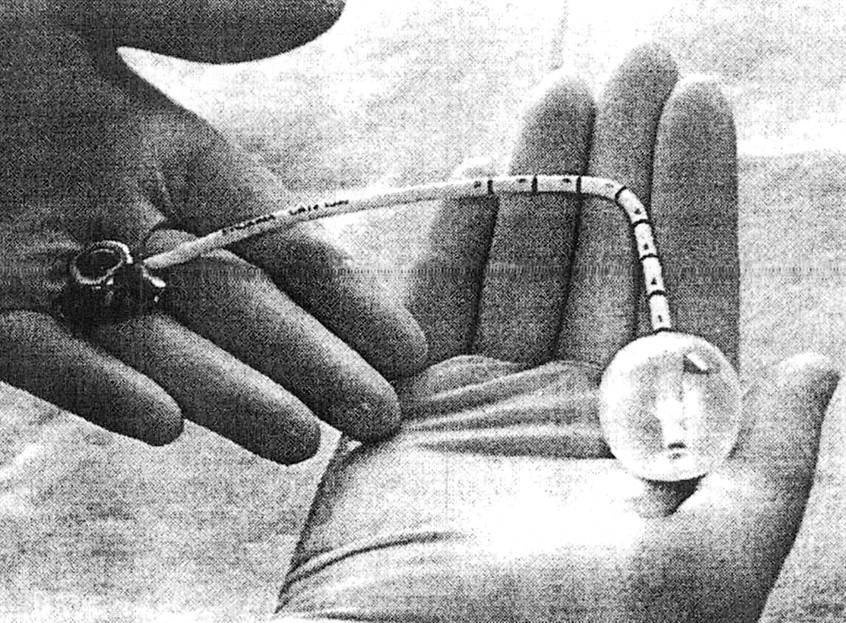
This letter is written to describe the GliaSite RTS balloon catheter system (Fig 1) used to deliver localized radiation therapy to patients after resection of malignant tumors. The device is temporarily implanted in resected brain tumor cavities, and the balloon portion is inflated with radioactive fluid. This delivers a prescribed dose of radiation to the tumor bed without the liquid coming into contact with the tissues.

The main components of the GliaSite device are an infusion port, a catheter, and a silicone balloon. The infusion port, composed of titanium, polysulfone, and silicone, provides access for inflating and deflating the balloon. The catheter consists of bilumen tubing composed of radiopaque silicone. The balloon portion has a dual configuration, with an inner balloon serving as a primary reservoir for the radioactive liquid, and an outer balloon serving as a backup in the event that the inner balloon breaks. Two radiopaque bands that consist of tungsten-loaded ink are located at the proximal and distal ends of the inner balloon, allowing for radiographic visualization of the balloon.

The components of the GliaSite allow for visualization on MR images, CT scans, and conventional radiographs. Before radiation therapy is administered, MR imaging, CT, or conventional radiography is used to verify device integrity and localization. When the balloon is inflated to the desired treatment volume with a solution of 75% normal saline and 25% nonionic contrast agent, MR imaging can further be used to ensure conformance of the surface of the balloon to the resection cavity wall.

The GliaSite RTS was used in an investigational protocol. The study had a total of 21 patients (including two at our institution) with recurrent high-grade primary brain tumors that were treated with the device. In addition, it is currently under investigational study for use in breast lumpectomies. The device uses Iotrex (iodine-125), which provides the source of radiation. The device delivers therapeutic radiation from 0.5 to 1 cm away from the balloon edge at a dose of 50 Gy (range, 40–60 Gy). The radioactive fluid is instilled into the GliaSite RTS device while the patient is in the radiation therapy suite. The balloon remains filled for 73–160 h, depending on the dose to be delivered.

The GliaSite RTS device was used to treat a patient with a ring-enhancing lesion with vasogenic edema in the left frontal lobe, which was proved at biopsy to be a glioblastoma multiforme (Fig 2A). An axial T1-weighted MR image showed that the spherical intracavitary device expanded to the tumor bed (Fig 2B). The contents were isointense with brain parenchyma, with a linear low-signal-intensity area, which is the catheter leading into the device balloon. An axial view CT scan without contrast enhancement showed the spherical intracavitary device expanded to the tumor bed (Fig 2C). The contents showed lower absorption coefficients than those of CSF in the ventricles. The spherical rim was hypoattenuating on CT scans, as was the catheter leading into the device. An anteroposterior conventional film radiograph of the skull showed a radiopaque intraaxial spherical device (Fig 2D). The injection port and catheter tubing were visualized and were external to the cranial bones.

fig 1.
  • Download figure
  • Open in new tab
  • Download powerpoint
fig 1.

The GliaSite RTS device

fig 1.
  • Download figure
  • Open in new tab
  • Download powerpoint
fig 1.

fig 2. Images in a patient with glioblastoma multiforme treated with the GliaSite RTS device.

A, Axial T1-weighted contrast-enhanced MR image of the brain shows a ring-enhancing lesion with vasogenic edema in the left frontal lobe, which was proved to be a glioblastoma multiforme at biopsy.

B, Axial T1-weighted contrast-enhanced MR image in the same patient after surgical implantation of the tumor bed radiation delivery device. Note that the liquid contents of the inflated device have signal characteristics similar to those of brain with this sequence.

C, Axial CT scan in the same patient shows the appearance of the device. The contents of the device have lower absorption coefficients than does ventricular fluid.

D, Device as seen on conventional radiograph. All elements are radiopaque on CT scans.

Footnotes

  • 1 Address reprint requests to Eric A. Dame, 8215 Anglers Point Drive, Tampa, FL 33637.

  • Copyright © American Society of Neuroradiology
PreviousNext
Back to top

In this issue

American Journal of Neuroradiology: 22 (9)
American Journal of Neuroradiology
Vol. 22, Issue 9
1 Oct 2001
  • Table of Contents
  • Index by author
Advertisement
Print
Download PDF
Email Article

Thank you for your interest in spreading the word on American Journal of Neuroradiology.

NOTE: We only request your email address so that the person you are recommending the page to knows that you wanted them to see it, and that it is not junk mail. We do not capture any email address.

Enter multiple addresses on separate lines or separate them with commas.
Imaging Appearance of a New Radiation Therapy Delivery Device for CNS Tumors
(Your Name) has sent you a message from American Journal of Neuroradiology
(Your Name) thought you would like to see the American Journal of Neuroradiology web site.
CAPTCHA
This question is for testing whether or not you are a human visitor and to prevent automated spam submissions.
Cite this article
Eric A. Dame, Gavin J.B. Putzer, James L. Pearlman, F. Reed Murtagh
Imaging Appearance of a New Radiation Therapy Delivery Device for CNS Tumors
American Journal of Neuroradiology Oct 2001, 22 (9) 1806-1807;

Citation Manager Formats

  • BibTeX
  • Bookends
  • EasyBib
  • EndNote (tagged)
  • EndNote 8 (xml)
  • Medlars
  • Mendeley
  • Papers
  • RefWorks Tagged
  • Ref Manager
  • RIS
  • Zotero
0 Responses
Respond to this article
Share
Bookmark this article
Imaging Appearance of a New Radiation Therapy Delivery Device for CNS Tumors
Eric A. Dame, Gavin J.B. Putzer, James L. Pearlman, F. Reed Murtagh
American Journal of Neuroradiology Oct 2001, 22 (9) 1806-1807;
del.icio.us logo Twitter logo Facebook logo Mendeley logo
  • Tweet Widget
  • Facebook Like
  • Google Plus One

Jump to section

  • Article
    • Footnotes
  • Figures & Data
  • Info & Metrics
  • Responses
  • References
  • PDF

Related Articles

  • No related articles found.
  • PubMed
  • Google Scholar

Cited By...

  • No citing articles found.
  • Crossref
  • Google Scholar

This article has not yet been cited by articles in journals that are participating in Crossref Cited-by Linking.

More in this TOC Section

  • Letter to the Editor regarding “Automated Volumetric Software in Dementia: Help or Hindrance to the Neuroradiologist?”
  • Reply:
  • Brain AVM’s Nidus: What if We Hadn’t Understood Anything?
Show more Letter

Similar Articles

Advertisement

Indexed Content

  • Current Issue
  • Accepted Manuscripts
  • Article Preview
  • Past Issues
  • Editorials
  • Editor's Choice
  • Fellows' Journal Club
  • Letters to the Editor
  • Video Articles

Cases

  • Case Collection
  • Archive - Case of the Week
  • Archive - Case of the Month
  • Archive - Classic Case

More from AJNR

  • Trainee Corner
  • Imaging Protocols
  • MRI Safety Corner
  • Book Reviews

Multimedia

  • AJNR Podcasts
  • AJNR Scantastics

Resources

  • Turnaround Time
  • Submit a Manuscript
  • Submit a Video Article
  • Submit an eLetter to the Editor/Response
  • Manuscript Submission Guidelines
  • Statistical Tips
  • Fast Publishing of Accepted Manuscripts
  • Graphical Abstract Preparation
  • Imaging Protocol Submission
  • Evidence-Based Medicine Level Guide
  • Publishing Checklists
  • Author Policies
  • Become a Reviewer/Academy of Reviewers
  • News and Updates

About Us

  • About AJNR
  • Editorial Board
  • Editorial Board Alumni
  • Alerts
  • Permissions
  • Not an AJNR Subscriber? Join Now
  • Advertise with Us
  • Librarian Resources
  • Feedback
  • Terms and Conditions
  • AJNR Editorial Board Alumni

American Society of Neuroradiology

  • Not an ASNR Member? Join Now

© 2025 by the American Society of Neuroradiology All rights, including for text and data mining, AI training, and similar technologies, are reserved.
Print ISSN: 0195-6108 Online ISSN: 1936-959X

Powered by HighWire