Advertisement

Main menu

  • Home
  • Content
    • Current Issue
    • Accepted Manuscripts
    • Article Preview
    • Past Issue Archive
    • Video Articles
    • AJNR Case Collection
    • Case of the Week Archive
    • Case of the Month Archive
    • Classic Case Archive
  • Special Collections
    • AJNR Awards
    • Low-Field MRI
    • Alzheimer Disease
    • ASNR Foundation Special Collection
    • Photon-Counting CT
    • View All
  • Multimedia
    • AJNR Podcasts
    • AJNR SCANtastic
    • Trainee Corner
    • MRI Safety Corner
    • Imaging Protocols
  • For Authors
    • Submit a Manuscript
    • Submit a Video Article
    • Submit an eLetter to the Editor/Response
    • Manuscript Submission Guidelines
    • Statistical Tips
    • Fast Publishing of Accepted Manuscripts
    • Graphical Abstract Preparation
    • Imaging Protocol Submission
    • Author Policies
  • About Us
    • About AJNR
    • Editorial Board
    • Editorial Board Alumni
  • More
    • Become a Reviewer/Academy of Reviewers
    • Subscribers
    • Permissions
    • Alerts
    • Feedback
    • Advertisers
    • ASNR Home

User menu

  • Alerts
  • Log in

Search

  • Advanced search
American Journal of Neuroradiology

American Journal of Neuroradiology

ASHNR American Society of Functional Neuroradiology ASHNR American Society of Pediatric Neuroradiology ASSR
  • Alerts
  • Log in

Advanced Search

  • Home
  • Content
    • Current Issue
    • Accepted Manuscripts
    • Article Preview
    • Past Issue Archive
    • Video Articles
    • AJNR Case Collection
    • Case of the Week Archive
    • Case of the Month Archive
    • Classic Case Archive
  • Special Collections
    • AJNR Awards
    • Low-Field MRI
    • Alzheimer Disease
    • ASNR Foundation Special Collection
    • Photon-Counting CT
    • View All
  • Multimedia
    • AJNR Podcasts
    • AJNR SCANtastic
    • Trainee Corner
    • MRI Safety Corner
    • Imaging Protocols
  • For Authors
    • Submit a Manuscript
    • Submit a Video Article
    • Submit an eLetter to the Editor/Response
    • Manuscript Submission Guidelines
    • Statistical Tips
    • Fast Publishing of Accepted Manuscripts
    • Graphical Abstract Preparation
    • Imaging Protocol Submission
    • Author Policies
  • About Us
    • About AJNR
    • Editorial Board
    • Editorial Board Alumni
  • More
    • Become a Reviewer/Academy of Reviewers
    • Subscribers
    • Permissions
    • Alerts
    • Feedback
    • Advertisers
    • ASNR Home
  • Follow AJNR on Twitter
  • Visit AJNR on Facebook
  • Follow AJNR on Instagram
  • Join AJNR on LinkedIn
  • RSS Feeds

AJNR Awards, New Junior Editors, and more. Read the latest AJNR updates

Research ArticleBRAIN

Importance of Angle Correction in the Measurement of Blood Flow Velocity with Transcranial Doppler Sonography

Jaroslaw Krejza, Zenon Mariak and Viken L. Babikian
American Journal of Neuroradiology October 2001, 22 (9) 1743-1747;
Jaroslaw Krejza
  • Find this author on Google Scholar
  • Find this author on PubMed
  • Search for this author on this site
Zenon Mariak
  • Find this author on Google Scholar
  • Find this author on PubMed
  • Search for this author on this site
Viken L. Babikian
  • Find this author on Google Scholar
  • Find this author on PubMed
  • Search for this author on this site
  • Article
  • Figures & Data
  • Info & Metrics
  • Responses
  • References
  • PDF
Loading

Article Figures & Data

Figures

  • Tables
  • fig 1.
    • Download figure
    • Open in new tab
    • Download powerpoint
    fig 1.

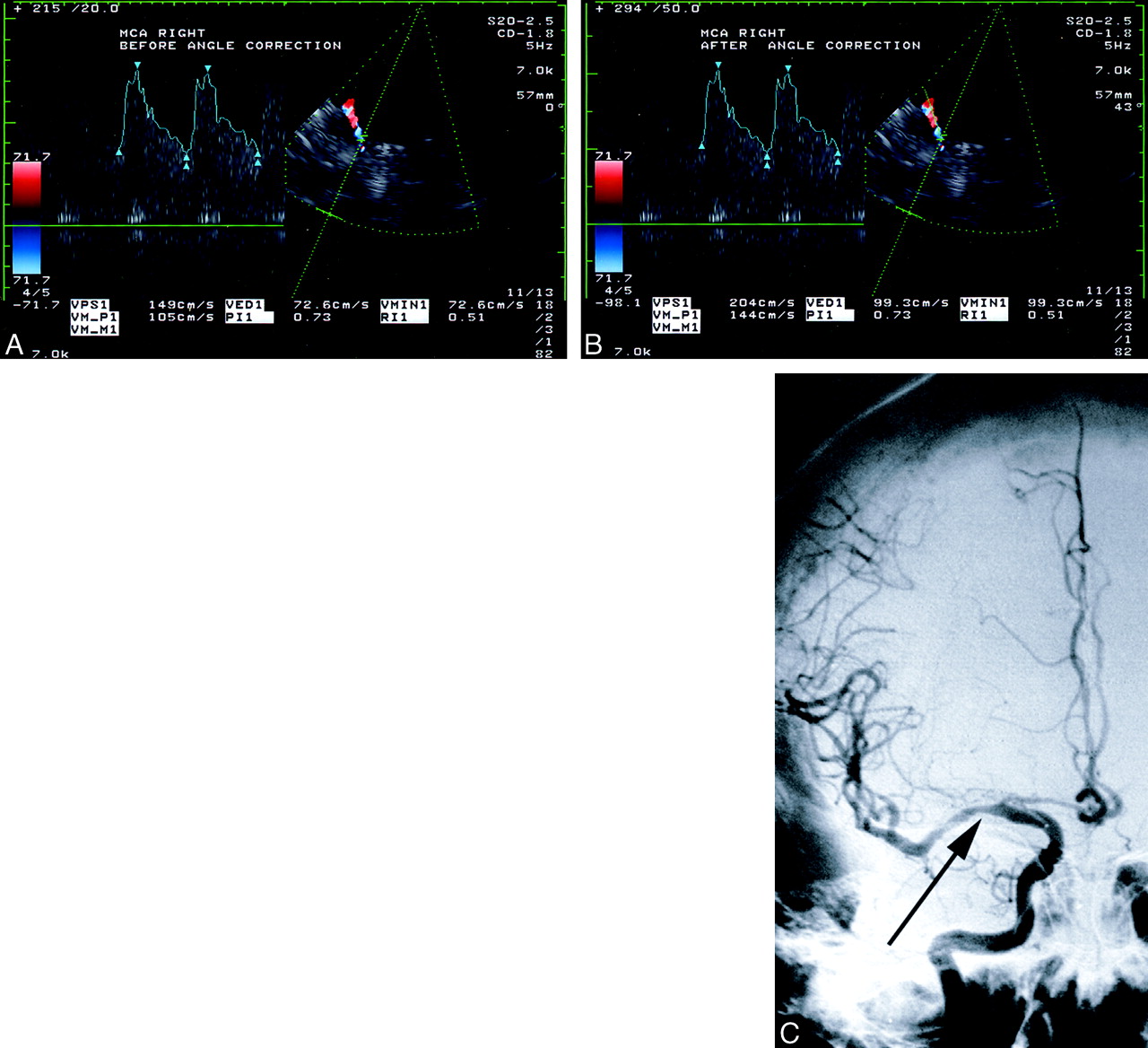
    Comparison between angle-corrected and uncorrected flow velocities and angiographic findings in a 54-year-old woman with MCA stenosis.

    A and B, At TCCD sonography, the sample volume is placed approximately 10 mm from the internal carotid artery bifurcation and within the color image of the right MCA. The velocity spectrum is adjacent to the left of the image of the artery. The uncorrected peak systolic velocity is 149 cm/s (B).

    C, Angiogram shows a lesion causing a stenosis of more than 50% (arrow) in the right MCA M1 segment. The angle-corrected peak systolic velocity is 204 cm/s.

  • fig 2.
    • Download figure
    • Open in new tab
    • Download powerpoint
    fig 2.

    Mass effect and arterial displacement from intracerebral hemorrhage.

    A, Brain CT scan in a 50-year-old man shows an intraparenchymal hematoma with a relatively limited mass effect on the basal cerebral cisterns.

    B, A 3D CT reconstruction illustrates the relationships between the MCA, the temporal sonographic window (W) and direction of the ultrasound beam direction (arrow), and the lesser sphenoid wing (arrowheads). When compared with the contralateral MCA, the hematoma has displaced the ipsilateral MCA toward the skull base.

Tables

  • Figures
  • Table1

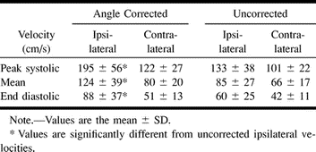
    Comparison of angle-corrected and uncorrected blood flow velocities in 18 patients with MCA stenosis of ≥50%

PreviousNext
Back to top

In this issue

American Journal of Neuroradiology: 22 (9)
American Journal of Neuroradiology
Vol. 22, Issue 9
1 Oct 2001
  • Table of Contents
  • Index by author
Advertisement
Print
Download PDF
Email Article

Thank you for your interest in spreading the word on American Journal of Neuroradiology.

NOTE: We only request your email address so that the person you are recommending the page to knows that you wanted them to see it, and that it is not junk mail. We do not capture any email address.

Enter multiple addresses on separate lines or separate them with commas.
Importance of Angle Correction in the Measurement of Blood Flow Velocity with Transcranial Doppler Sonography
(Your Name) has sent you a message from American Journal of Neuroradiology
(Your Name) thought you would like to see the American Journal of Neuroradiology web site.
CAPTCHA
This question is for testing whether or not you are a human visitor and to prevent automated spam submissions.
Cite this article
Jaroslaw Krejza, Zenon Mariak, Viken L. Babikian
Importance of Angle Correction in the Measurement of Blood Flow Velocity with Transcranial Doppler Sonography
American Journal of Neuroradiology Oct 2001, 22 (9) 1743-1747;

Citation Manager Formats

  • BibTeX
  • Bookends
  • EasyBib
  • EndNote (tagged)
  • EndNote 8 (xml)
  • Medlars
  • Mendeley
  • Papers
  • RefWorks Tagged
  • Ref Manager
  • RIS
  • Zotero
0 Responses
Respond to this article
Share
Bookmark this article
Importance of Angle Correction in the Measurement of Blood Flow Velocity with Transcranial Doppler Sonography
Jaroslaw Krejza, Zenon Mariak, Viken L. Babikian
American Journal of Neuroradiology Oct 2001, 22 (9) 1743-1747;
del.icio.us logo Twitter logo Facebook logo Mendeley logo
  • Tweet Widget
  • Facebook Like
  • Google Plus One

Jump to section

  • Article
    • Abstract
    • Methods
    • Results
    • Discussion
    • Footnotes
    • References
  • Figures & Data
  • Info & Metrics
  • Responses
  • References
  • PDF

Related Articles

  • No related articles found.
  • PubMed
  • Google Scholar

Cited By...

  • Sickle Cell Disease: Reference Values and Interhemispheric Differences of Nonimaging Transcranial Doppler Blood Flow Parameters
  • Sickle Cell Disease and Transcranial Doppler Imaging: Inter-Hemispheric Differences in Blood Flow Doppler Parameters
  • Consensus Recommendations for Transcranial Color-Coded Duplex Sonography for the Assessment of Intracranial Arteries in Clinical Trials on Acute Stroke
  • Transcranial Color-Coded Sonography for the Detection of Middle Cerebral Artery Stenosis
  • Effect of Age on Cerebral Blood Flow Velocity in Patients After Aneurysmal Subarachnoid Hemorrhage
  • Crossref
  • Google Scholar

This article has not yet been cited by articles in journals that are participating in Crossref Cited-by Linking.

More in this TOC Section

  • Enhanced Axonal Metabolism during Early Natalizumab Treatment in Relapsing-Remitting Multiple Sclerosis
  • Progression of Microstructural Damage in Spinocerebellar Ataxia Type 2: A Longitudinal DTI Study
  • SWI or T2*: Which MRI Sequence to Use in the Detection of Cerebral Microbleeds? The Karolinska Imaging Dementia Study
Show more BRAIN

Similar Articles

Advertisement

Indexed Content

  • Current Issue
  • Accepted Manuscripts
  • Article Preview
  • Past Issues
  • Editorials
  • Editor's Choice
  • Fellows' Journal Club
  • Letters to the Editor
  • Video Articles

Cases

  • Case Collection
  • Archive - Case of the Week
  • Archive - Case of the Month
  • Archive - Classic Case

More from AJNR

  • Trainee Corner
  • Imaging Protocols
  • MRI Safety Corner
  • Book Reviews

Multimedia

  • AJNR Podcasts
  • AJNR Scantastics

Resources

  • Turnaround Time
  • Submit a Manuscript
  • Submit a Video Article
  • Submit an eLetter to the Editor/Response
  • Manuscript Submission Guidelines
  • Statistical Tips
  • Fast Publishing of Accepted Manuscripts
  • Graphical Abstract Preparation
  • Imaging Protocol Submission
  • Evidence-Based Medicine Level Guide
  • Publishing Checklists
  • Author Policies
  • Become a Reviewer/Academy of Reviewers
  • News and Updates

About Us

  • About AJNR
  • Editorial Board
  • Editorial Board Alumni
  • Alerts
  • Permissions
  • Not an AJNR Subscriber? Join Now
  • Advertise with Us
  • Librarian Resources
  • Feedback
  • Terms and Conditions
  • AJNR Editorial Board Alumni

American Society of Neuroradiology

  • Not an ASNR Member? Join Now

© 2025 by the American Society of Neuroradiology All rights, including for text and data mining, AI training, and similar technologies, are reserved.
Print ISSN: 0195-6108 Online ISSN: 1936-959X

Powered by HighWire