
Article Figures & Data
Figures
- fig 1.
Square regions of interest, 1.0 cm2 in size, were centered bilaterally on basal ganglia (1), thalami (2), optic radiations (3), calcarine gray matter (4), corticospinal tracts (5), posterior white matter (6), and frontal white matter (7). MR spectroscopic imaging spectra were reconstructed to be centered on each location
- fig 2.
Representative MR spectroscopic imaging data for a premature and a term neonate.
A, Images and spectral array from the brain of a premature, 30-week postconceptional age neonate with normal outcome.
B, Images and spectral array from the brain of a term, 41-week postconceptional age neonate with normal neurologic outcome.
- fig 2.
Continued
- fig 3.
Representative spectra from 1-cm3 voxels from three regions in the brain of a premature (postconceptional age, 30 weeks) and a term (postconceptional age, 40 weeks) neonate. Note higher relative N-acetylaspartate levels in the thalamus and especially the basal ganglia of the term infant. Note also a similar metabolite pattern in the frontal white matter, which is consistent with later maturation of this region
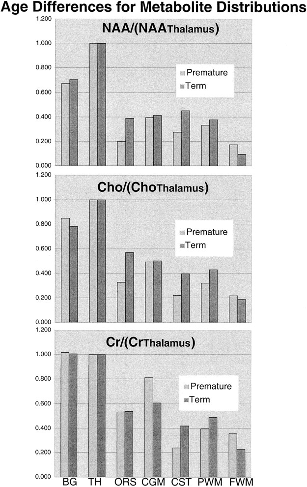
- fig 4.
Graphic representation of the topological variation of N-acetylaspartate (NA), choline (Ch), and creatine (Cr) peak areas relative to thalamus values. Higher levels of all metabolites were observed in the basal ganglia (BG) and thalamus (TH). N-acetylaspartate levels were higher in gray matter regions in both groups; however, relative to the calcarine gray matter (CGM), the difference decreased with age for the optic radiations (ORS), corticospinal tracts (CST), and parietal white matter (PWM), suggesting neuronal maturation in these regions. The minor relative changes in frontal watershed white matter (FWM) metabolite levels are consistent with late maturation
- fig 5.
Graphic representation of the differences between premature and term N-acetylaspartate (top), choline (middle), and creatine (bottom) peak areas for each anatomic region relative to thalamus values
- fig 6.
Graphic representation of the differences between premature and term N-acetylaspartate:choline (top), N-acetylaspartate:creatine (middle), and choline:creatine (bottom) peak area ratios for each anatomic region


Tables

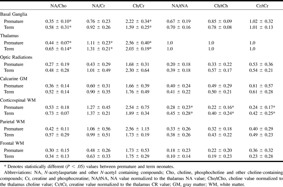
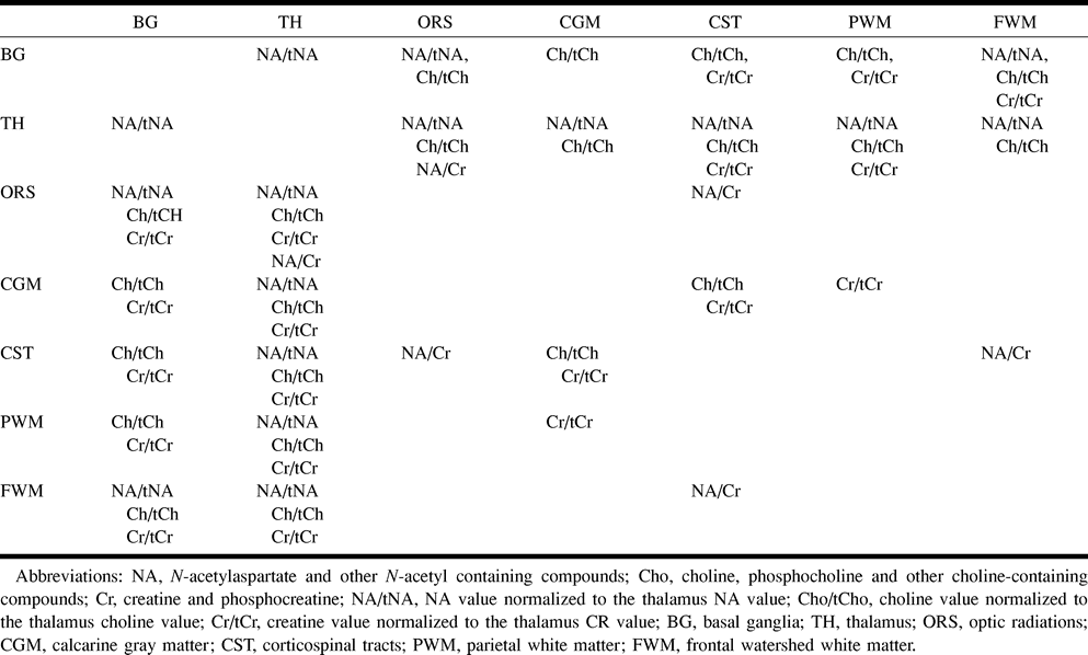
- TABLE 2:
Premature neonates—MR spectroscopy imaging ratios significantly (P < .05) different between anatomic regions
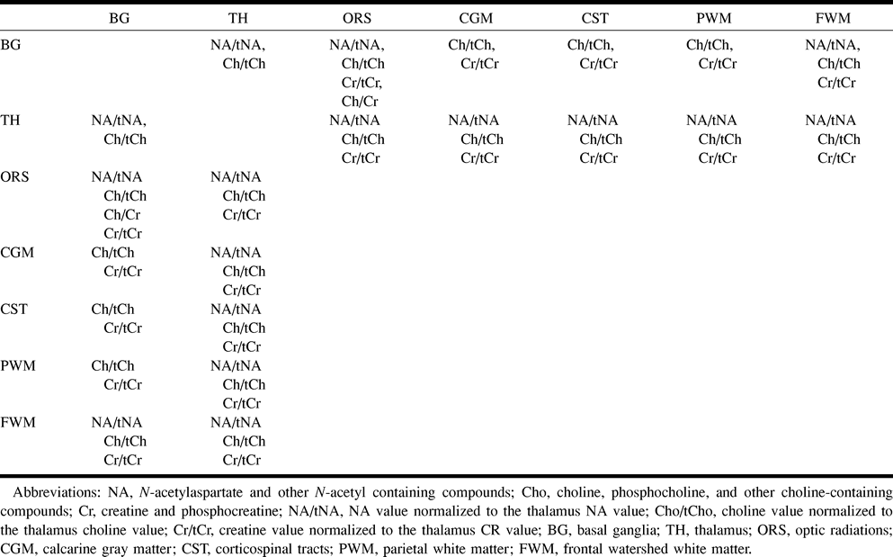
- TABLE 3:
Term neonates—MR spectroscopy imaging ratios significantly (P < .05) different between anatomic regions


