Advertisement

Main menu

  • Home
  • Content
    • Current Issue
    • Accepted Manuscripts
    • Article Preview
    • Past Issue Archive
    • Video Articles
    • AJNR Case Collection
    • Case of the Week Archive
    • Case of the Month Archive
    • Classic Case Archive
  • Special Collections
    • AJNR Awards
    • Low-Field MRI
    • Alzheimer Disease
    • ASNR Foundation Special Collection
    • Photon-Counting CT
    • View All
  • Multimedia
    • AJNR Podcasts
    • AJNR SCANtastic
    • Trainee Corner
    • MRI Safety Corner
    • Imaging Protocols
  • For Authors
    • Submit a Manuscript
    • Submit a Video Article
    • Submit an eLetter to the Editor/Response
    • Manuscript Submission Guidelines
    • Statistical Tips
    • Fast Publishing of Accepted Manuscripts
    • Graphical Abstract Preparation
    • Imaging Protocol Submission
    • Author Policies
  • About Us
    • About AJNR
    • Editorial Board
    • Editorial Board Alumni
  • More
    • Become a Reviewer/Academy of Reviewers
    • Subscribers
    • Permissions
    • Alerts
    • Feedback
    • Advertisers
    • ASNR Home

User menu

  • Alerts
  • Log in

Search

  • Advanced search
American Journal of Neuroradiology

American Journal of Neuroradiology

ASHNR American Society of Functional Neuroradiology ASHNR American Society of Pediatric Neuroradiology ASSR
  • Alerts
  • Log in

Advanced Search

  • Home
  • Content
    • Current Issue
    • Accepted Manuscripts
    • Article Preview
    • Past Issue Archive
    • Video Articles
    • AJNR Case Collection
    • Case of the Week Archive
    • Case of the Month Archive
    • Classic Case Archive
  • Special Collections
    • AJNR Awards
    • Low-Field MRI
    • Alzheimer Disease
    • ASNR Foundation Special Collection
    • Photon-Counting CT
    • View All
  • Multimedia
    • AJNR Podcasts
    • AJNR SCANtastic
    • Trainee Corner
    • MRI Safety Corner
    • Imaging Protocols
  • For Authors
    • Submit a Manuscript
    • Submit a Video Article
    • Submit an eLetter to the Editor/Response
    • Manuscript Submission Guidelines
    • Statistical Tips
    • Fast Publishing of Accepted Manuscripts
    • Graphical Abstract Preparation
    • Imaging Protocol Submission
    • Author Policies
  • About Us
    • About AJNR
    • Editorial Board
    • Editorial Board Alumni
  • More
    • Become a Reviewer/Academy of Reviewers
    • Subscribers
    • Permissions
    • Alerts
    • Feedback
    • Advertisers
    • ASNR Home
  • Follow AJNR on Twitter
  • Visit AJNR on Facebook
  • Follow AJNR on Instagram
  • Join AJNR on LinkedIn
  • RSS Feeds

AJNR Awards, New Junior Editors, and more. Read the latest AJNR updates

Research ArticleHead and Neck Imaging

CT Findings Associated with Eagle Syndrome

Ryan D. Murtagh, Jamie T. Caracciolo and Gaspar Fernandez
American Journal of Neuroradiology August 2001, 22 (7) 1401-1402;
Ryan D. Murtagh
  • Find this author on Google Scholar
  • Find this author on PubMed
  • Search for this author on this site
Jamie T. Caracciolo
  • Find this author on Google Scholar
  • Find this author on PubMed
  • Search for this author on this site
Gaspar Fernandez
  • Find this author on Google Scholar
  • Find this author on PubMed
  • Search for this author on this site
  • Article
  • Figures & Data
  • Info & Metrics
  • Responses
  • References
  • PDF
Loading

Abstract

Summary: Eagle syndrome is an aggregate of symptoms caused by an elongated ossified styloid process, the cause of which remains unclear. This is a rare finding that often goes undetected in the absence of radiographic studies. In this case, we present the diagnostic CT and lateral view plain film radiography findings of a 39-year-old woman with clinical evidence of Eagle syndrome.

Eagle syndrome can occur unilaterally or bilaterally and most frequently results in symptoms of dysphagia, headache, pain on rotation of the neck, pain on extension of the tongue, change in voice, and a sensation of hypersalivation (1, 2). We present rare and diagnostic radiographic evidence of this on both plain film radiographs and CT scans. Although well documented in otolaryngology literature and dentistry literature, this syndrome has not been reported in the radiology literature.

Case Report

A 39-year-old woman presented at the Otolaryngology Clinic complaining of dysphagia and vague throat discomfort that she had experienced continuously for slightly more than a year. She also complained of occasional “shooting” pains to her left ear that lasted only a couple of minutes and then disappeared. Her history was uneventful for any significant trauma, but she did recall that she had undergone a tonsillectomy approximately 7 years previously.

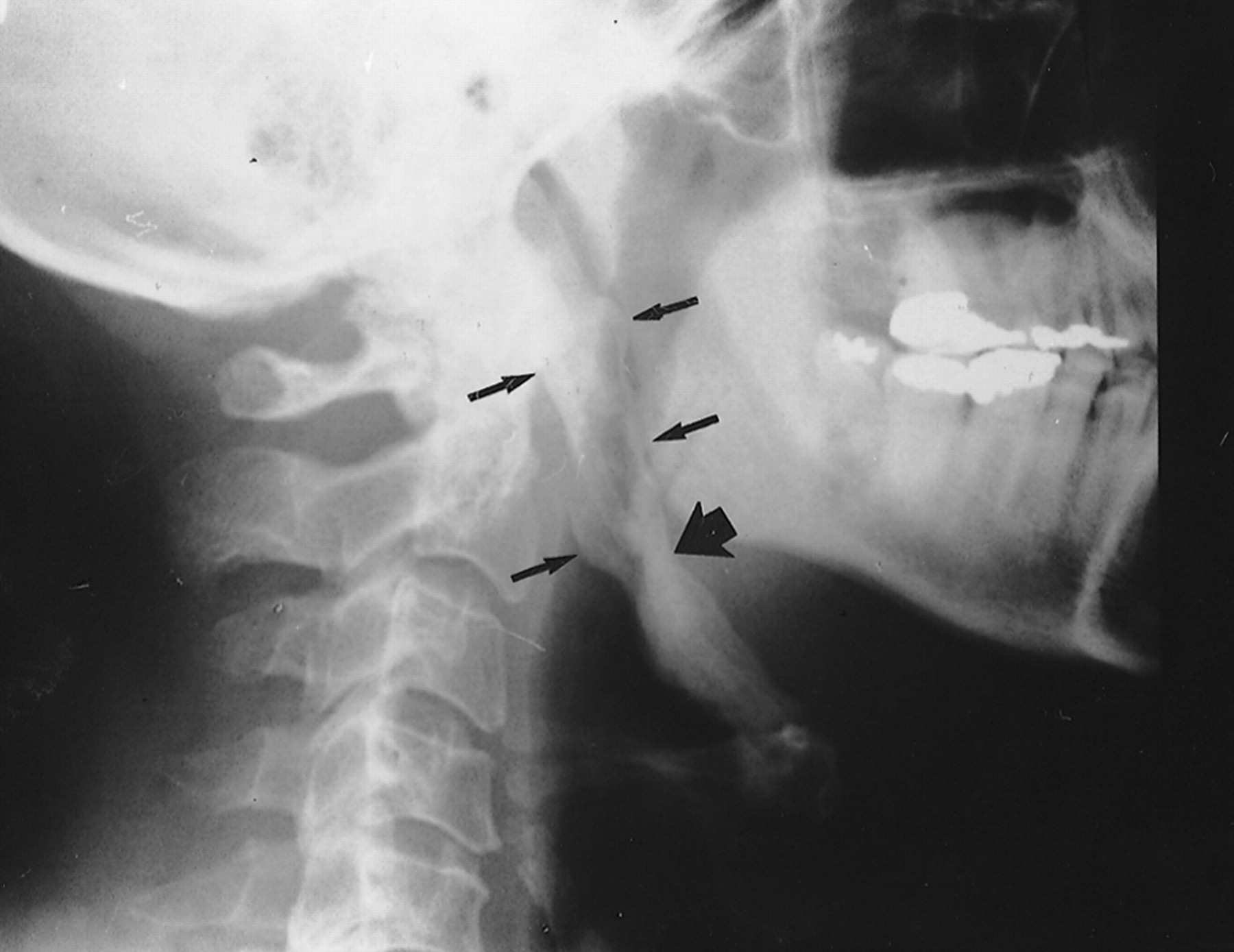
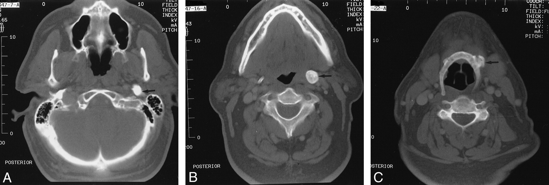
During the physical examination, a solid mass was felt on palpation of the left tonsillar fossa, and radiographic studies were ordered. Lateral view plain film radiographs showed an elongated ossified styloid process, with the left larger than the right (Fig 1). CT provides complimentary information to that of plain radiography, including definition of the relationship of calcifications to surrounding neck soft tissue structures in the axial plane (Fig 2). On the left, the thick calcified process extended from the stylomastoid foramen to the hyoid bone.

fig 1.
  • Download figure
  • Open in new tab
  • Download powerpoint
fig 1.

Lateral view plain radiograph of the cervical spine shows a large, heavily ossified structure extending from the base of the skull anterolaterally and caudally to the hyoid bone (small arrows)

fig 2.
  • Download figure
  • Open in new tab
  • Download powerpoint
fig 2.

Unenhanced high resolution bone window CT scans of the neck show the heavily ossified styloid process in the axial plane.

A, Origin is seen just in front of the stylomastoid foramen (arrow).

B, Midportion of calcified styloid process, at the widest aspect (arrow), can be seen.

C, Distal insertion of the calcified styloid into the anterolateral hyoid bone (arrow) can be seen.

The patient underwent surgical removal of a 4.7-cm styloid bone through an external approach. Surgery and hospital stay were uneventful, and there was no compression of surrounding nerves. The patient remained symptom free 9 months after surgery.

Discussion

Eagle syndrome is an aggregate of symptoms that includes recurrent throat pain, foreign body sensation, dysphagia, and/or facial pain as a direct result of an elongated styloid process or calcified stylohyoid ligament (3). Although approximately 4% of the population is thought to have an elongated styloid process, only a small percentage (between 4% and 10.3%) of this group is thought to actually be symptomatic (4). No data could be found to correlate degree of elongation of the styloid to severity of symptoms.

Diagnosis is made both radiographically and by physical examination. Palpation of the styloid process in the tonsillar fossa is indicative of elongated styloid in that processes of normal length are not normally palpable. Palpation of the tip of the styloid should exacerbate existing symptoms (4). If highly suspicious for Eagle syndrome, confirmation can be made by radiographic studies (4). Most frequently, a panoramic radiograph is used to determine whether the styloid process is elongated. In reviewing these radiographs, it should be noted that the normal length of the styloid in an adult is approximately 2.5 cm (5) whereas an elongated styloid is generally >3 cm in length (6). Although Eagle syndrome is thought to be caused by an elongated styloid process or calcified stylohyoid ligament, the presence of an elongated styloid process is not pathognomonic for Eagle syndrome because many patients with incidental findings of an elongated styloid process are asymptomatic. Lateral view radiographs of the skull can be substituted for panoramic radiographs (as in this case), and an anteroposterior view radiograph should be obtained to determine whether there is any lateral deviation of the styloid. As stated earlier, CT is useful in that it provides complementary information to that provided by plain radiographic studies.

The actual cause of the elongation is a poorly understood process. Several theories have been proposed: 1) congenital elongation of the styloid process due to persistence of a cartilaginous analog of the stylohyal (one of the embryologic precursors of the styloid), 2) calcification of the stylohyoid ligament by an unknown process, and 3) growth of osseous tissue at the insertion of the stylohyoid ligament (3).

The pathophysiological mechanism of symptoms is debated as well. Theories include the following: 1) traumatic fracture of the styloid process causing proliferation of granulation tissue, which places pressure on the surrounding structures (7); 2) compression of adjacent nerves, the glossopharyngeal, lower branch of the trigeminal, or chorda tympani; 3) degenerative and inflammatory changes in the tendonous portion of the stylohyoid insertion, called insertion tendonitis; 4) irritation of the pharyngeal mucosa by direct compression or post-tonsillectomy scarring (involves cranial nerves V, VII, IX, and X); and 5) impingement of the carotid vessels, producing irritation of the sympathetic nerves in the arterial sheath (3).

Treatment of Eagle syndrome is both surgical and nonsurgical. Nonsurgical treatments include reassurance, nonsteroidal anti-inflammatory medications, and steroid injections (1). Surgical treatment is by one of two methods. Otolaryngologist W. Eagle preferentially used a transpharyngeal approach through which the elongated portion of the styloid process was removed (8). Although this technique does avoid external scarring, it has been heavily criticized because of the increased risk of deep space neck infection and poor visualization of the surgical field (must be performed through the mouth) (2, 8). Alternatively, the elongated portion can be removed by an extraoral approach. Although both procedures are effective in removing an elongated styloid process, the extraoral approach is thought to be superior because of the decreased risk of deep space neck infection and better visualization of the surgical field (2, 8).

Footnotes

  • 1 Address reprint requests to Ryan D. Murtagh.

References

  1. ↵
    Baugh RF, Stocks RM. Eagle's syndrome: a reappraisal. Ear Nose Throat J 1993;72:341-344
    PubMed
  2. ↵
    Strauss M, Zohar Y, Laurian N. Elongated styloid process syndrome: intraoral versus external approach for styloid surgery. Laryngoscope 1979;95:976-979
  3. ↵
    Balbuena L, Hayes D, Ramirez SG, Johnson R. Eagle's syndrome (elongated styloid process). South Med J 1997;90:331-334
    CrossRefPubMed
  4. ↵
    Rechtweg JS, Wax MK. Eagle's syndrome: a review. Am J Otolaryngol 1998;19:316-321
    CrossRefPubMed
  5. ↵
    Eagle WW. Elongated styloid process: symptoms and treatment. Arch Otolaryngol 1958;64:172-176
  6. ↵
    Keur JJ, Campbell JP, McCarthy JF, Ralph WJ. The clinical significance of the elongated styloid process. Oral Surg Oral Med Oral Pathol 1986;61:399-404
    CrossRefPubMed
  7. ↵
    Balasubramanian S. The ossification of the stylohyoid ligament and its relation to facial pain. Br Dent J 1964;116:108-111
  8. ↵
    Chase DC, Zarmen A, Bigelow WC, McCoy JM. Eagle's syndrome: a comparison of intraoral versus extraoral surgical approaches. Oral Surg Oral Med Oral Path 1986;62:625-629
    CrossRefPubMed
  • Received July 7, 2000.
  • Copyright © American Society of Neuroradiology
View Abstract
PreviousNext
Back to top

In this issue

American Journal of Neuroradiology: 22 (7)
American Journal of Neuroradiology
Vol. 22, Issue 7
1 Aug 2001
  • Table of Contents
  • Index by author
Advertisement
Print
Download PDF
Email Article

Thank you for your interest in spreading the word on American Journal of Neuroradiology.

NOTE: We only request your email address so that the person you are recommending the page to knows that you wanted them to see it, and that it is not junk mail. We do not capture any email address.

Enter multiple addresses on separate lines or separate them with commas.
CT Findings Associated with Eagle Syndrome
(Your Name) has sent you a message from American Journal of Neuroradiology
(Your Name) thought you would like to see the American Journal of Neuroradiology web site.
CAPTCHA
This question is for testing whether or not you are a human visitor and to prevent automated spam submissions.
Cite this article
Ryan D. Murtagh, Jamie T. Caracciolo, Gaspar Fernandez
CT Findings Associated with Eagle Syndrome
American Journal of Neuroradiology Aug 2001, 22 (7) 1401-1402;

Citation Manager Formats

  • BibTeX
  • Bookends
  • EasyBib
  • EndNote (tagged)
  • EndNote 8 (xml)
  • Medlars
  • Mendeley
  • Papers
  • RefWorks Tagged
  • Ref Manager
  • RIS
  • Zotero
0 Responses
Respond to this article
Share
Bookmark this article
CT Findings Associated with Eagle Syndrome
Ryan D. Murtagh, Jamie T. Caracciolo, Gaspar Fernandez
American Journal of Neuroradiology Aug 2001, 22 (7) 1401-1402;
del.icio.us logo Twitter logo Facebook logo Mendeley logo
  • Tweet Widget
  • Facebook Like
  • Google Plus One

Jump to section

  • Article
    • Abstract
    • Case Report
    • Discussion
    • Footnotes
    • References
  • Figures & Data
  • Info & Metrics
  • Responses
  • References
  • PDF

Related Articles

  • No related articles found.
  • PubMed
  • Google Scholar

Cited By...

  • Eagle syndrome and carotid artery dissection: a rare skull base cause of stroke
  • Eagle Syndrome: an underdiagnosed cause of orofacial pain
  • Eagle Syndrome: A Unique Cause of Carotid Bleeding
  • Horners syndrome secondary to carotid artery dissection caused by Eagle syndrome
  • Massively enlarged styloid process presenting as a submandibular mass
  • Dysphagia and Submandibular Swelling
  • Crossref
  • Google Scholar

This article has not yet been cited by articles in journals that are participating in Crossref Cited-by Linking.

More in this TOC Section

  • MRI of the Parasellar Ligaments
  • ASL Sensitivity for Head and Neck Paraganglioma
  • Post SRS Peritumoral Hyperintense Signal of VSs
Show more HEAD AND NECK IMAGING

Similar Articles

Advertisement

Indexed Content

  • Current Issue
  • Accepted Manuscripts
  • Article Preview
  • Past Issues
  • Editorials
  • Editor's Choice
  • Fellows' Journal Club
  • Letters to the Editor
  • Video Articles

Cases

  • Case Collection
  • Archive - Case of the Week
  • Archive - Case of the Month
  • Archive - Classic Case

More from AJNR

  • Trainee Corner
  • Imaging Protocols
  • MRI Safety Corner
  • Book Reviews

Multimedia

  • AJNR Podcasts
  • AJNR Scantastics

Resources

  • Turnaround Time
  • Submit a Manuscript
  • Submit a Video Article
  • Submit an eLetter to the Editor/Response
  • Manuscript Submission Guidelines
  • Statistical Tips
  • Fast Publishing of Accepted Manuscripts
  • Graphical Abstract Preparation
  • Imaging Protocol Submission
  • Evidence-Based Medicine Level Guide
  • Publishing Checklists
  • Author Policies
  • Become a Reviewer/Academy of Reviewers
  • News and Updates

About Us

  • About AJNR
  • Editorial Board
  • Editorial Board Alumni
  • Alerts
  • Permissions
  • Not an AJNR Subscriber? Join Now
  • Advertise with Us
  • Librarian Resources
  • Feedback
  • Terms and Conditions
  • AJNR Editorial Board Alumni

American Society of Neuroradiology

  • Not an ASNR Member? Join Now

© 2025 by the American Society of Neuroradiology All rights, including for text and data mining, AI training, and similar technologies, are reserved.
Print ISSN: 0195-6108 Online ISSN: 1936-959X

Powered by HighWire