Advertisement

Main menu

  • Home
  • Content
    • Current Issue
    • Accepted Manuscripts
    • Article Preview
    • Past Issue Archive
    • Video Articles
    • AJNR Case Collection
    • Case of the Week Archive
    • Case of the Month Archive
    • Classic Case Archive
  • Special Collections
    • AJNR Awards
    • Low-Field MRI
    • Alzheimer Disease
    • ASNR Foundation Special Collection
    • Photon-Counting CT
    • View All
  • Multimedia
    • AJNR Podcasts
    • AJNR SCANtastic
    • Trainee Corner
    • MRI Safety Corner
    • Imaging Protocols
  • For Authors
    • Submit a Manuscript
    • Submit a Video Article
    • Submit an eLetter to the Editor/Response
    • Manuscript Submission Guidelines
    • Statistical Tips
    • Fast Publishing of Accepted Manuscripts
    • Graphical Abstract Preparation
    • Imaging Protocol Submission
    • Author Policies
  • About Us
    • About AJNR
    • Editorial Board
    • Editorial Board Alumni
  • More
    • Become a Reviewer/Academy of Reviewers
    • Subscribers
    • Permissions
    • Alerts
    • Feedback
    • Advertisers
    • ASNR Home

User menu

  • Alerts
  • Log in

Search

  • Advanced search
American Journal of Neuroradiology

American Journal of Neuroradiology

ASHNR American Society of Functional Neuroradiology ASHNR American Society of Pediatric Neuroradiology ASSR
  • Alerts
  • Log in

Advanced Search

  • Home
  • Content
    • Current Issue
    • Accepted Manuscripts
    • Article Preview
    • Past Issue Archive
    • Video Articles
    • AJNR Case Collection
    • Case of the Week Archive
    • Case of the Month Archive
    • Classic Case Archive
  • Special Collections
    • AJNR Awards
    • Low-Field MRI
    • Alzheimer Disease
    • ASNR Foundation Special Collection
    • Photon-Counting CT
    • View All
  • Multimedia
    • AJNR Podcasts
    • AJNR SCANtastic
    • Trainee Corner
    • MRI Safety Corner
    • Imaging Protocols
  • For Authors
    • Submit a Manuscript
    • Submit a Video Article
    • Submit an eLetter to the Editor/Response
    • Manuscript Submission Guidelines
    • Statistical Tips
    • Fast Publishing of Accepted Manuscripts
    • Graphical Abstract Preparation
    • Imaging Protocol Submission
    • Author Policies
  • About Us
    • About AJNR
    • Editorial Board
    • Editorial Board Alumni
  • More
    • Become a Reviewer/Academy of Reviewers
    • Subscribers
    • Permissions
    • Alerts
    • Feedback
    • Advertisers
    • ASNR Home
  • Follow AJNR on Twitter
  • Visit AJNR on Facebook
  • Follow AJNR on Instagram
  • Join AJNR on LinkedIn
  • RSS Feeds

AJNR Awards, New Junior Editors, and more. Read the latest AJNR updates

Research ArticleHead and Neck Imaging

Imaging of Hydrogel Episcleral Buckle Fragmentation as a Late Complication After Retinal Reattachment Surgery

John I. Lane, James G. Randall, Norbert G. Campeau, Paul K. Overland, Colin A. McCannel and Thomas A. Matsko
American Journal of Neuroradiology June 2001, 22 (6) 1199-1202;
John I. Lane
  • Find this author on Google Scholar
  • Find this author on PubMed
  • Search for this author on this site
James G. Randall
  • Find this author on Google Scholar
  • Find this author on PubMed
  • Search for this author on this site
Norbert G. Campeau
  • Find this author on Google Scholar
  • Find this author on PubMed
  • Search for this author on this site
Paul K. Overland
  • Find this author on Google Scholar
  • Find this author on PubMed
  • Search for this author on this site
Colin A. McCannel
  • Find this author on Google Scholar
  • Find this author on PubMed
  • Search for this author on this site
Thomas A. Matsko
  • Find this author on Google Scholar
  • Find this author on PubMed
  • Search for this author on this site
  • Article
  • Figures & Data
  • Info & Metrics
  • Responses
  • References
  • PDF
Loading

Article Figures & Data

Figures

  • Tables
  • fig 1.
    • Download figure
    • Open in new tab
    • Download powerpoint
    fig 1.

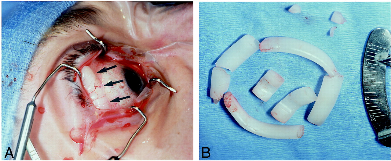
    Case 4. A, Intraoperative photograph shows extruded, swollen band surrounding right eye (arrows).B, Surgical specimen shows expanded fragments of the hydrolyzed buckle material

  • fig 2.
    • Download figure
    • Open in new tab
    • Download powerpoint
    fig 2.

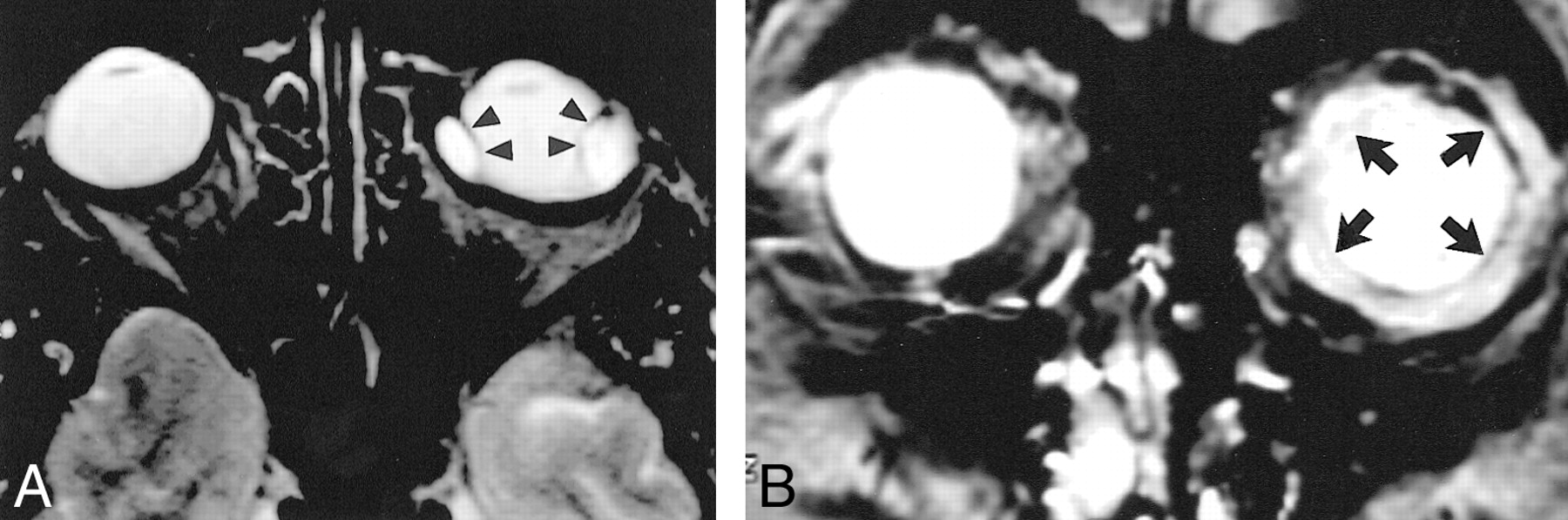
    Case 3. A and B, Contrast-enhanced axial (A) and coronal (B) CT scans show circumferential soft tissue mass surrounding the globe with focal, dystrophic calcification and a peripherally enhancing rim (arrows)

  • fig 3.
    • Download figure
    • Open in new tab
    • Download powerpoint
    fig 3.

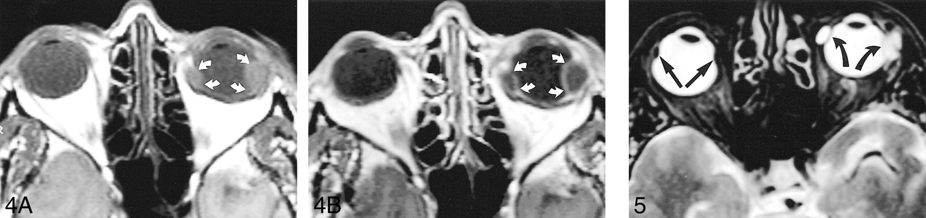
    A and B, T2-weighted axial (A, case 2) and coronal (B, case 4) MR images (2000/80/1) show circumferential, hyperintense mass (arrowheads, arrows) surrounding the globe. Increased T2 signal reflects increased water content and swelling of hydrolyzed buckle material

  • fig 4.
    • Download figure
    • Open in new tab
    • Download powerpoint
    fig 4.

    Case 2.

    A and B, Noncontrast (A) and contrast-enhanced (B) T1-weighted axial images (500/15/1) show soft tissue mass isointense with muscle, with peripheral rim of enhancement (arrows) corresponding to thick, fibrous capsule and foreign body reaction seen at pathologic examination.fig 5. Case 1. T2-weighted axial image (2000/80/1) shows normal profile of an uncomplicated encircling silicone band in the right orbit (straight arrows) as compared with hydrolyzed, fragmented hydrogel buckle in the left orbit (curved arrows). An uncomplicated hydrogel band would be expected to have a similar profile to that of the silicone band

Tables

  • Figures
  • Table1

    Hydrogel fragmentation: clinical and imaging data

PreviousNext
Back to top

In this issue

American Journal of Neuroradiology: 22 (6)
American Journal of Neuroradiology
Vol. 22, Issue 6
1 Jun 2001
  • Table of Contents
  • Index by author
Advertisement
Print
Download PDF
Email Article

Thank you for your interest in spreading the word on American Journal of Neuroradiology.

NOTE: We only request your email address so that the person you are recommending the page to knows that you wanted them to see it, and that it is not junk mail. We do not capture any email address.

Enter multiple addresses on separate lines or separate them with commas.
Imaging of Hydrogel Episcleral Buckle Fragmentation as a Late Complication After Retinal Reattachment Surgery
(Your Name) has sent you a message from American Journal of Neuroradiology
(Your Name) thought you would like to see the American Journal of Neuroradiology web site.
CAPTCHA
This question is for testing whether or not you are a human visitor and to prevent automated spam submissions.
Cite this article
John I. Lane, James G. Randall, Norbert G. Campeau, Paul K. Overland, Colin A. McCannel, Thomas A. Matsko
Imaging of Hydrogel Episcleral Buckle Fragmentation as a Late Complication After Retinal Reattachment Surgery
American Journal of Neuroradiology Jun 2001, 22 (6) 1199-1202;

Citation Manager Formats

  • BibTeX
  • Bookends
  • EasyBib
  • EndNote (tagged)
  • EndNote 8 (xml)
  • Medlars
  • Mendeley
  • Papers
  • RefWorks Tagged
  • Ref Manager
  • RIS
  • Zotero
0 Responses
Respond to this article
Share
Bookmark this article
Imaging of Hydrogel Episcleral Buckle Fragmentation as a Late Complication After Retinal Reattachment Surgery
John I. Lane, James G. Randall, Norbert G. Campeau, Paul K. Overland, Colin A. McCannel, Thomas A. Matsko
American Journal of Neuroradiology Jun 2001, 22 (6) 1199-1202;
del.icio.us logo Twitter logo Facebook logo Mendeley logo
  • Tweet Widget
  • Facebook Like
  • Google Plus One

Jump to section

  • Article
    • Abstract
    • Case Reports
    • Discussion
    • Footnotes
    • References
  • Figures & Data
  • Info & Metrics
  • Responses
  • References
  • PDF

Related Articles

  • No related articles found.
  • PubMed
  • Google Scholar

Cited By...

  • No citing articles found.
  • Crossref
  • Google Scholar

This article has not yet been cited by articles in journals that are participating in Crossref Cited-by Linking.

More in this TOC Section

  • FDG Uptake and Thyroid Cancer Post-BRAF Therapy
  • Normal Facial Nerve Enhancement on VIBE Sequence
  • Comprehensive Review of Inner Ear Anatomy on Photon-Counting CT
Show more Head and Neck Imaging

Similar Articles

Advertisement

Indexed Content

  • Current Issue
  • Accepted Manuscripts
  • Article Preview
  • Past Issues
  • Editorials
  • Editor's Choice
  • Fellows' Journal Club
  • Letters to the Editor
  • Video Articles

Cases

  • Case Collection
  • Archive - Case of the Week
  • Archive - Case of the Month
  • Archive - Classic Case

More from AJNR

  • Trainee Corner
  • Imaging Protocols
  • MRI Safety Corner

Multimedia

  • AJNR Podcasts
  • AJNR Scantastics

Resources

  • Turnaround Time
  • Submit a Manuscript
  • Submit a Video Article
  • Submit an eLetter to the Editor/Response
  • Manuscript Submission Guidelines
  • Statistical Tips
  • Fast Publishing of Accepted Manuscripts
  • Graphical Abstract Preparation
  • Imaging Protocol Submission
  • Evidence-Based Medicine Level Guide
  • Publishing Checklists
  • Author Policies
  • Become a Reviewer/Academy of Reviewers
  • News and Updates

About Us

  • About AJNR
  • Editorial Board
  • Editorial Board Alumni
  • Alerts
  • Permissions
  • Not an AJNR Subscriber? Join Now
  • Advertise with Us
  • Librarian Resources
  • Feedback
  • Terms and Conditions
  • AJNR Editorial Board Alumni

American Society of Neuroradiology

  • Not an ASNR Member? Join Now

© 2025 by the American Society of Neuroradiology All rights, including for text and data mining, AI training, and similar technologies, are reserved.
Print ISSN: 0195-6108 Online ISSN: 1936-959X

Powered by HighWire