
Article Figures & Data
Figures
- fig 1.
The petrosquamosal sinus.
A, Posterior coronal section of the temporal bone. Petrosquamosal suture between the squama (laterally) (2) and the petrous bone (medially) (3). PPS (1) is illustrated in the upper portion of the petrosquamosal suture. Residual arterial and venous vascular tracts (4) probably are the cause of bleeding during transmastoid or translabyrinthine procedures.
B, Axial view showing the origin of the PSS and its relation with the middle meningeal venous network (from Padget). 1, transverse sinus; 2, sigmoid sinus; 3, inferior petrosal sinus; 4, superior petrosal sinus; 5, petrosquamosal sinus; 6, middle meningeal sinus draining into the cavernous sinus and the pterygoid venous plexus via the foramen ovale (small arrow); 7, retromandibular vein draining into the external jugular vein; 8, sinus communicans.
- fig 2.
Case 1. Axial HRCT section passing through the upper portion of the TMJ and the external auditory canal shows a convex canal (arrowhead) in the premeatic squama that ends at the superior aspect of the TMJ (arrow)
- fig 3.
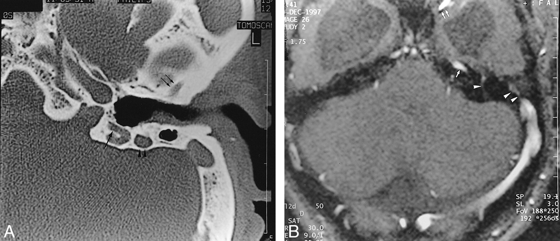
Case 3.
A, Axial HRCT section passing through the attici shows a sulcus (arrows) about 2 mm thick, originating from the anterior portion of the transverse sinus and passing anteriorly through the lateral portion of the petrous bone. Also shown are middle ear hypoplasia and posterior semicircular canal aplasia.
B, Axial CT section 5 mm below A shows the PSS entering into a retroarticulare foramen. Note the dehiscence of the genu of the facial nerve (white arrowhead).
C, HRCT 10 mm below B shows aplasia of the jugular foramen. Posterior condylar and mastoid canals (arrow) are illustrated.
- fig 4.
Case 4.
A, Axial HRCT shows anterior (double arrows) and posterior (large arrow) PSS sulcus of the temporal bone. The mastoid portion of the facial canal is enlarged (arrowheads).
B, MR venography shows absence of the right transverse sinus. A large left contralateral transverse sinus is present with flow signals consistent with the superior petrosal sinus (arrow), pterygoid plexus (double arrows), transpetrous vein (arrowhead) and PPS (double arrowheads). Enlargement of the mastoid portion of the facial canal relates to an excessively developed facial vein (not shown). Note the presence of hydrocephalus and “kissing carotid arteries.”
- fig 5.
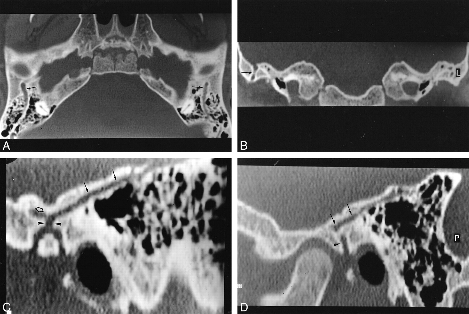
Case 5.
A, Axial HRCT section shows bilateral PSS sulci (arrows) located on the upper portion of the petrous bone, coursing posteriorly from the union of the transverse and sigmoid sinuses to the superior portion of the TMJ.
B, Coronal CT section shows a vertical transquamous pathway of the right PSS (arrow).
C and D, Sagittal reformatted CT sections show the pathway of the PSS ending in the supraglenoid foramen (arrowheads) on the left side (C) and the foramen retroarticulare (arrowhead) on the right side. There is a left anterior collateral pathway (open arrow), suggestive of a sinus communicans.
- fig 6.
Case 5.
A, Upper axial HRCT section passing through the attici shows an anterior, well-defined hole (arrow) located in the petrosquamosal suture (arrow).
B, Sagittal reformatted section shows the pathway of the petrosquamosal sinus(arrow) located on the upper portion of the temporal bone.
C, Postcontrast, T1-weighted image shows a tubular enhancement (white arrowhead) of the petrosquamosal sinus with signal intensity similar to that of other venous structures.


Tables

Clinical and anatomic characteristics of patients with a PSS


