
Article Figures & Data
Figures
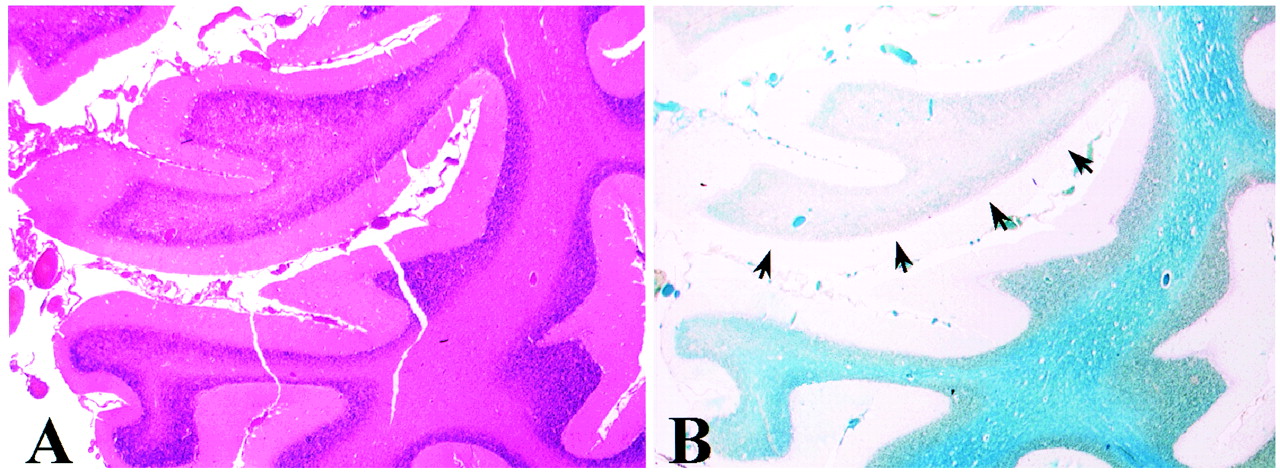
- fig 1.
Hematoxylin-eosin (A) and luxol fast blue (B) staining of the lesion seen in the cerebellum of case 2. Hematoxylin-eosin staining shows intact gray matter and some degenerative changes of the white matter. Luxol fast blue staining of myelin (blue) shows a restricted, focal area of demyelination (arrows)
- fig 2.
PD (3000/40/1 [TR/TE/TI]) (first image) and non-contrast FLAIR (9002/200/2200/2 [TR/TE/TI/excitations]) MR images of case 1 at indicated times after admission to the hospital. The first scan, performed at 2 days, does not reveal any intraparenchymal lesions. In the second scan done 1 week after admission, when the patient was unconscious and connected to a respirator, only two weakly high-signal areas are located in the basal ganglia, which by 3 weeks have grown (arrows). Despite significant recovery, several new lesions are seen in the periventricular white matter 1 month after admission (arrows). By 2 months, almost all the lesions are resolved
- fig 3.
Non-contrast FLAIR (9002/200/2200/2) MR images of case 2 at 3 and 6 weeks after admission. MR imaging performed at 3 weeks does not show any focal lesions, whereas the last MR scan shows a few high-density lesions located in the deep periventricular white matter (arrows) in addition to a subcortical infarct in the left frontoparietal lobe (arrowhead). A large, high-density lesion is located in the right cerebellar hemisphere
- fig 4.
T2-weighted (4000/115/1 [TR/TE/excitation]) MR images of case 3. The first MR scan taken 2 days after admission shows a large lesion in the right centrum semiovale (arrows), as well as in the periventricular white matter and basal ganglia (arrowheads). By 2 weeks, these lesions have grown despite steroid treatment and slight improvement in the patient's condition. By 1 month, some petecchial hemorrhage is seen in the lesions (arrows). On the last MR scan, performed 4 months after admission to the hospital, the lesions are somewhat decreased in size. Note the mild cortical pseudoatrophy likely caused by the steroid treatment
- fig 5.
Non-contrast FLAIR (9002/200/2200/2) images of case 4. The first MR scan shows a few focal ischemic white matter lesions located in the right frontal and left occipital lobes. The second scan, performed 2 months after admission, reveals several new lesions located in the deep periventricular white matter. These lesions do not show any major resolution in the follow-up scans performed at 4 and 8 months


Tables
In this issue


Jump to section
Related Articles
Cited By...
- Serial Imaging of Virus-Associated Necrotizing Disseminated Acute Leukoencephalopathy (VANDAL) in COVID-19
- Acute disseminated encephalomyelitis in 228 patients: A retrospective, multicenter US study
- Acute disseminated encephalomyelitis (ADEM) following a H3N3 parainfluenza virus infection in a pregnant asthmatic woman with respiratory failure
- Teaching NeuroImages: MRI time lag with acute disseminated encephalomyelitis
- Multiple cerebral enhancing lesions in an acutely ill child
- Acute disseminated encephalomyelitis